. 2009 Jul 15;17(14):5189-97.
doi: 10.1016/j.bmc.2009.05.047. Epub 2009 May 27.

Computational selection of inhibitors of Abeta aggregation and neuronal toxicity

Affiliations

Computational selection of inhibitors of Abeta aggregation and neuronal toxicity

Deliang Chen et al. Bioorg Med Chem. .

Abstract

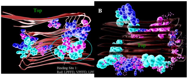
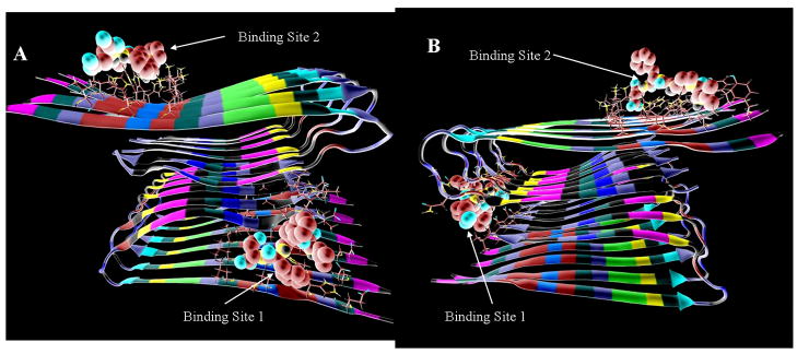
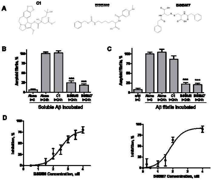
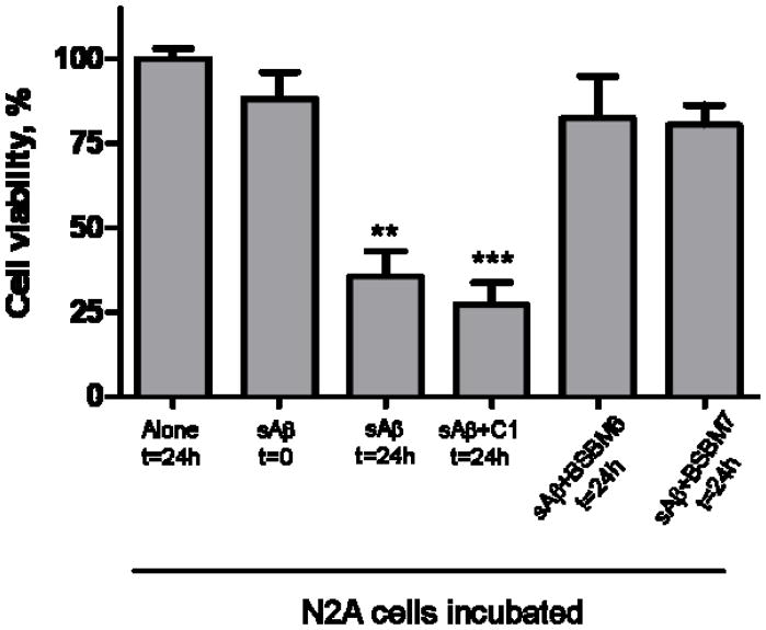
Alzheimer's disease (AD) is characterized by the cerebral accumulation of misfolded and aggregated amyloid-beta protein (Abeta). Disease symptoms can be alleviated, in vitro and in vivo, by 'beta-sheet breaker' pentapeptides that reduce plaque load. However the peptide nature of these compounds, made them biologically unstable and unable to penetrate membranes with high efficiency. The main goal of this study was to use computational methods to identify small molecule mimetics with better drug-like properties. For this purpose, the docked conformations of the active peptides were used to identify compounds with similar activities. A series of related beta-sheet breaker peptides were docked to solid state NMR structures of a fibrillar form of Abeta. The lowest energy conformations of the active peptides were used to design three dimensional (3D)-pharmacophores, suitable for screening the NCI database with Unity. Small molecular weight compounds with physicochemical features and a conformation similar to the active peptides were selected, ranked by docking and biochemical parameters. Of 16 diverse compounds selected for experimental screening, 2 prevented and reversed Abeta aggregation at 2-3microM concentration, as measured by Thioflavin T (ThT) fluorescence and ELISA assays. They also prevented the toxic effects of aggregated Abeta on neuroblastoma cells. Their low molecular weight and aqueous solubility makes them promising lead compounds for treating AD.


Figures

Fig. 1
Fig. 1
Docking positions of 30 β-sheet breaker pentapeptides on two fibril structures of Aβ, Aβ40m2_1.pdb (left), and Aβ40p2_1.pdb (right), colored according to their activities in preventing Aβ aggregation (red, highest relative activities > 70% of LPFFD activity, or ln(Act) > 4.25; green, lowest relative activities <=7% or ln(Act) < 2.0; yellow have intermediate activities (70> Relative activity > 8; or 4.25 > (ln(Act) < 2). The top of the fibril is indicated for orientation purposes.
Fig. 2
Fig. 2
Fig. 2. A, B) Preferred docking sites for LPFFD (shown with CPK coloring) to the fibril stuctures A) Aβ40m2_1.pdb and B) Aβ40p2_1.pdb.
Fig. 3
Fig. 3
Pharmacophores designed based on the docking model of LPFFD to A, B) docking sites 1, 2 of the fibril structure Aβ40m2_1. C,D) docking sites 1,2 of the fibril structure Aβ40p2_1.
Fig. 4
Fig. 4
Structures and NCI number of NCI compounds selected for bioassays.
Fig. 5
Fig. 5
In vitro activity of selected compounds on Aβ fibrillogenesis. A: Chemical structure of two putative β-sheet breaker mimetics: β-sheet breaker mimetic 6 (BSBM6) and β-sheet breaker mimetic 7 (BSBM7), and the inactive C1 control compound. B: The effect of selected compounds on Aβ amyloid formation was studied by incubation of soluble Aβ1–42 in the absence or the presence of an equimolar concentration of the molecules. Amyloid formation was measured by ThT, as described in Methods. Results are expressed as a percentage of fibrils formed by the peptide incubated alone for 24h. The data was analyzed by student-t test by comparing each result with the control of Aβ incubated alone. ***, P<0.001. C: The ability of the compounds to disassemble pre-form fibrils was assessed by incubation of the molecules with Aβ aggregates made by pre-incubation of Aβ1–42 alone. The amount of fibrils before and after incubation with the compounds was studied by ThT. Results are expressed as a percentage of fibrils remaining after incubation alone for 24h. The data was analyzed by student-t test by comparing with the control of fibrils incubated alone. ***, P<0.001. D: The concentration-dependent effect of BSBM6 and BSBM7 on Aβ aggregation was studied by incubating soluble Aβ1–42 with various quantities of the compounds for 24h at 37°C. Formation of aggregates was quantified by sedimentation assay, followed by ELISA, as described in Methods. The data in panels B, C and D corresponds to the average ± standard error of three different experiments.
Fig. 6
Fig. 6
The activity of selected compounds on preventing Aβ neurotoxicity was studied in cell cultures. N2A cells were treated with soluble Aβ1–42 (3.3 μM) which was pre-incubated for 48h either alone or in the presence of 3.3 μM of BSBM6, BSBM7 or C1. After 24h incubation with the mixture peptide/compounds, cell viability was evaluated by the MTS assay. Data represent the average ± standard error of 3 determinations and is expressed as the percentage of viability obtained when cells were incubated alone for 24h. Data for the compounds was statistically compared with cells incubated alone suing the student-t test. ** P<0.01; *** P<0.001.
Figure 7
Figure 7
Comparison, based on the the best ranked conformations (lowest binding scores) from AutoDock, of the type of hydrogen bonds formed between atoms of the bad aggregation inhibitors (A,B) and good ones (C,D) and the fibril. The side chains of the pentapeptides LTicFFD (inactive) and LPFFD (active) are not shown. The hydrogen bonds between ligands and fibril are indicated by red arrows (those that would be expected to break or weaken the β sheet hydrogen bond network of the fibril) and blue arrows (those expected to enhance the stability of the e β sheet hydrogen bond network). Yellow arrows indicate those close interactions that should not affect β sheet hydrogen bond network.

References

    1. Hendrie HC. Epidemiology of dementia and Alzheimer’s disease. Am J Geriatr Psychiatry. 1998;6:S3–18. - PubMed
    1. Alzheimer A, Stelzmann RA, Schnitzlein HN, Murtagh FR. An English translation of Alzheimer’s 1907 paper, “Uber eine eigenartige Erkankung der Hirnrinde”. Clin Anat. 1995;8(6):429–431. - PubMed
    1. Thal DR, Rub U, Orantes M, Braak H. Phases of A beta-deposition in the human brain and its relevance for the development of AD. Neurology. 2002;58(12):1791–1800. - PubMed
    1. Braak H, Braak E. Staging of Alzheimer’s disease-related neurofibrillary changes. Neurobiol Aging. 1995;16(3):271–278. - PubMed
    1. Selkoe DJ. Toward a comprehensive theory for Alzheimer’s disease. Hypothesis: Alzheimer’s disease is caused by the cerebral accumulation and cytotoxicity of amyloid beta-protein. Ann N Y Acad Sci. 2000;924:17–25. - PubMed

Publication types

MeSH terms

LinkOut - more resources