. 2009 Jul 2;5(1):97-110.
doi: 10.1016/j.stem.2009.05.023. Epub 2009 Jun 18.

Gene targeting of a disease-related gene in human induced pluripotent stem and embryonic stem cells

Affiliations

Gene targeting of a disease-related gene in human induced pluripotent stem and embryonic stem cells

Jizhong Zou et al. Cell Stem Cell. .

Abstract

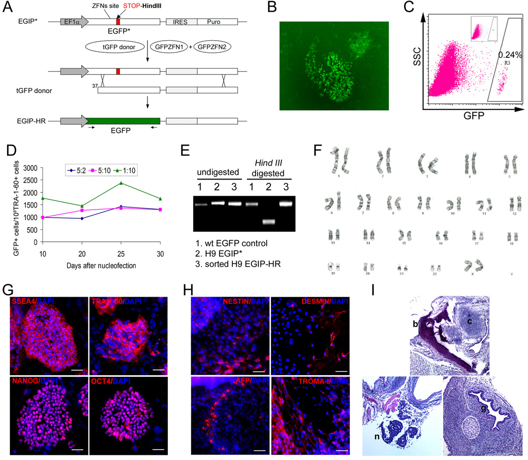
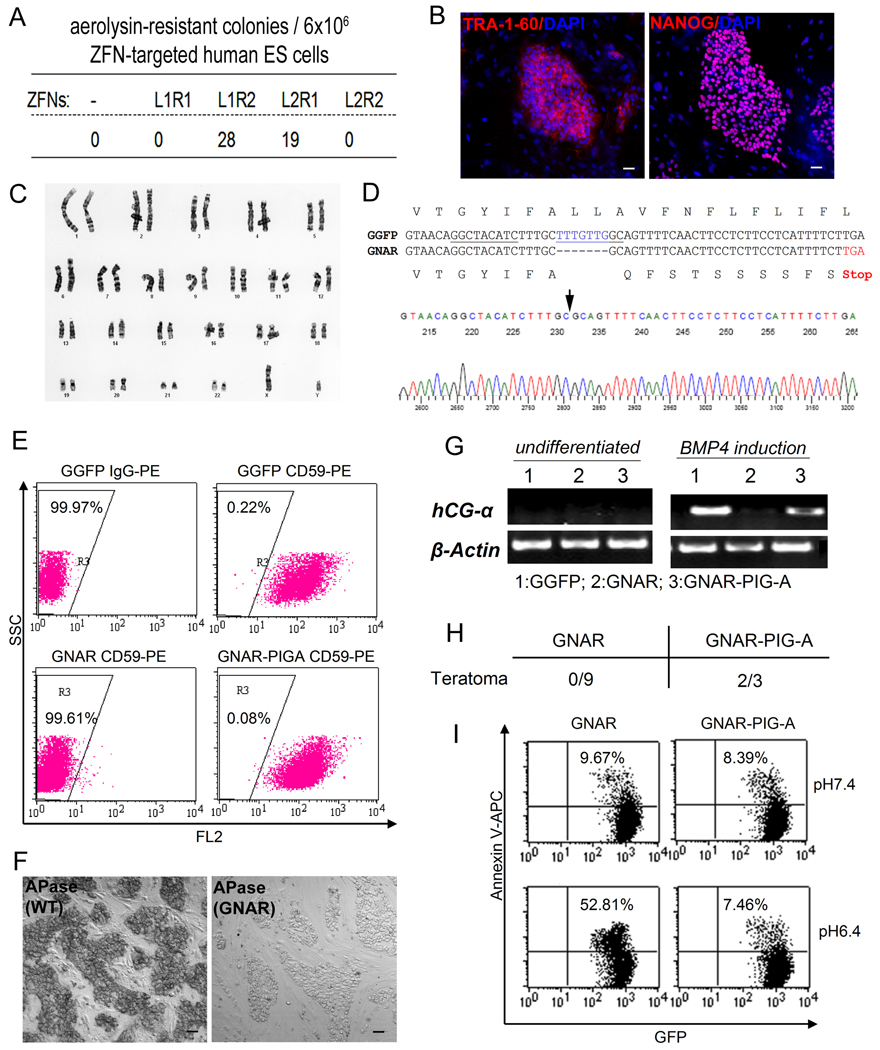
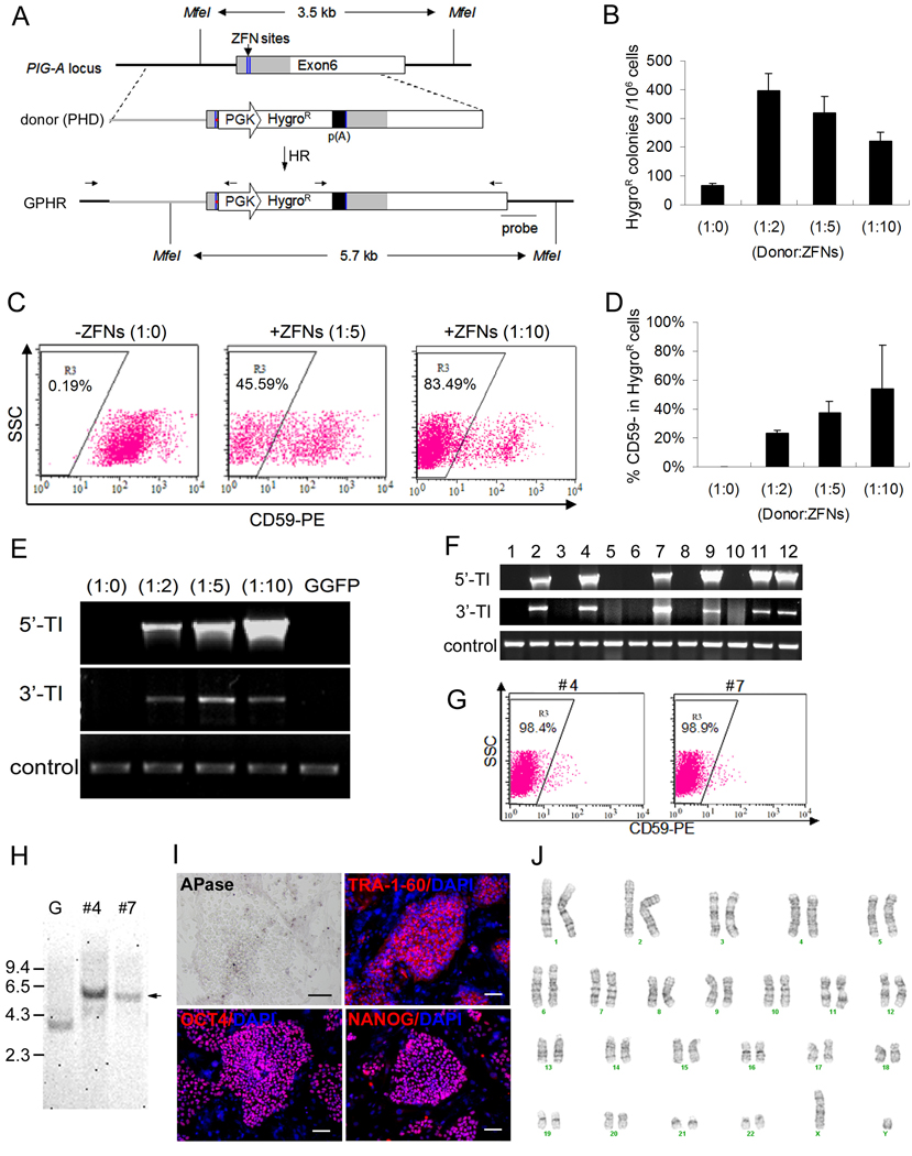
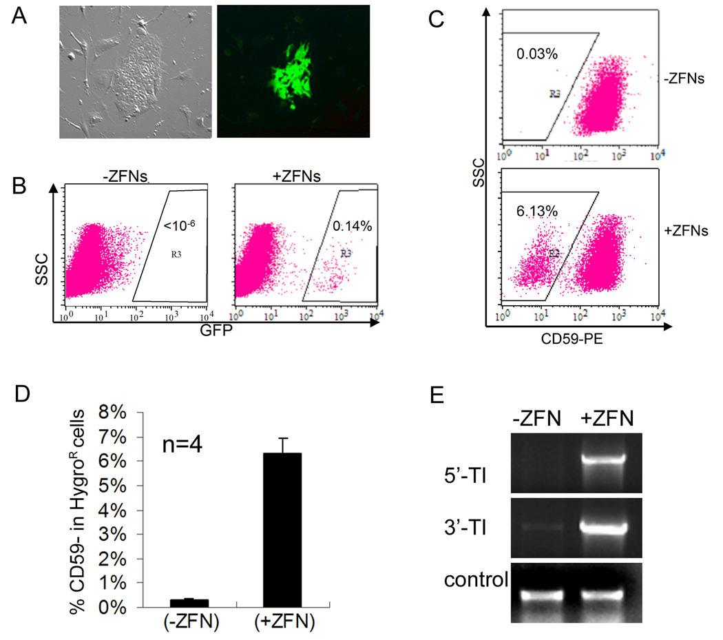
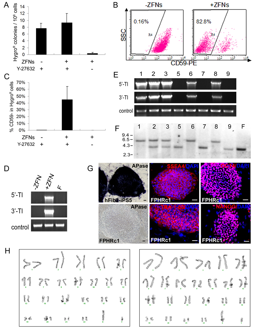
We report here homologous recombination (HR)-mediated gene targeting of two different genes in human iPS cells (hiPSCs) and human ES cells (hESCs). HR-mediated correction of a chromosomally integrated mutant GFP reporter gene reaches efficiencies of 0.14%-0.24% in both cell types transfected by donor DNA with plasmids expressing zinc finger nucleases (ZFNs). Engineered ZFNs that induce a sequence-specific double-strand break in the GFP gene enhanced HR-mediated correction by > 1400-fold without detectable alterations in stem cell karyotypes or pluripotency. Efficient HR-mediated insertional mutagenesis was also achieved at the endogenous PIG-A locus, with a > 200-fold enhancement by ZFNs targeted to that gene. Clonal PIG-A null hESCs and iPSCs with normal karyotypes were readily obtained. The phenotypic and biological defects were rescued by PIG-A transgene expression. Our study provides the first demonstration of HR-mediated gene targeting in hiPSCs and shows the power of ZFNs for inducing specific genetic modifications in hiPSCs, as well as hESCs.


Figures

Figure 1
Figure 1. Correction of a mutant EGFP reporter gene by ZFN-mediated gene targeting
(A) Scheme of the EGFP gene correction strategy. An enhanced GFP gene was mutated (EGFP*) by the insertion of a 35-bp fragment containing a translational stop codon and a Hind III site, positioned 12-bp downstream of the GFP ZFN target site. The defective EGFP(*) transgene was delivered and integrated into human cells by a lentiviral vector called EGIP*. For EGFP gene correction, a repair donor (tGFP) containing 5’-truncated EGFP coding sequence was co-transfected with two plasmids expressing a pair of GFP-targeting ZFNs (GFPZFN1 & GFPZFN2). If HR-mediated repair occurs, expression of the wild-type EGFP gene will be restored. Arrows below the corrected EGFP gene represent primers used to detect the restored full-length EGFP gene. (B) Fluorescence microscopy of EGFP gene correction with ZFNs in H9-EGIP* human ES cells at day 7 post Nucleofection. (C) FACS analysis showing that the efficiency of ZFN-mediated EGFP gene correction in H9-EGIP* is as high as 0.24% (gated on TRA-1-60+ human ES cells). Inset shows that no GFP+ cells are detected in 106 cells collected, in the absence of GFP-specific ZFNs. (D) Numbers of corrected GFP+ cells (per million of TRA-1-60 positive human ES cells) were monitored by FACS over 30 days. Three different ratios of Donor:ZFNs were used at 5:2, 5:10, and 1:10. A ratio (w/w) of 1:10 is equivalent to a molar ratio of 1 donor DNA for 6.1 molecules of each ZFN. (E) PCR amplification of wild-type EGFP gene using primers indicated in (A). H9-EGIP* template contains a Hind III site therefore PCR product can be digested by Hind III (sample #2). After ZFNs-mediated HR, GFP+ cells were sorted and showed restoration of the wild-type EGFP gene without the Hind III site (samples #1 and #3). (F) H9 EGIP cells after ZFN-mediated HR show a normal karyotype (46,XY). (G) H9 EGIP cells after ZFN-mediated HR maintain expression of pluripotency markers such as SSEA4, TRA-1-60, NANOG and OCT4 after expansion by long-term culture. Scale bar = 50 µm. (H) Immuno-staining of embryoid body (EB) formed by H9 EGIP cells after ZFN-mediated HR. Day 14 EB shows ectoderm (NESTIN), mesoderm (DESMIN), endoderm (AFP) and trophectoderm (TROMA-I) markers. Scale bar = 50 µm. (I) Teratoma formation of H9 EGIP cells after ZFN-mediated HR. H&E staining indicates in vivo differentiation of ectodermal (neuroepithelial, n), mesodermal (bone, b; cartilage, c) and endodermal (glandular epithelial, g) structures.
Figure 2
Figure 2. Generation of PIG-A null mutants by ZFN-mediated NHEJ in H1 human ES cells
(A) Four combinations of ZFN pairs targeting the PIG-A gene were tested for their abilities to induce mutagenic NHEJ in H1 (XY) human ES cells that constitutively express GFP (GGFP). GGFP human ES cells were first transfected by Nucleofection with one of the four ZFN combinations (L1R1, L1R2, L2R1, L2R2) and expanded. 17 days after nucleofection, expanded cells were treated with 0.5 nM aerolysin and allowed to form colonies for 7 days (Chen et al., 2008). The Number of surviving colonies following aerolysin selection was shown. (B). One of the aerolysin-resistant colonies derived following expression of the L1 and R2 ZFN pairs was isolated and termed as GNAR. After expansion, GNAR cells display unique human ES cell morphology and markers such as TRA-1-60 and NANOG. (C) GNAR cells exhibit a normal karyotype (46,XY). (D) DNA sequence in PIG-A exon 6 in GNAR cells, compared with that from parental GGFP (H1) human ES cells. A 7-bp deletion (blue) in the coding sequence was found, resulting in frame shift and a premature translational stop codon (red). The PIG-A ZFN target sites are underlined. A sequencing chromatogram of GNAR cells is shown below the sequence alignment, with an arrow indicating the deletion site. (E) FACS analysis showing that GNAR cells have lost the expression of GPI-APs such as CD59. This defect can be rescued by introduction of exogenous PIG-A cDNA (GNAR-PIGA). Dot plot was gated on GFP+ human ES cells. (F) GNAR cells show loss of GPI-APs such as alkaline phosphatase (APase) on the cell surface. Scale bar = 50 µm. (G) BMP4-induced trophoblast lineage differentiation defect in GNAR cells indicated by loss of hCG-α expression. BMP4 treatment was carried out as described previously (Chen et al., 2008). (H) Rate of teratoma formation of GNAR ES cells with or without PIG-A transgene expression. Zero out of 9 injections showed teratoma formation monitored up to 4 months. These defects can be largely rescued by exogenous PIG-A cDNA expression. (I) Differential sensitivities of GNAR and GNAR-PIG-A human ES cells. GNAR cells lack complement inhibitory proteins such as CD59 and CD55, while PIG-A cDNA expression restored these GPI-APs on the cell-surface. Under lower pH (pH=6.4) that activates complement-mediated cell lysis, GNAR showed an significantly increased cell death monitored by Annexin V staining, compared with the GNAR-PIG-A cells or ES cells under normal pH (7.4, upper rows).
Figure 3
Figure 3. Knockout of the endogenous PIG-A gene in human ES cells by ZFN-mediated homologous recombination (HR)
(A) Scheme of PIG-A gene knockout using ZFNs targeting the coding sequence (gray) within exon 6. The DNA sequence of the two half ZFN sites (blue lines) were shown in Figure S10. The HR donor (PHD) vector contains a PGK-HygroR-poly(A) expression cassette flanked by two arms of PIG-A homology sequences (2 kb left + 2kb right). In the PHD vector, a frame shift mutation and stop codon (red dot) upstream of the PGK-HygroR-poly(A) cassette were introduced to replace the spacer sequence between the two ZFN sites. Two sets of PCR primers indicated above the anticipated HR product (GPHR) were used to confirm the junctions of targeted insertion. A DNA probe further downstream of the PIG-A locus and outside the right arm of PHD was used for Southern blot analysis. (B) After Nucleofection and drug selection, the number of HygroR colonies per million input cells was calculated and compared among different ratios of Donor:ZFNs. The ratio (w/w) of 1:5 is equivalent to 1 donor for 4.2 DNA molecules of each ZFN. Six experiments were performed with the ratio of 1:5 and three experiments for the remaining ratios. (C) Representative FACS analysis of CD59, a GPI-AP, expressed in the HygroR cell population after gene targeting. Dot plot was gated on GFP+ human ES cells. (D) Summary of CD59- population in the HygroR cells from two experiments, as analyzed in (C). (E) PCR analysis of genomic DNA confirms that targeted integration (TI) occurred in HygroR cells only when ZFNs were used and when GPI-AP deficient cells were obtained. TI was detected by primer sets specific for 5’ and 3’ integration junctions as indicated in (A). An arbitrary genomic region (near PIG-A exon 4) was amplified as a positive DNA control. (F) Twelve GPHR colonies were manually picked after HygroR selection following ZFN-mediated PIG-A knockout in GGFP cells. 6 out of 12 showed targeted integration (TI). (G) Two of the 12 colonies, #4 and #7, were shown to have nearly complete loss of CD59. (H) Southern blot analysis of genomic DNA after Mfe I digestion confirmed these two clones (#4 and #7) contain expected targeted insertion at the PIG-A locus. Arrow indicates a longer Mfe I band (5.7-kb, due to the PGK-HygroR insertion) instead of 3.5-kb band in parental GGFP (see Figure 3A). G: GGFP parental human ES cells. (I) Staining of one of the two clones, #4 (GPHRc4). This clone lacks APase, a GPI-AP, on the cell surface, but maintains undifferentiated human ES cell markers such as TRA-1-60, OCT4 and NANOG. Scale bar = 50 µm. Similar data with clone #7 are shown in Figure S5. (J) GPHRc4 (clone #4) cells exhibit a normal karyotype (46,XY). Similar data with clone #7 are shown in Figure S5.
Figure 4
Figure 4. HR-mediated gene targeting of EGFP and PIG-A genes in female human MP2 iPS cells
(A) Microscopy of corrected (GFP+) MP2 iPS cells harboring the EGIP* reporter, 5 days after Nucleofection with GFP-specific ZFNs. (B) FACS analysis of EGFP gene correction in MP2 iPS cells with or without GFP-specific ZFNs. Dot plots were gated on TRA-1-60+ cells. (C) PIG-A gene targeting in MP2 iPS cells with or without PIG-A specific ZFNs was performed as described in Figure 3A using the 1:5 ratio of Donor:ZFN DNA. After Nucleofection and hygromycin B selection, TRA-1-60+ (human iPS/ES) cells were analyzed for the presence or absence of CD59 (a GPI-AP). Representative FACS dot plots are shown. (D) Summary of FACS analysis of CD59- MP2 iPS cell populations after PIG-A gene targeting from 4 independent experiments. (E) PCR confirms the targeted integration into the PIG-A locus in MP2 iPS cells.
Figure 5
Figure 5. HR-mediated PIG-A gene-targeting in male human iPS cells, generating clonal PIG-A null iPS cells
(A) PIG-A knockout was performed in hFib2-iPS5 cells derived from adult (male) fibroblasts, using the strategy shown in Figure 3A. A Nucleofection protocol similar to that used for human ES cells was carried out, except ROCK inhibitor (Y27632) was added. Mean and SEM of HygroR colonies per million of input cells from 3 independent experiments are presented. (B) FACS analysis of PIG-A knockout in hFib2-iPS5 cells with or without ZFNs in the presence of ROCK inhibitor Y27632. TRA-1-60+ (human iPS) cells were gated and analyzed for the presence or absence of CD59. Representative dot plots are shown here. (C) Comparisons of percentages of CD59-negative cells in HygroR hFib2-iPS5 cells after PIG-A gene targeting using various treatments as described above. N=3 (D) PCR analysis confirms that HygroR hFib2-iPS5 cells contain targeted integration after ZFN-mediated HR treatment. F: parental hFib2-iPS5 cells. (E) PCR analysis shows targeted integration in five out of eight hFib2-iPS5 colonies we expanded and examined. (F) Southern blot analysis detects the presence of the targeted (5.7-kb) and the wildtype (3.5-kb) PIG-A alleles (see a diagram in Figure 3A). The expected HR product (the targeted insertion) is found in the same five colonies that are positive by PCR detection (E). Three of them (c1, c6 and c8) appear to be homogeneous PIG-A knockout hFib2-iPS5 clones (FPHR) showing only the targeted allele (5.7-kb). Two (c2 and c3) contains both the targeted and the wildtype alleles. Because the hFib2-iPS5 cells (male, XY) have only one PIG-A allele in the X-chromosome, this is likely due to the fact that colony picked after hygromycin selection is not from a single clone and instead from mixed cells. The remaining 3 HygroR colonies result from off-target insertion showing only the wildtype PIG-A allele as the parental hFib2-iPS5 cells (F). The southern blot data are highly consistent with the pattern of GPI-AP expression in the 8 analyzed colonies (Figure S6B). (G) Staining of the FPHRc1 (PIG-A knockout) iPS clone shows loss of APase (a GPI-AP) on the cell surface, compared to the parental hFib2-iPS5 cells, but maintains the expression of pluripotency markers (TRA-1-60, SSEA-4, OCT4 and NANOG). Scale bar = 50 µm. (H) FPHRc1 (left) and FPHRc6 (right) clones retain a normal karyotype (46,XY).

Similar articles

Cited by

References

    1. Cai L, Ye Z, Zhou BY, Mali P, Zhou C, Cheng L. Promoting human embryonic stem cell renewal or differentiation by modulating Wnt signal and culture conditions. Cell Res. 2007;17:62–72. - PubMed
    1. Carroll D. Progress and prospects: Zinc-finger nucleases as gene therapy agents. Gene Ther. 2008;15:1463–1468. - PMC - PubMed
    1. Cathomen T, Joung KJ. Zinc-finger nucleases: the next generation emerges. Mol Ther. 2008;16:1200–1207. - PubMed
    1. Chen G, Ye Z, Yu X, Zou J, Mali P, Brodsky RA, Cheng L. Trophoblast differentiation defect in human embryonic stem cells lacking PIG-A and GPI-anchored cell-surface proteins. Cell Stem Cell. 2008;2:345–355. - PMC - PubMed
    1. Costa M, Dottori M, Sourris K, Jamshidi P, Hatzistavrou T, Davis R, Azzola L, Jackson S, Lim SM, Pera M, et al. A method for genetic modification of human embryonic stem cells using electroporation. Nat Protoc. 2007;2:792–796. - PubMed

Publication types

Substances