. 2009 Sep 1;333(1):14-25.
doi: 10.1016/j.ydbio.2009.06.016. Epub 2009 Jun 18.

Lmx1a maintains proper neurogenic, sensory, and non-sensory domains in the mammalian inner ear

Affiliations

Lmx1a maintains proper neurogenic, sensory, and non-sensory domains in the mammalian inner ear

Soo Kyung Koo et al. Dev Biol. .

Abstract

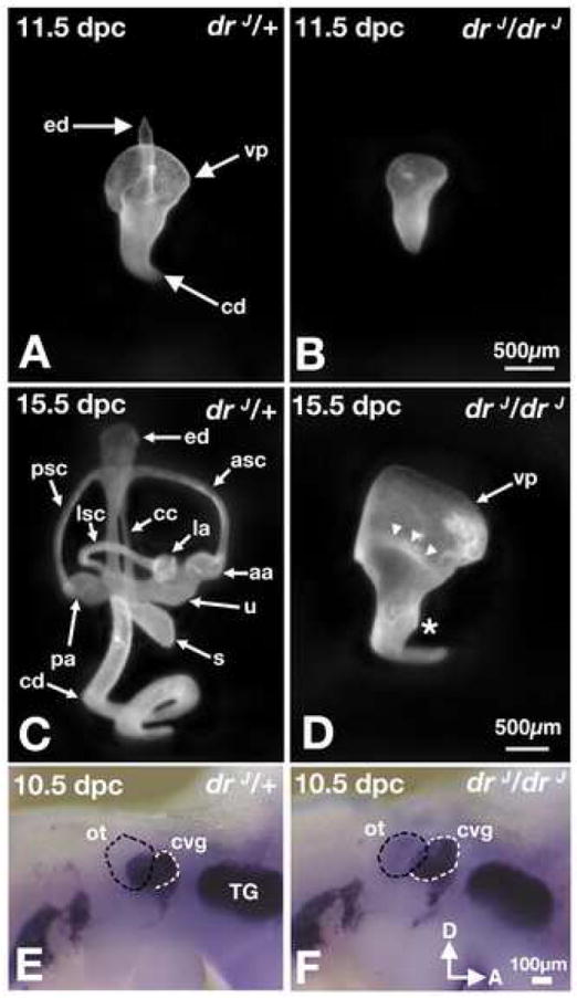
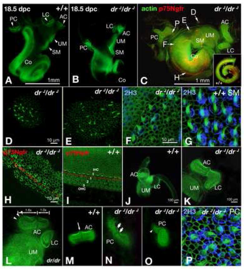
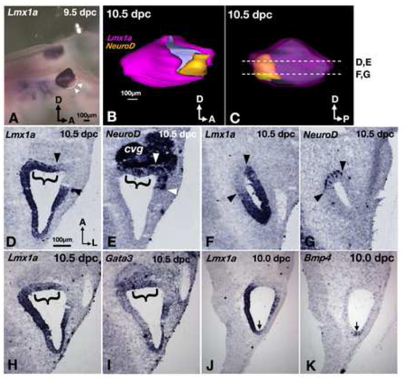
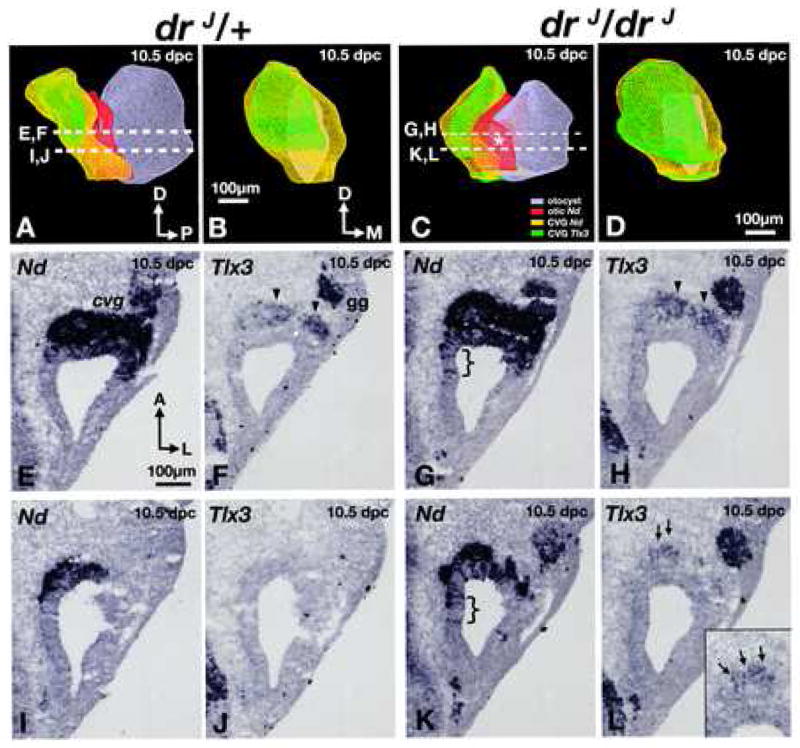
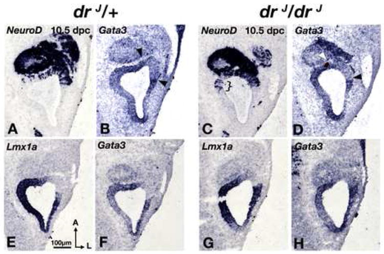
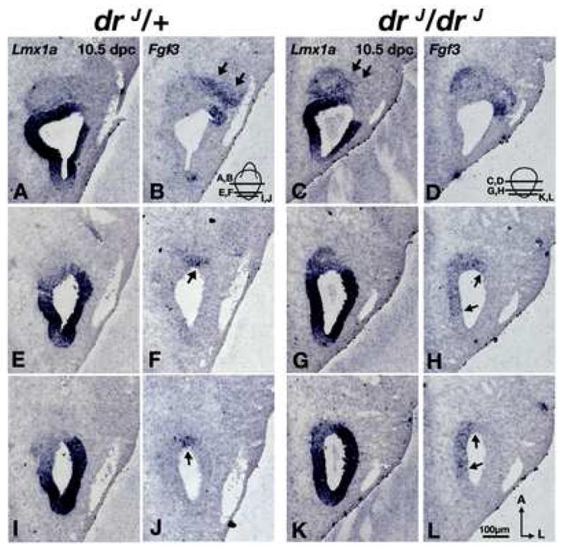
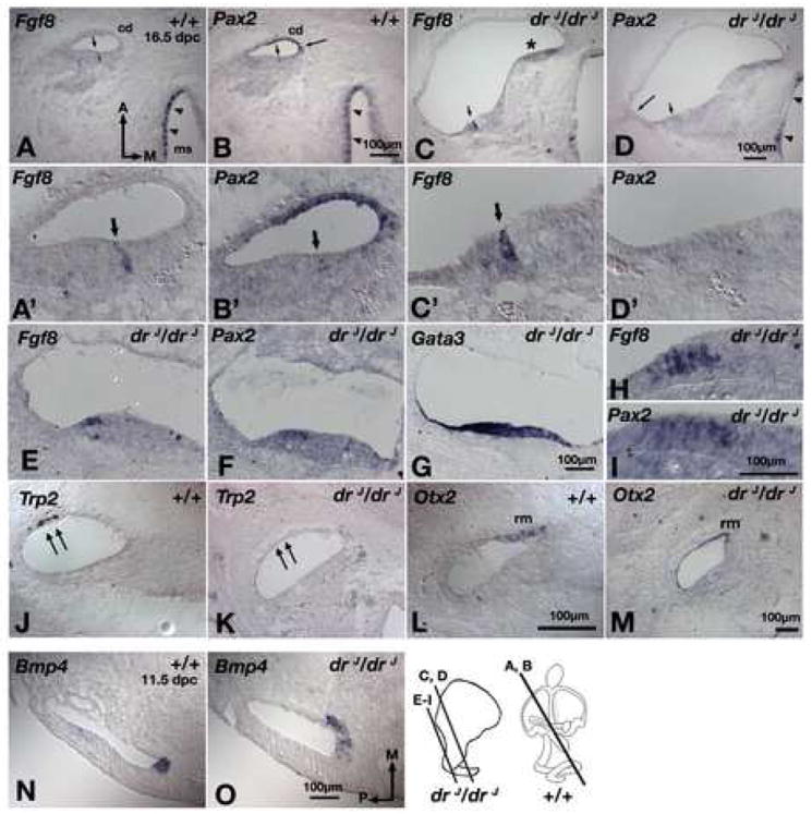
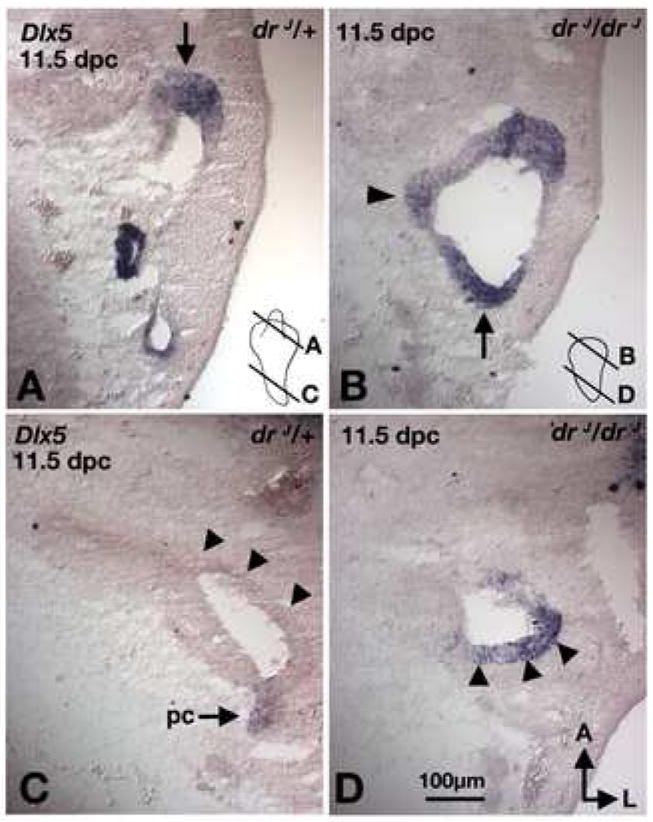
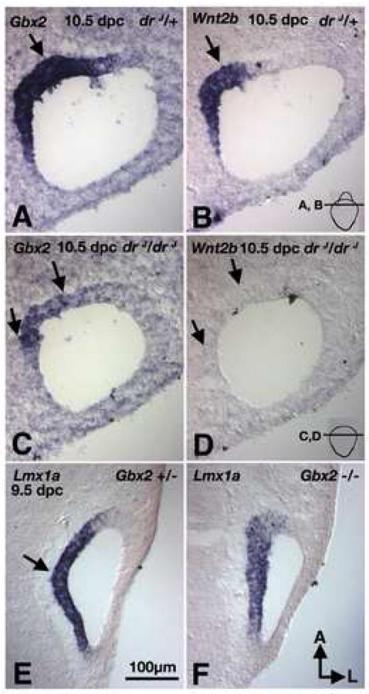
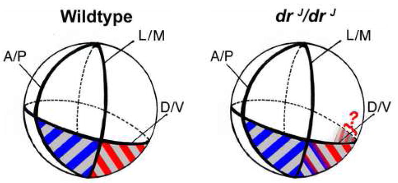
Lmx1a is a LIM homeodomain-containing transcription factor, which is required for the formation of multiple organs. Lmx1a is broadly expressed in early stages of the developing inner ear, but its expression is soon restricted to the non-sensory regions of the developing ear. In an Lmx1a functional null mutant, dreher (dr(J)/dr(J)), the inner ears lack a non-sensory structure, the endolymphatic duct, and the membranous labyrinth is poorly developed. These phenotypes are consistent with Lmx1a's role as a selector gene. More importantly, while all three primary fates of the inner ear - neural, sensory, and non-sensory - are specified in dr(J)/dr(J), normal boundaries among these tissues are often violated. For example, the neurogenic domain of the ear epithelium, from which cells delaminate to form the cochleovestibular ganglion, is expanded. Within the neurogenic domain, the demarcation between the vestibular and auditory neurogenic domains is most likely disrupted as well, based on the increased numbers of vestibular neuroblasts and ectopic expression of Fgf3, which normally is associated specifically with the vestibular neurogenic region. Furthermore, aberrant and ectopic sensory organs are observed; most striking among these is vestibular-like hair cells located in the cochlear duct.


Figures

Fig. 1
Fig. 1. Paint-filled and whole-mount inner ears of drJ/drJ mutants
Lateral views of drJ/+ (A,C) and drJ/drJ (B,D) inner ears. (A,B) At 11.5 dpc, inner ears of drJ/drJ mutants lack the endolymphatic duct. The canal pouches are smaller in size and the cochlear duct has limited extension. (C,D) At 15.5 dpc, the morphologies of drJ/drJ inner ears remain rudimentary compared to those in drJ/+ embryos. The vertical (vp) and lateral (arrowheads) canal pouches show no sign of resorption, and extension of the cochlear duct is limited. Asterisk indicates the rudimentary saccule. (E,F) Whole-mount drJ/+ (E) drJ/drJ (F) embryos probed for NeuroD transcripts. An increase in the size of CVG (white outline) is apparent adjacent to the drJ/drJ otocyst (black outline), showing NeuroD-positive neuroblasts within the otic epithelium that have not yet delaminated and are not part of the CVG. aa, anterior ampulla; asc, anterior semicircular canal; cd, cochlear duct; ed, endolymphatic duct; la, lateral ampulla; pa, posterior ampulla; ot, otocyst; psc, posterior semicircular canal; s, saccule; TG, trigeminal ganglion; u, utricle; vp, vertical canal pouch.
Fig. 2
Fig. 2. Abnormal sensory organs in drJ/drJ inner ears
(A-P) Whole-mount sensory epithelia labeled with FITC-phalloidin. Some specimens are double-labeled with anti-2H3 for neurofilament (F,G,P) or anti-p75Ngfr for pillar cells (C,H,I). (A,B) Sensory organs are housed in separate chambers in wildtype inner ears, but they are located within a common chamber in drJ/drJ mutants. (C) Image of a flattened drJ/drJ inner ear. The cristae are abnormal in shape, the cochlear duct only has a ¾ turn, and p75Ngfr labeling is absent in the basal turn (F). Inset shows normal p75Ngfr labeling in a wildtype cochlear duct. (D,E,F,H,P) are higher power views of specific regions in (C). (D,E) Ectopic sensory patches located adjacent to the maculae of the utricle and saccule (UM, SM). (F) Presence of hair cells in the p75Ngfr-negative, basal turn area of drJ/drJ cochlear duct. The hair bundles resemble those in a normal macula, but the cell bodies are not surrounded by 2H3-positive calyxes as in a normal macula (G). (H) Middle turn of drJ/drJ cochlear duct showing disorganized hair cells that are separated by a row of p75Ngfr-positive pillar cells. (I) Middle turn of wildtype cochlea showing three rows of outer hair cells and one row of inner hair cells, separated by a row of p75Ngfr labeling. (J) Wildtype inner ear showing the anterior crista, lateral crista, and utricular macula in separate chambers. (K) Fusion of the anterior and lateral cristae in drJ/drJ mutants. (L) Asymmetrical shape of the anterior crista in drJ/drJ. The medial half of the AC is 1.6 times longer than the lateral half. (M) Symmetrical shape of AC in wildtype. (N,O) Abnormal posterior cristae in drJ/drJ mutants with one consisting of two cruciata (double arrows) and another with asymmetrical halves (arrowhead). (P) Posterior crista of drJ/drJ showing normal hair bundles and calyxes. AC, anterior crista; LC, lateral crista; PC, posterior crista; UM, utricular macula; SM, saccular macula; Co, cochlea. Scale bars: (A,B);(D,E);(F,G,P);(H,I);(K-O).
Fig. 3
Fig. 3. Lmx1a expression influences neural subtype specification
(A) Lmx1a expression in a whole-mount embryo. Lmx1a is not expressed in the antero-ventral region of the otocyst (arrowheads). (B) Lateral and (C) medial views of a 3-D reconstructed inner ear showing the relationships of Lmx1a (pink) and NeuroD (yellow) expression domains. The pink in (C) is rendered transparent to reveal the yellow area underneath. Asterisk indicates the neurogenic region that is devoid of Lmx1a expression. (D-G) Representative sections from ear shown in (B) and (C). Brackets indicate the neurogenic regions that do express Lmx1a. Arrowheads mark the neurogenic regions that do not express Lmx1a. (H,I) adjacent sections probed for Lmx1a (H) and Gata3 (I). (J,K) adjacent sections probed for Lmx1a (J), and Bmp4 (K), showing Lmx1a expression in the Bmp4-positive posterior crista (arrows). Sections are rotated 90° clockwise from orientation in (C). Orientation and scale bar in D apply to E–K.
Fig. 4
Fig. 4. Ganglion malformations in drJ/drJ mutants
(A,C) Antero-medial views of 3-D reconstructed, right inner ears (grey) of (A) heterozygous and (C) homozygous drJ/drJ mutants. NeuroD expression within the otocyst is red, and expression of NeuroD and Tlx3 in the CVG are yellow and green, respectively. Asterisk represents a medial expansion of the NeuroD domain in the dreher otocyst. (B,D) Anterior views of the CVG in (A) and (C), respectively. (E,F) and (I,J) are representative sections from the drJ/+ specimen shown in (A). Comparable sections from the drJ/drJ specimen in (C) are shown in (G,H) and (K,L). (E-H) At this level, there is an expansion of the neurogenic domain (G; bracket) and more Tlx3 expression in the CVG of drJ/drJ compared to drJ/+ (H; arrowheads). (I-J) Slightly ventral, where there is only residual neurogenic domain that is Tlx3 negative in drJ/+, there is a broader neurogenic domain in drJ/drJ mutants (K; bracket) and some Tlx3-positive neuroblasts can be detected (L; arrows; inset). Sections are rotated 90° clockwise from orientation in (A or C). gg, geniculate ganglion. Orientation and scale bar in E apply to F-L.
Fig. 5
Fig. 5. Gata3 expression domain is unchanged in drJ/drJ mutants
(A,B) and (C,D) are comparable midsections of drJ /+ and drJ/drJ otocysts, respectively. There is no obvious difference in Gata3 expression domains between the two genotypes. However, there is a clear medial expansion of NeuroD expression in the drJ/drJ mutant (C; bracket). (E,F) and (G,H) are comparable midsections of drJ /+ and drJ/drJ otocysts, respectively. There is no difference in the expression pattern of Lmx1a or Gata3 between the two genotypes. Sections chosen in this figure are at comparable levels to those shown in Fig. 4 (E-H).
Fig. 6
Fig. 6. Expansion of the vestibular neurogenic region in drJ/drJ mutants
(A,B) and (C,D) are comparable midsections of drJ /+ and drJ/drJ otocysts, respectively. There is no obvious difference between drJ /+ and drJ/drJ in their Lmx1a (A,C) and Fgf3 (B,D) expression domains except there is more Lmx1a expression in delaminated neuroblasts of drJ/drJ mutants (C; arrows). At a more ventral level (E-L), Fgf3 expression is expanded medially in drJ/drJ mutants (H,L; arrows). Orientation and scale bar apply to all.
Fig. 7
Fig. 7. Abnormal gene expression patterns in the cochlear duct and cristae of drJ/drJ mutants
(A,B) Adjacent sections of wildtype inner ears showing Fgf8 (A) is expressed in the inner hair cells of the organ of Corti (arrow) and hair cells of the macula of the saccule (ms; arrowheads). Pax2 (B) is expressed in the stria vascularis (long arrow), hair cells of the vestibule (arrowheads), but expression in the hair cells of the organ of Corti is barely detectable (short arrow). (C,D) Adjacent sections of the apical region of drJ/drJ cochlear duct showing Fgf8 (C) is expressed in the inner hair cells of the organ of Corti, but Pax2 (D) is in neither the stria vascularis (long arrow) nor sensory hair cells (short arrow). Asterisk indicates an area where the tissue is folded. (A’-D’) Higher magnification of sections shown in (A-D). (E,F,G) Adjacent sections of the basal region of the drJ/drJ cochlear duct showing high numbers of hair cells expressing Fgf8 (E) and Pax2 (F) in a Gata3-positive, cochlear region (G). (H,I) Higher magnification of sections shown in (E) and (F), respectively. (J,L) In wildtype cochlear duct, Trp2 (J) and Otx2 (L) are expressed in the stria vascularis (double arrows) and Reissner’s membrane (rm), respectively. In drJ/drJ cochlear duct, Pax2 (D, long arrow) and Trp2 (K) in the stria vascularis are not detected, but Otx2 expression in the Reissner’s membrane is evident (M). (N,O) Bmp4 expression in the presumptive anterior crista is expanded medially in drJ/drJ inner ears (O) compared to wildtype (N). cd, cochlear duct. Scale bars: (A,B);(C,D);(E-G);(H,I);(J-L);(N,O)
Fig. 8
Fig. 8. Expression patterns of Dlx5 in drJ/drJ mutants
Sections taken from comparable regions of drJ/+ (A,C) and drJ/drJ (B,D) inner ears. (A,B) In the dorsal otic region, Dlx5 expression is located in the rim of the canal pouches in both drJ/+ and drJ/drJ ears (arrows). (C,D) Dlx5 is only expressed in the lateral side of the cochlear duct region in drJ/drJ but not drJ/+ ears (arrowheads). Orientation and scale bar apply to all.
Fig. 9
Fig. 9. Gene expression analyses of the endolymphatic duct phenotype in drJ/drJ and Gbx2-/-mutants
(A,B) Adjacent sections of drJ/+ inner ears probed for Gbx2 (A) and Wnt2b (B) transcripts. Gbx2 and Wnt2b are co-expressed in the base of the endolymphatic duct region (arrow). (C,D) Adjacent sections of drJ/drJ inner ears from a comparable region as (A,B) and probed for Gbx2 (C) and Wnt2b (D) transcripts. Gbx2 expression is present in drJ/drJ mutants but not Wnt2b. (E,F) In Gbx2 null embryos Lmx1a is present in the medial region (F), similar to the Gbx2 +/- embryos (E). Orientation and scale bar apply to all.
Fig. 10
Fig. 10. Lmx1a maintains specified domains
The globes represent the developing otocyst in wildtype and drJ/drJ mutants. The ventral neurogenic region is shaded gray. The ventro-lateral putative vestibular neurogenic region (blue stripes) is Lmx1a-negative, Fgf3-positive, and Gata3-negative. The ventro-medial, presumably auditory neurogenic region (red stripes) is Lmx1a-positive, Fgf3-negative, and Gata3-positive. In the developing otocysts of wildtype mice there is a distinct border between the Fgf3 and Lmx1a expression domains. However, in drJ/drJ, non-functional Lmx1a cannot maintain the proper Fgf3–positive region and the Fgf3 expression domain expands medially (blue-red gradient). Lmx1a expression domain does not change, but the ventro-medial neurogenic region (gray shading in drJ/drJ) is expanded posteriorly in the mutant otocyst. Because Gata3 is normally expressed at low levels in the medial otic epithelium, it is unclear whether the Gata3 domain is expanded in that region of drJ/drJ otocyst (red question mark).

Similar articles

Cited by

References

    1. Anagnostopoulos AV. A compendium of mouse knockouts with inner ear defects. Trends Genet. 2002;18:499. - PubMed
    1. Arques CG, Doohan R, Sharpe J, Torres M. Cell tracing reveals a dorsoventral lineage restriction plane in the mouse limb bud mesenchyme. Development. 2007;134:3713–22. - PubMed
    1. Bell D, Streit A, Gorospe I, Varela-Nieto I, Alsina B, Giraldez F. Spatial and temporal segregation of auditory and vestibular neurons in the otic placode. Dev Biol. 2008;322:109–20. - PubMed
    1. Bok J, Chang W, Wu DK. Patterning and morphogenesis of the vertebrate inner ear. Int J Dev Biol. 2007a;51:521–33. - PubMed
    1. Bok J, Dolson DK, Hill P, Ruther U, Epstein DJ, Wu DK. Opposing gradients of Gli repressor and activators mediate Shh signaling along the dorsoventral axis of the inner ear. Development. 2007b;134:1713–22. - PubMed

Publication types

MeSH terms

LinkOut - more resources