

Cell lines from the Egyptian fruit bat are permissive for modified vaccinia Ankara
- PMID: 19540275
- PMCID: PMC7172177
- DOI: 10.1016/j.virusres.2009.06.007
Cell lines from the Egyptian fruit bat are permissive for modified vaccinia Ankara
Abstract
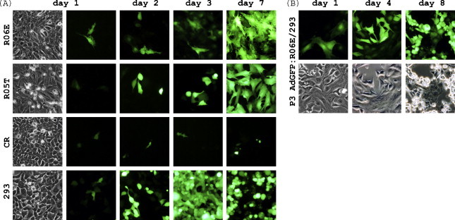
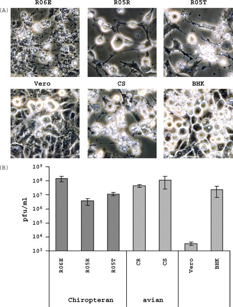
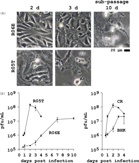
Bats are reservoir hosts for a spectrum of infectious diseases. Some pathogens (such as Hendra, Nipah and Marburg viruses) appear to use mainly fruit bats as reservoir. We describe designed immortalization of primary fetal cells from the Egyptian fruit bat (Rousettus aegyptiacus) to facilitate isolation and characterization of pathogens associated with these mammals. Three cell lines with different properties were recovered and successful immortalization was confirmed by continuous cultivation for over 18 months. Surprisingly, the cell lines are fully permissive for a highly attenuated poxvirus, modified vaccinia Ankara (MVA). MVA is a safe and well characterized vaccine vector that cannot replicate in most mammalian cells. High permissivity of Rousettus cell lines could justify testing bats for susceptibility to MVA as a replication competent vector with low zoonotic potential to induce herd immunity in bat colonies against viruses causing rabies or haemorrhagic fevers.
Figures

Similar articles
-
Authentication of the R06E fruit bat cell line.Viruses. 2012 May;4(5):889-900. doi: 10.3390/v4050889. Epub 2012 May 23. Viruses. 2012. PMID: 22754654 Free PMC article.
-
Modified vaccinia virus Ankara multiplies in rat IEC-6 cells and limited production of mature virions occurs in other mammalian cell lines.J Gen Virol. 2006 Jan;87(Pt 1):21-27. doi: 10.1099/vir.0.81479-0. J Gen Virol. 2006. PMID: 16361414
-
Host range and cytopathogenicity of the highly attenuated MVA strain of vaccinia virus: propagation and generation of recombinant viruses in a nonhuman mammalian cell line.Virology. 1997 Nov 24;238(2):198-211. doi: 10.1006/viro.1997.8845. Virology. 1997. PMID: 9400593
-
Modified Vaccinia virus Ankara: innate immune activation and induction of cellular signalling.Vaccine. 2013 Sep 6;31(39):4231-4. doi: 10.1016/j.vaccine.2013.03.017. Epub 2013 Mar 21. Vaccine. 2013. PMID: 23523404 Review.
-
Modified Vaccinia Virus Ankara: History, Value in Basic Research, and Current Perspectives for Vaccine Development.Adv Virus Res. 2017;97:187-243. doi: 10.1016/bs.aivir.2016.07.001. Epub 2016 Aug 1. Adv Virus Res. 2017. PMID: 28057259 Free PMC article. Review.
Cited by
-
A Deleted Deletion Site in a New Vector Strain and Exceptional Genomic Stability of Plaque-Purified Modified Vaccinia Ankara (MVA).Virol Sin. 2020 Apr;35(2):212-226. doi: 10.1007/s12250-019-00176-3. Epub 2019 Dec 12. Virol Sin. 2020. PMID: 31833037 Free PMC article.
-
A model system for in vitro studies of bank vole borne viruses.PLoS One. 2011;6(12):e28992. doi: 10.1371/journal.pone.0028992. Epub 2011 Dec 16. PLoS One. 2011. PMID: 22194969 Free PMC article.
-
Ebolavirus polymerase uses an unconventional genome replication mechanism.Proc Natl Acad Sci U S A. 2019 Apr 23;116(17):8535-8543. doi: 10.1073/pnas.1815745116. Epub 2019 Apr 8. Proc Natl Acad Sci U S A. 2019. PMID: 30962389 Free PMC article.
-
Establishment of a cell line from the hematophagous Bat Desmodus rotundus susceptible to Lyssavirus rabies.Braz J Microbiol. 2025 Jun;56(2):1311-1320. doi: 10.1007/s42770-025-01651-8. Epub 2025 Mar 4. Braz J Microbiol. 2025. PMID: 40038190
-
Molecular characterization of human pathogenic bunyaviruses of the Nyando and Bwamba/Pongola virus groups leads to the genetic identification of Mojuí dos Campos and Kaeng Khoi virus.PLoS Negl Trop Dis. 2014 Sep 4;8(9):e3147. doi: 10.1371/journal.pntd.0003147. eCollection 2014 Sep. PLoS Negl Trop Dis. 2014. PMID: 25188437 Free PMC article.
References
-
- Alexopoulou L., Holt A.C., Medzhitov R., Flavell R.A. Recognition of double-stranded RNA and activation of NF-kappaB by Toll-like receptor 3. Nature. 2001;413(6857):732–738. - PubMed
Publication types
MeSH terms
Substances
LinkOut - more resources
Full Text Sources
Other Literature Sources
Research Materials
