. 2009 Aug 31;168(1-2):19-25.
doi: 10.1016/j.resp.2009.06.011. Epub 2009 Jun 18.

Multiple pontomedullary mechanisms of respiratory rhythmogenesis

Affiliations

Multiple pontomedullary mechanisms of respiratory rhythmogenesis

A P L Abdala et al. Respir Physiol Neurobiol. .

Abstract

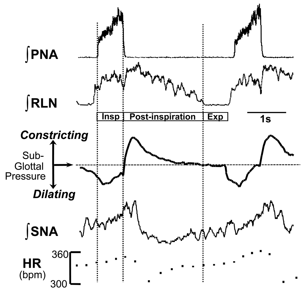
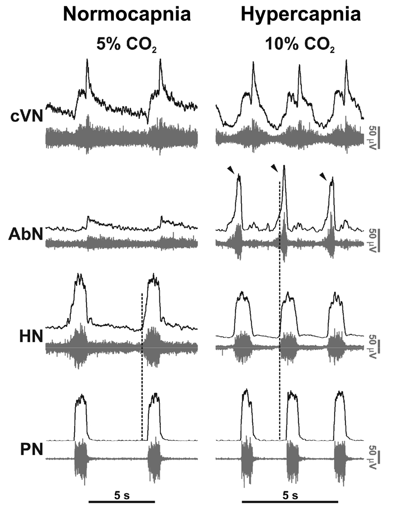
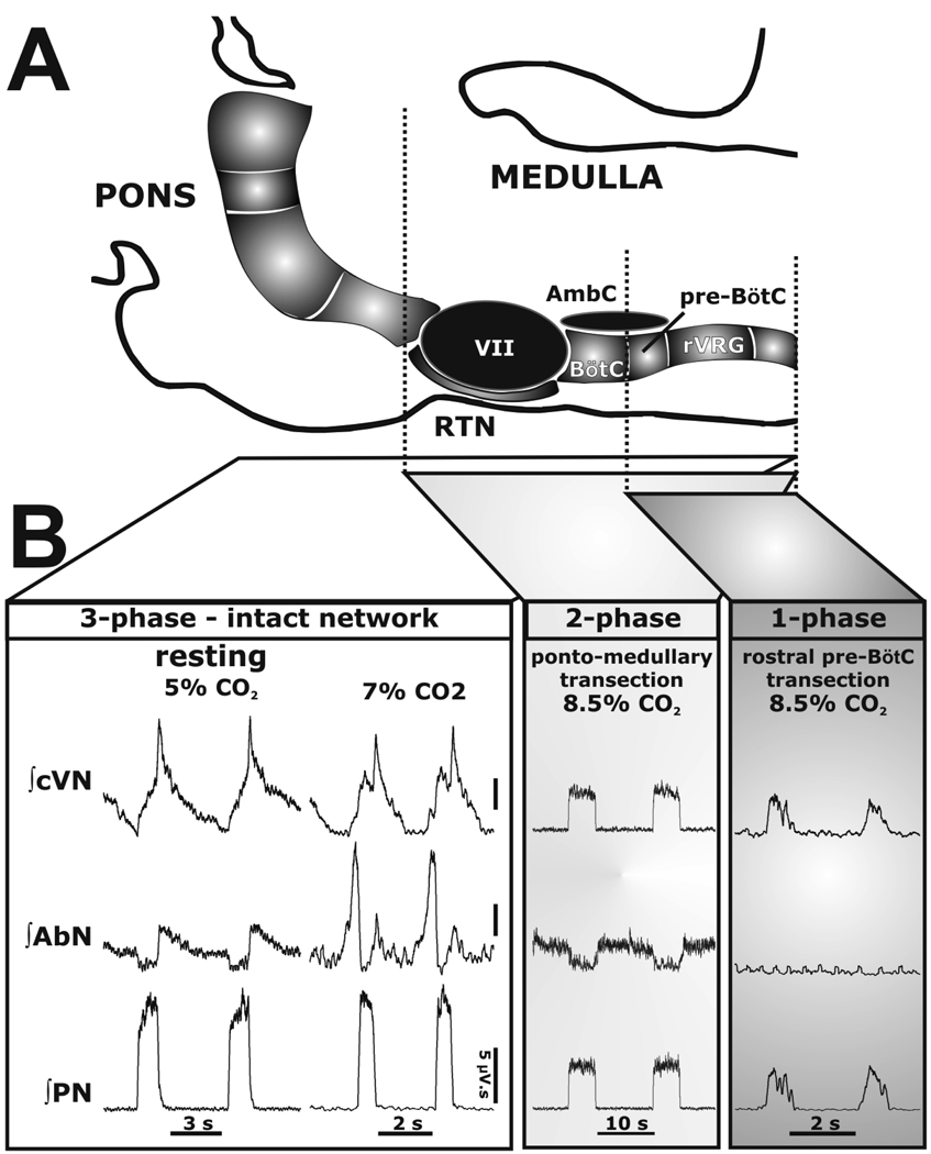
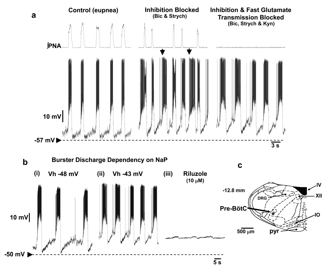
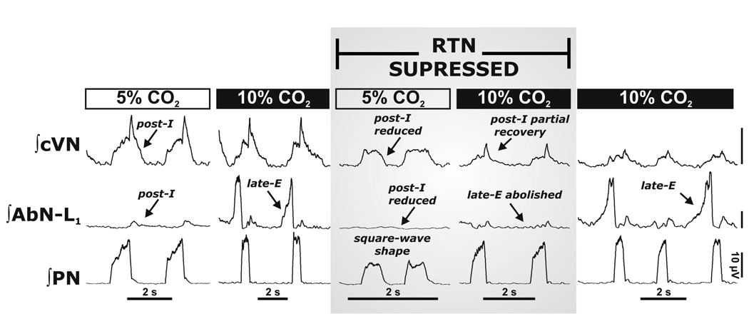
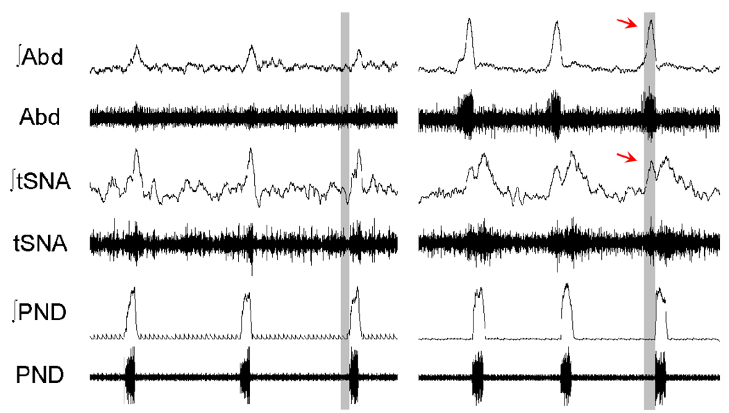
Mammalian central pattern generators producing rhythmic movements exhibit robust but flexible behavior. However, brainstem network architectures that enable these features are not well understood. Using precise sequential transections through the pons to medulla, it was observed that there was compartmentalization of distinct rhythmogenic mechanisms in the ponto-medullary respiratory network, which has rostro-caudal organization. The eupneic 3-phase respiratory pattern was transformed to a 2-phase and then to a 1-phase pattern as the network was physically reduced. The pons, the retrotrapezoid nucleus and glycine mediated inhibition are all essential for expression of the 3-phase rhythm. The 2-phase rhythm depends on inhibitory interactions (reciprocal) between Bötzinger and pre-Bötzinger complexes, whereas the 1-phase-pattern is generated within the pre-Bötzinger complex and is reliant on the persistent sodium current. In conditions of forced expiration, the RTN region was found to be essential for the expression of abdominal late expiratory activity. However, it is unknown whether the RTN generates or simply relays this activity. Entrained with the central respiratory network is the sympathetic nervous system, which exhibits patterns of discharge coupled with the respiratory cycle (in terms of both gain and phase of coupling) and dysfunctions in this coupling appear to underpin pathological conditions. In conclusion, the respiratory network has rhythmogenic capabilities at multiple levels of network organization, allowing expression of motor patterns specific for various physiological and pathophysiological respiratory behaviors.


Figures

Figure 1
Figure 1. Cardio-laryngo-sympathetic-respiratory coupling recorded in situ
A montage showing integrated activities of the phrenic nerve (PNA), recurrent laryngeal nerve (RLN) and sympathetic nerve activity (SNA; thoracic chain) to show coordination of cranial and spinal cardio-respiratory motor outflows in an arterially perfused rat preparation without pulmonary stretch receptor feedback. Note the three phase rhythm (inspiration, Insp; post-inspiration; expiration , Exp) corresponding to that defined by Richter (1982). During central inspiratory activity indexed by phrenic (Insp), heart rate (HR) increases (due to central synaptic inhibition of cardiac vagal motoneurons and increased sympathetic discharge) and the glottis dilates due to activation of laryngeal abductors. During early expirations (so called, post-inspiration), heart rate falls as the inspiratory related inhibition of the cardiac vagal motoneurons is removed. At this time the laryngeal adductors fire causing a transient constriction of the glottis; the latter stalls expiratory air flow so giving adequate time for gas exchange. Note, that the SNA is respiratory phase locked and peaks in the post-inspiratory phase. Modified from Paton & Nolan (2000).
Figure 2
Figure 2. Eupneic pattern of respiratory motor activity in situ
In control conditions (normocapnia) phrenic (PN) and hypoglossal (HN) have an inspiratory ramp shaped activity envelope whereas abdominal nerve (AbN; lumbar segment 1) exhibits small amplitude post-inspiratory activity. Using 10% carbon dioxide (i.e. 5% above normocapnic conditions), central respiratory drive was raised. This resulted in generation of augmenting expiratory activity in the AbN outflow (Late-E; arrowed) and advanced the onset of pre-inspiratory HN activity relative to PN; the latter indicating reduced airway resistance during both the forced expiration and inspiration. The pattern of PN now showed an abrupt onset in discharge (see Abdala et al. 2009).
Figure 3
Figure 3. 3-2-1 phase hypothesis of respiration
A: Schematic drawing depicting the spatial arrangement of the ventral respiratory column viewed sagittally. Microtransections of the brainstem are indicated by the vertical dashed lines. Abbreviations: AmbC: compact nucleus ambiguus; BötC: Bötzinger Complex; LRt: lateral reticular nucleus; Pn: pontine nucleus; pre-BötC: pre- Bötzinger Complex; RTN/vlPF: retrotrapezoid nucleus and ventrolateral parafacial regions; rVRG: rostral ventral respiratory group; V: trigeminal motor nucleus; VII: facial motor nucleus. B: Activity patterns of phrenic (PN), abdominal (AbN), and central vagus (cVN) nerves from an intact preparation (3-phase pattern) during eucapnia (5% CO2) and hypercapnia (7% CO2), and after a ponto-medullary transection. The latter resulted in a 2-phase pattern in which late-E abdominal bursts are abolished during hypercapnia (8.5% CO2). After a transection at the rostral boundary of the pre-BötC, a 1-phase inspiratory pattern was evoked and all expiratory motor activity was abolished.
Figure 4
Figure 4. Respiratory burster neurons recorded from a ponto-medullary intact in situ rat preparation
Prior to their synaptic isolation, burster neurons were characterized as inspiratory and held relatively hyperpolarized during eupnea (a). Bursters depolarized subsequent to blockade of glycine and GABAA receptors (1µM strychnine, 20 µM bicuculline) when ectopic bursts that did not correspond to a phrenic burst were evident (arrowed). Blockade of ionotropic glutamate receptors arrested phrenic discharge and resulted in intrinsic bursting which was voltage-dependent (bi,ii) and sensitive to blockade of persistent sodium current. Some bursters were found in the pre-BötC (c) while others were in the rostral ventral respiratory group region (see St-John et al. 2009 for details).
Figure 5
Figure 5. Reversible inactivation of the RTN abolished the 3-phase respiratory pattern and hypercapnia evoked Late-E abdominal nerve activity
The RTN/vlPF was inactivated by bilateral microinjections of isoguvacine hydrochloride (GABAA receptor agonist). This resulted in a depression of post-inspiratory motor output on both AbN and central vagus (cVN) nerves, and the phrenic nerve pattern (PN) was transformed to a “square-wave” shape. RTN/vlPF suppression also abolished late-E AbN bursts during hypercapnia (10% CO2). Note that hypercapnia partially reinstated post-inspiratory activity during RTN/vlPF inactivation. Late-E activity recovered after isoguvacine washed out (∼1 hr; right panel). All traces show integrated nerve activities.
Figure 6
Figure 6. Chronic intermittent hypoxia causes long term plasticity in respiratory modulation of sympathetic activity
Cardiovascular and respiratory control systems can only work efficiently when coupled. In control conditions, rats show inspiratory/post-inspiratory modulation of thoracic sympathetic nerve activity (tSNA) relative to phrenic discharge (PND). This modulation is chronically altered in juvenile rats exposed to chronic intermittent hypoxia (CIH) for 10 days. In CIH treated rats an additional burst of sympathetic activity emerges, which correlated with the development of a Late-E burst in the abdominal motor outflow (Abd). The latter may well contribute to the hypertension generated in the CIH model of sleep apnoea.

References

    1. Abbott SB, Stornetta RL, Fortuna MG, Depuy SD, West GH, Harris TE, Guyenet PG. Photostimulation of retrotrapezoid nucleus phox2b–expressing neurons in vivo produces long-lasting activation of breathing in rats. J Neurosci. 2009;29:5806–5819. - PMC - PubMed
    1. Abdala APL, Rybak IA, Smith JC, Paton JFR. Abdominal expiratory activity in the rat brainstem-spinal cord in situ: patterns, origins, and implications for respiratory rhythm generation. J. Physiol. 2009 (in press) - PMC - PubMed
    1. Anrep GV, Pascual W, Rossler R. Respiratory variations of the heart rate. II. The central mechanism of the sinus arrhythmia and the inter-relationships between central and reflex mechanism. Proc. Royal Soc. Lond. 1936;119:218–230. (Series B)
    1. Bianchi AL, Gestreau C. The respiratory network: An overview of a half century research. Respir. Physiol. Neurobiol. 2009 ?, ? - ? (This issue) - PubMed
    1. Büsselberg D, Bischoff AM, Paton JFR, Richter DW. Loss of glycinergic inhibition reveals two modes of respiratory rhythm generation. Pflügers Arch. 2001;441:444–449. - PubMed

Publication types