

Growth plate mechanics and mechanobiology. A survey of present understanding
- PMID: 19540500
- PMCID: PMC2739053
- DOI: 10.1016/j.jbiomech.2009.05.021
Growth plate mechanics and mechanobiology. A survey of present understanding
Abstract
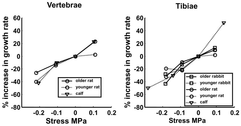
The longitudinal growth of long bones occurs in growth plates where chondrocytes synthesize cartilage that is subsequently ossified. Altered growth and subsequent deformity resulting from abnormal mechanical loading is often referred to as mechanical modulation of bone growth. This phenomenon has key implications in the progression of infant and juvenile musculoskeletal deformities, such as adolescent idiopathic scoliosis, hyperkyphosis, genu varus/valgus and tibia vara/valga, as well as neuromuscular diseases. Clinical management of these deformities is often directed at modifying the mechanical environment of affected bones. However, there is limited quantitative and physiological understanding of how bone growth is regulated in response to mechanical loading. This review of published work addresses the state of knowledge concerning key questions about mechanisms underlying biomechanical modulation of bone growth. The longitudinal growth of bones is apparently controlled by modifying the numbers of growth plate chondrocytes in the proliferative zone, their rate of proliferation, the amount of chondrocytic hypertrophy and the controlled synthesis and degradation of matrix throughout the growth plate. These variables may be modulated to produce a change in growth rate in the presence of sustained or cyclic mechanical load. Tissue and cellular deformations involved in the transduction of mechanical stimuli depend on the growth plate tissue material properties that are highly anisotropic, time-dependent, and that differ in different zones of the growth plate and with developmental stages. There is little information about the effects of time-varying changes in volume, water content, osmolarity of matrix, etc. on differentiation, maturation and metabolic activity of chondrocytes. Also, the effects of shear forces and torsion on the growth plate are incompletely characterized. Future work on growth plate mechanobiology should distinguish between changes in the regulation of bone growth resulting from different processes, such as direct stimulation of the cell nuclei, physico-chemical stimuli, mechanical degradation of matrix or cellular components and possible alterations of local blood supply.
Figures
Similar articles
-
Enlargement of growth plate chondrocytes modulated by sustained mechanical loading.J Bone Joint Surg Am. 2002 Oct;84(10):1842-8. doi: 10.2106/00004623-200210000-00016. J Bone Joint Surg Am. 2002. PMID: 12377917
-
Growth plate cartilage shows different strain patterns in response to static versus dynamic mechanical modulation.Biomech Model Mechanobiol. 2016 Aug;15(4):933-46. doi: 10.1007/s10237-015-0733-6. Epub 2015 Oct 9. Biomech Model Mechanobiol. 2016. PMID: 26452368
-
Computational modeling of the mechanical modulation of the growth plate by sustained loading.Theor Biol Med Model. 2012 Sep 25;9:41. doi: 10.1186/1742-4682-9-41. Theor Biol Med Model. 2012. PMID: 23009361 Free PMC article.
-
Part 2. Review and meta-analysis of studies on modulation of longitudinal bone growth and growth plate activity: A micro-scale perspective.J Orthop Res. 2021 May;39(5):919-928. doi: 10.1002/jor.24992. Epub 2021 Jan 29. J Orthop Res. 2021. PMID: 33458882 Review.
-
Chondrocytes and longitudinal bone growth: the development of tibial dyschondroplasia.Poult Sci. 2000 Jul;79(7):994-1004. doi: 10.1093/ps/79.7.994. Poult Sci. 2000. PMID: 10901201 Review.
Cited by
-
Biomechanical evaluation of predictive parameters of progression in adolescent isthmic spondylolisthesis: a computer modeling and simulation study.Scoliosis. 2012 Jan 18;7(1):2. doi: 10.1186/1748-7161-7-2. Scoliosis. 2012. PMID: 22257363 Free PMC article.
-
Effects of loratadine, a histamine H1 receptor antagonist, on the skeletal system of young male rats.Drug Des Devel Ther. 2019 Sep 23;13:3357-3367. doi: 10.2147/DDDT.S215337. eCollection 2019. Drug Des Devel Ther. 2019. PMID: 31576110 Free PMC article.
-
Hypoxia promotes maintenance of the chondrogenic phenotype in rat growth plate chondrocytes through the HIF-1α/YAP signaling pathway.Int J Mol Med. 2018 Dec;42(6):3181-3192. doi: 10.3892/ijmm.2018.3921. Epub 2018 Oct 9. Int J Mol Med. 2018. PMID: 30320354 Free PMC article.
-
Pneumatic microfluidic cell compression device for high-throughput study of chondrocyte mechanobiology.Lab Chip. 2018 Jul 10;18(14):2077-2086. doi: 10.1039/c8lc00320c. Lab Chip. 2018. PMID: 29897088 Free PMC article.
-
Achondroplasia: Development, pathogenesis, and therapy.Dev Dyn. 2017 Apr;246(4):291-309. doi: 10.1002/dvdy.24479. Epub 2017 Mar 2. Dev Dyn. 2017. PMID: 27987249 Free PMC article. Review.
References
-
- Abbaszade I, Liu RQ, Yang F, Rosenfeld SA, Ross OH, Link JR, Ellis DM, Tortorella MD, Pratta MA, Hollis JM, Wynn R, Duke JL, George HJ, Hillman MC, Jr, Murphy K, Wiswall BH, Copeland RA, Decicco CP, Bruckner R, Nagase H, Itoh Y, Newton RC, Magolda RL, Trzaskos JM, Burn TC, et al. Cloning and characterization of ADAMTS11, an aggrecanase from the ADAMTS family. J Biol Chem. 1999;274(33):23443–50. - PubMed
-
- Akyuz E, Braun JT, Brown NA, Bachus KN. Static versus dynamic loading in the mechanical modulation of vertebral growth. Spine. 2006;31(25):E952–8. - PubMed
-
- Alberty A, Peltonen J, Ritsila V. Effects of distraction and compression on proliferation of growth plate chondrocytes. A study in rabbits. Acta Orthop Scand. 1993;64(4):449–55. - PubMed
-
- Alvarez J, Balbin M, Santos F, Fernandez M, Ferrando S, Lopez JM. Different bone growth rates are associated with changes in the expression pattern of types II and X collagens and collagenase 3 in proximal growth plates of the rat tibia. J Bone Miner Res. 2000;15(1):82–94. - PubMed
-
- Apte SS, Kenwright J. Physeal distraction and cell proliferation in the growth plate. J Bone Joint Surg Br. 1994;76(5):837–43. - PubMed
Publication types
MeSH terms
Grants and funding
LinkOut - more resources
Full Text Sources
