. 2009 Aug;46(13):2493-504.
doi: 10.1016/j.molimm.2009.05.022. Epub 2009 Jun 21.

Requirement of TLR4 and CD14 in dendritic cell activation by Hemagglutinin B from Porphyromonas gingivalis

Affiliations

Requirement of TLR4 and CD14 in dendritic cell activation by Hemagglutinin B from Porphyromonas gingivalis

Dalia E Gaddis et al. Mol Immunol. 2009 Aug.

Abstract

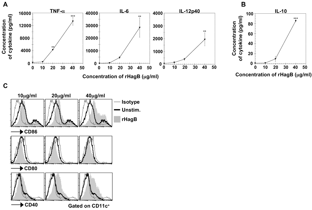
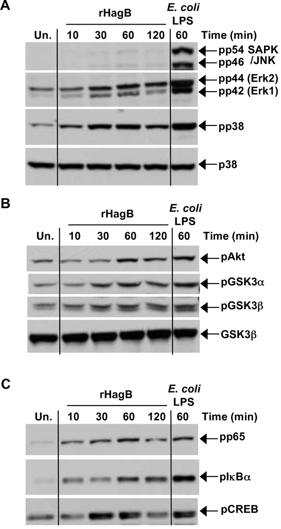
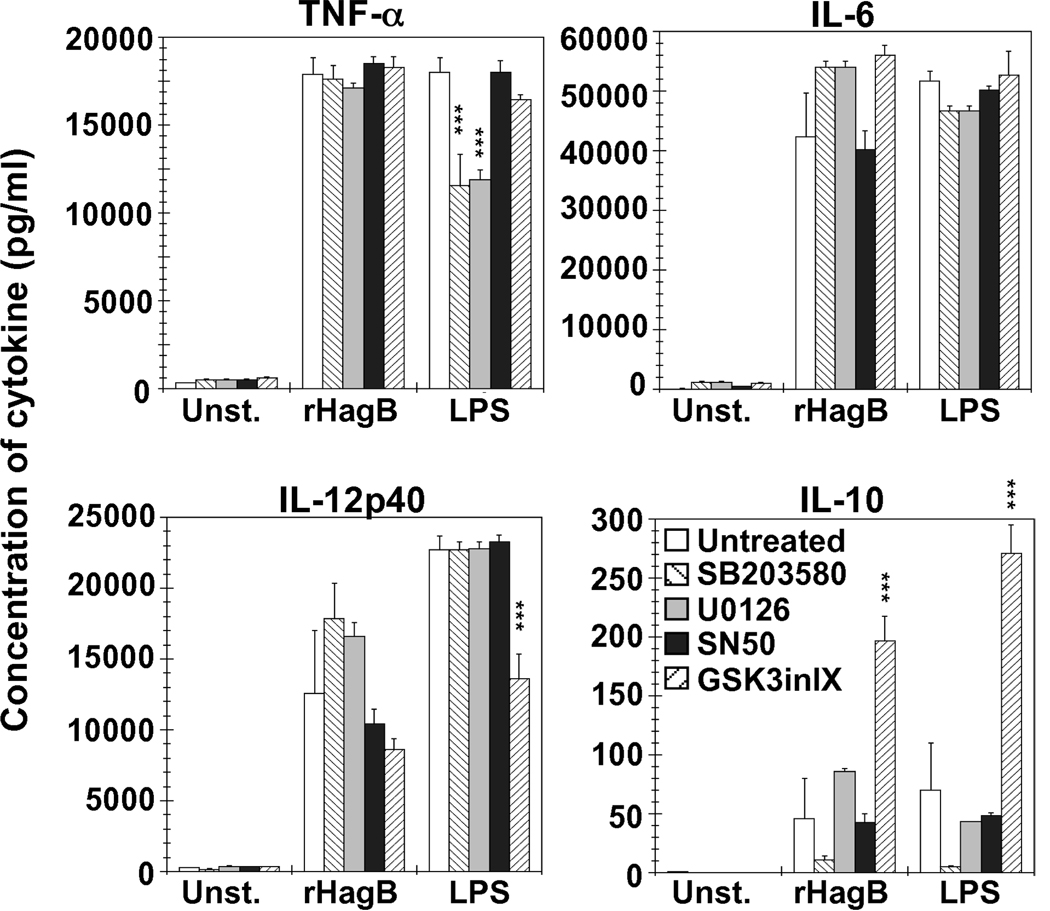
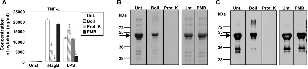
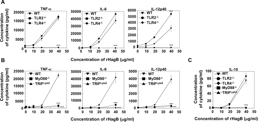
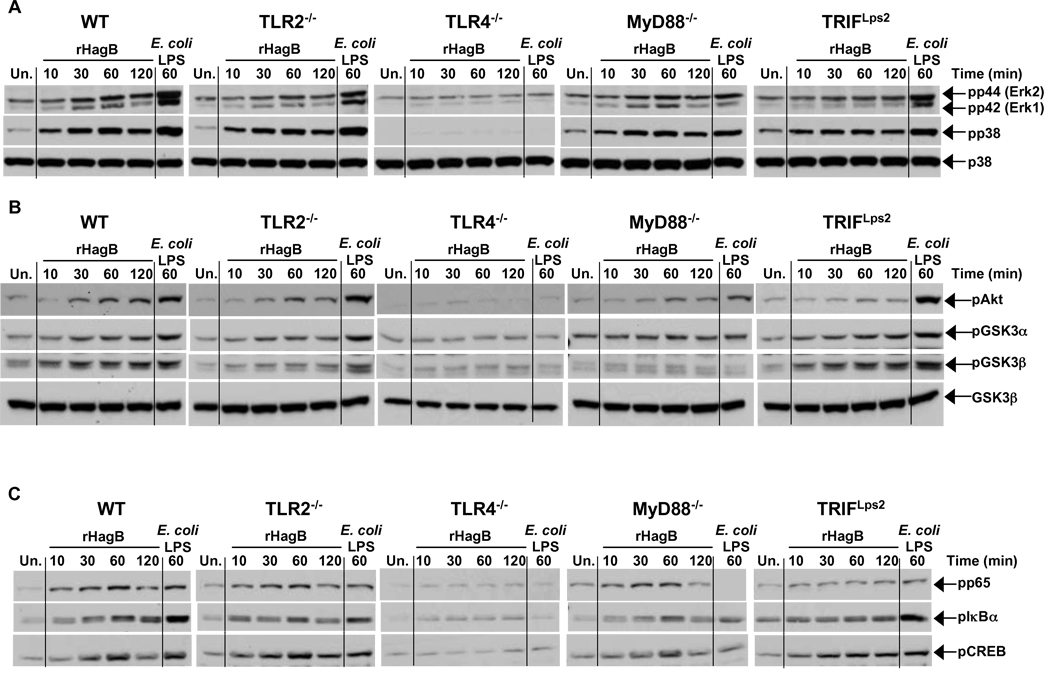
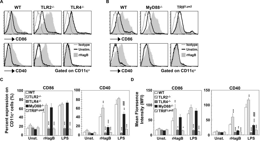
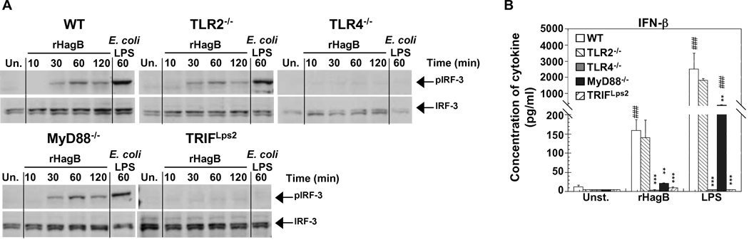
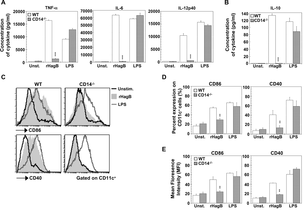
Porphyromonas gingivalis is a Gram-negative anaerobic bacterium that is one of the causative agents of chronic adult periodontal disease. Among the potential virulence factors of P. gingivalis are the hemagglutinins. Recombinant Hemagglutinin B (rHagB) from P. gingivalis has been shown to activate the immune system by inducing specific antibodies that protect against experimental periodontal bone loss following P. gingivalis infection. Since different microbial products can stimulate dendritic cells (DC) through Toll-like receptors (TLRs), subsequently leading to T cell activation and antibody production, we wanted to investigate the immunostimulatory effect of rHagB on DC and the role of TLR signaling in this process. Using an endotoxin free rHagB preparation, our results show that stimulation of murine bone marrow-derived DC with rHagB leads to upregulation of the costimulatory molecules CD86 and CD40, activation of p38 and ERK MAP kinases, transcription factors NF-kappaB, CREB and IRF-3 and the production of IL-6, TNF-alpha, IL-12p40 and to a lesser extent IL-10 and IFN-beta. This activation process was absolutely dependent on TLR4 and CD14. While upregulation of CD86 was independent of the adaptor molecule MyD88, CD40 upregulation and optimal cytokine (IL-6, TNF-alpha, IL-12p40, IL-10 and IFN-beta) production required both MyD88 and TRIF molecules. These results are of importance since they are the first to provide insights into the interaction of rHagB with DC and TLRs. The information from this study will aid in the design of effective vaccines strategies against chronic adult periodontal disease.


Figures

Fig. 1
Fig. 1
Stimulation of DC with rHagB results in the production of cytokines and the upregulation of costimulatory molecules. Bone marrow-derived DC (2×10⁵) from WT mice were either unstimulated (0 µg/ml; negative control) or stimulated with 10, 20 or 40 µg/ml rHagB. Culture supernatants were harvested 24 h post-stimulation and assessed for the levels of the pro-inflammatory cytokines TNF-α, IL-6 and IL-12p40 (A) and the anti-inflammatory cytokine IL-10 (B) by ELISA. Results are expressed as the mean ± standard error of triplicate cultures from one of three independent experiments. ***, ** Significant differences at P < 0.001 and P < 0.01, respectively, compared to unstimulated cultures. (C) DC (2×10⁵) from WT mice were stimulated with 10, 20 and 40 µg/ml rHagB for 16 h (shaded histograms) or left unstimulated as negative controls (thick lines). Cells were harvested and stained with fluorescent-labeled antibodies against CD11c, CD80, CD86, CD40 or matched isotype controls (thin lines). Histogram plots were gated on CD11c+ cells. Results represent one of three independent experiments.
Fig. 2
Fig. 2
Signaling pathways activated following rHagB stimulation of DC. DC were stimulated with 40 µg/ml rHagB for 10, 30, 60 or 120 min. Following stimulation, cells were lysed and whole cell lysates were assessed for phosphorylation of (A) JNK, ERK1/2 and p38, (B) Akt and GSK3α/β and (C) NF-κBp65, IκBα and CREB by Western blot. Total p38 (A) and GSK3β (B) were used as loading controls. Unstimulated DC (far left lane) or DC stimulated with 100 ng/ml E. coli K12 LPS for 60 min (far right lane) were used as controls. Results represent one of three independent experiments.
Fig. 3
Fig. 3
Cytokine production by DC stimulated with rHagB is dependent on more than one signaling pathway, while GSK3 signaling regulates IL-10. DC (2×10⁵) from WT mice were treated with 10 µM of U0126 (left striped bar), SB203580 (grey bar), NF-κB SN50 (black bar), or GSK-3 Inhibitor IX (right striped bar) (ERK1/2, p38, NF-κB, or GSK3 specific inhibitors, respectively) for 2 h. Untreated cells were used as a negative control (white bars). The cultures were then stimulated with 40 µg/ml rHagB, 100 ng/ml E. coli K12 LPS, or left unstimulated. Culture supernatants were harvested 24 h post-stimulation and assessed for the production of TNF-α, IL-6, IL-12p40 and IL-10 by ELISA. Results are expressed as the mean ± standard error of triplicate cultures from one of two independent experiments. *** Significant differences at P < 0.001, compared to untreated cultures stimulated with rHagB or LPS.
Fig. 4
Fig. 4
Activity of rHagB is not due to endotoxin contamination. (A) DC from WT mice (2×10⁵ ) were stimulated with 40 µg/ml rHagB, 100 ng/ml E. coli K12 LPS, or left unstimulated. Prior to stimulation, samples were untreated (white bars), boiled for 30 min (striped bars) or treated with proteinase K (grey bars) or polymyxin B (PMB) (black bars). Culture supernatants were harvested 24 h post-stimulation with rHagB or LPS and assessed for TNF-α production. Results are expressed as the mean ± standard error of triplicate cultures from one of three independent experiments. *** Significant differences at P < 0.001 compared to untreated cultures stimulated with rHagB or LPS. (B) Stained SDS-PAGE and (C) Western blot probed with specific HRP conjugated antibody against Penta.His for equivalent amounts of untreated and treated rHagB protein samples. Arrows represent rHagB protein band that runs at ~ 49 kDa. Results represent one of three independent experiments.
Fig. 5
Fig. 5
Cytokine production by rHagB activated DC is mediated through TLR4 signaling and requires both MyD88 and TRIF. DC (2×10⁵) from WT, TLR2−/− and TLR4−/− mice (A, C) or WT, MyD88−/− and TRIF Lps2 mice (B, C) were stimulated with 10, 20 or 40 µg/ml rHagB for 24 h. Culture supernatants were then harvested and assessed for TNF-α, IL-6, IL-12p40 (A, B) and IL-10 production (C) by ELISA. Results are expressed as the mean ± standard error of triplicate cultures from one of four independent experiments. *** Significant differences at P < 0.001 compared to WT cultures stimulated with rHagB.
Fig. 6
Fig. 6
Phosphorylation of p38, ERK1/2, Akt/GSK3 and activation of NF-κB and CREB by rHagB stimulated DC is dependent on TLR4. DC from WT, TLR2−/−, TLR4−/−, MyD88−/− and TRIFLps2 mice were stimulated with 40 µg/ml rHagB for 10, 30, 60 or 120 min. Following stimulation, cells were lysed and whole cell lysates were assessed for (A) ERK1/2 and p38 and (B) Akt and GSK3α/β (C) p65 NF-κB (Ser 536), IκBα and CREB phosphorylation by Western blot. Total p38 (A) and GSK3β (B) were used as loading controls. Unstimulated DC (far left lanes) or DC stimulated with 100 ng/ml E. coli K12 LPS for 60 min (far right lanes) were used as controls. Results are representative of two independent experiments.
Fig. 7
Fig. 7
Upregulation of CD86 and CD40 with rHagB stimulation is mediated through TLR4 and TRIF signaling. DC (2×10⁵) from WT, TLR2−/− and TLR4−/− mice (A, C and D), or WT, MyD88−/− and TRIF Lps2 mice (B, C and D) were stimulated with 40 µg/ml rHagB (shaded histograms), 100 ng/ml E. coli K12 LPS or left unstimulated (thick lines) for 16 h. Cells were harvested and stained with fluorescent-labeled antibodies to CD11c, CD86, CD40 or matched isotype controls (thin lines). Histogram plots were gated on CD11c+ cells. Data in (C) and (D) are expressed as the percentage of CD86 and CD40 positive cells and the mean florescence on CD11c+, respectively. Results are expressed as the mean ± standard error of four independent experiments. ***, ** and * Significant differences at P < 0.001, P < 0.01 and P < 0.05, respectively, compared to WT cultures stimulated with rHagB or LPS. ### Significant differences at P < 0.001 compared to unstimulated MyD88−/− cultures.
Fig. 8
Fig. 8
Phosphorylation of IRF-3 and production of IFN-β by rHagB stimulated DC is dependent on TLR4 signaling and the adaptor molecule TRIF. (A) DC from WT, TLR2−/−, TLR4−/−, MyD88−/− and TRIF Lps2 mice were stimulated with 40 µg/ml rHagB for 10, 30, 60 or 120 min. Following stimulation, cells were lysed and whole cell lysates were tested for phosphorylation of IRF-3 by Western blot. Total IRF-3 was used as a loading control. Unstimulated DC (far left lanes) or DC stimulated with 100 ng/ml E. coli K12 LPS for 60 min (far right lanes) were used as controls. Results are representative of two independent experiments. (B) DC (2×10⁵) were stimulated with 40 µg/ml rHagB, 100 ng/ml of E. coli K12 LPS or left untreated. Culture supernatants were harvested 24 h post-stimulation and assayed for IFN-β production by ELISA. Results are expressed as the mean ± standard error of duplicate cultures from three independent experiments. *** and ** Significant differences at P < 0.001 and P < 0.01, respectively, compared to WT cultures stimulated with rHagB or LPS. ### Significant differences at P < 0.001 compared to control unstimulated cultures.
Fig. 9
Fig. 9
CD14 is required for rHagB to activate DC. DC (2×10⁵ ) from WT (open bars) and CD14−/− (grey dotted bars) mice were stimulated with 40 µg/ml rHagB (shaded histograms), 100 ng/ml of E. coli K12 LPS (grey lines) or left untreated (black lines). (A and B) Culture supernatants were harvested 24 h post-stimulation and assayed for levels of TNF-α, IL-6, IL-12p40 and IL-10 by ELISA. Results are expressed as the mean ± standard error of triplicate cultures from one of three representative experiments. *** Significant differences at P < 0.001 compared to WT cultures stimulated with rHagB. (C, D and E) Cells were harvested 16 h post-stimulation and stained with fluorescent-labeled antibodies against CD11c, CD86 and CD40. Histogram plots were gated on CD11c+ cells (C) and bar graphs represent the percentage expression of CD86 and CD40 on CD11c+ cells (D) or the mean florescence intensity on CD11c+ cells (E). Results are expressed as the mean ± standard error of three independent experiments. ** Significant difference at P < 0.01 compared to WT cultures stimulated with rHagB.

References

    1. Akashi S, Saitoh S, Wakabayashi Y, Kikuchi T, Takamura N, Nagai Y, Kusumoto Y, Fukase K, Kusumoto S, Adachi Y, Kosugi A, Miyake K. Lipopolysaccharide interaction with cell surface Toll-like receptor 4-MD-2: higher affinity than that with MD-2 or CD14. J. Exp. Med. 2003;198:1035–1042. - PMC - PubMed
    1. Aosai F, Rodriguez Pena MS, Mun HS, Fang H, Mitsunaga T, Norose K, Kang HK, Bae YS, Yano A. Toxoplasma gondii-derived heat shock protein 70 stimulates maturation of murine bone marrow-derived dendritic cells via Toll-like receptor 4. Cell Stress Chaperones. 2006;11:13–22. - PMC - PubMed
    1. Ardeshna KM, Pizzey AR, Devereux S, Khwaja A. The PI3 kinase, p38 SAP kinase, and NF-kappaB signal transduction pathways are involved in the survival and maturation of lipopolysaccharide-stimulated human monocyte-derived dendritic cells. Blood. 2000;96:1039–1046. - PubMed
    1. Argueta-Donohue J, Carrillo N, Valdes-Reyes L, Zentella A, Aguirre-Garcia M, Becker I, Gutierrez-Kobeh L. Leishmania mexicana: participation of NF-kappaB in the differential production of IL-12 in dendritic cells and monocytes induced by lipophosphoglycan (LPG) Exp. Parasitol. 2008;120:1–9. - PubMed
    1. Arrighi JF, Rebsamen M, Rousset F, Kindler V, Hauser C. A critical role for p38 mitogen-activated protein kinase in the maturation of human blood-derived dendritic cells induced by lipopolysaccharide, TNF-alpha, and contact sensitizers. J. Immunol. 2001;166:3837–3845. - PubMed

Publication types

MeSH terms

Substances