Review
. 2010 May;1804(5):1180-9.
doi: 10.1016/j.bbapap.2009.06.011. Epub 2009 Jun 21.

Mechanism and evolution of DNA primases

Affiliations
Review

Mechanism and evolution of DNA primases

Robert D Kuchta et al. Biochim Biophys Acta. 2010 May.

Abstract

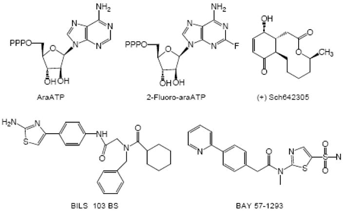
DNA primase synthesizes short RNA primers that replicative polymerases further elongate in order to initiate the synthesis of all new DNA strands. Thus, primase owes its existence to the inability of DNA polymerases to initiate DNA synthesis starting with 2 dNTPs. Here, we discuss the evolutionary relationships between the different families of primases (viral, eubacterial, archael, and eukaryotic) and the catalytic mechanisms of these enzymes. This includes how they choose an initiation site, elongate the growing primer, and then only synthesize primers of defined length via an inherent ability to count. Finally, the low fidelity of primases along with the development of primase inhibitors is described.


Figures

Figure 1
Figure 1
Structures of the RNA polymerase domains (RPD) of bacterial and phage primases. A. Solvent accessible surface of the E. coli DnaG RPD, emphasizing the overall almond-like shape of these enzymes. A short piece of single-stranded DNA (shown as stick model) occupies the putative template binding groove. The RPD’s of E. coli DnaG primase (B; 3B39.pdb), A. aeolicus DnaG primase (C; 2AU3.pdb) and bacteriophage T7 primase (D; 1NUI.pdb) are displayed as ribbon models using identical colors for structurally homologous domains. The toprim fold and the N-terminal RPD subdomain 1 are presented in grey and green, respectively. The C-terminal RPD subdomain 3 is highlighted in orange. The oxygens of putative active site residues are shown as red sticks. The structures of A. aeolicus and T7 feature a zinc binding domain (ZBD; purple) that is also present in E. coli but so far has not been resolved by X crystallography. Interestingly, the ZBD of A. aeolicus binds tightly to the rear of the associated polymerase domain, whereas the ZBD of T7 adopts an uncoupled and extended conformation.
Figure 2
Figure 2
Archaeal primase fold. Ribbon presentations of the small primase subunits of Sulfolobus solfataricus (Sso, pdb entry: 1ZTZ) and Pyrococcus horikoshii (Pho, pdb entry: 1V34). The β sheets are colored blue, the helical regions grey. Pho has a helical domain (magenta) that Sso is mostly lacking. The Pho structure contains a UTP (orange sticks), Zn2+ ions are shown in green. The expected catalytic triads are depicted as grey sticks with oxygens in red (Sso: D101, D103 and D235; Pho: D95, D97 and D280).
Figure 3
Figure 3
Model of the T7 primase-helicase hexamer with a Zn+2 binding domain of primase-helicase interacting with the neighboring primase-helicase at a 3′-CTG initiation site.
Figure 4
Figure 4
Hinge model for counting by primase where the two subunits either begin in a “closed” complex and transition to an open complex (Top) or vice versa (Bottom).
Figure 5
Figure 5
Model of one subunit of the hexameric E. coli helicase and one subunit of the trimeric primase bound to DNA at an initiation site via a trans interaction between the Zn+2 binding domain of one primase molecule and the polymerase domain of a neighboring primase monomer.
Figure 6
Figure 6
The subunit contacts in pol α-primase.
Figure 7
Figure 7
The three-point hand off in transferring primers from E. coli primase to the processivity factor and polymerase. Primase binds and then synthesizes a primer on ssb coated DNA (black line). The γ-complex then loads the β clamp, followed by DNA polymerase binding.
Chart 1
Chart 1
Examples of bases that human and herpes primase do not efficiently polymerize.
Chart 2
Chart 2
Examples of bases that herpes and human primase efficiently polymerize.
Chart 3
Chart 3
Structures of human primase inhibitors (Top) and herpes primase inhibitors (Bottom).

References

    1. Kornberg A, Baker T. DNA Replication. 2. W. H. Freeman & Co; San Francisco: 1992.
    1. Zhang SS, Grosse F. Accuracy of DNA Primase. Journal of Molecular Biology. 1990;216:475–479. - PubMed
    1. Hauschka PV. Analysis of Nucleotide Pools in Animal Cells. Methods in Cell Biology. 1973;3:362–462. - PubMed
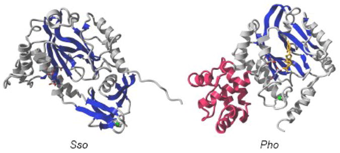
    1. LeBreton M, Henneke G, Norais C, Flament D, Myllykallio H, Querellou J, Raffin JP. The Heterodimeric Primase from the Euryarchaeon Pyrococcus abyssi: a Multifunctional Enzyme for Initiation and Repair? J Mol Biol. 2007;374:1172–1185. - PubMed
    1. Lao-Sireix SH, Bell SD. The Heterodimeric Primase of the Hyperthermophilic Archaeon Sulfolobus solfataricus Possesses DNA and RNA Primase, Polymerase, and 3′-terminal Nucleotidyl Transferase Activities. J Mol Biol. 2004;344:1251–1263. - PubMed

Publication types

LinkOut - more resources