Review
. 2009 Jun;19(3):263-74.
doi: 10.1016/j.conb.2009.06.001. Epub 2009 Jun 21.

Semaphorin function in neural plasticity and disease

Affiliations
Review

Semaphorin function in neural plasticity and disease

R Jeroen Pasterkamp et al. Curr Opin Neurobiol. 2009 Jun.

Abstract

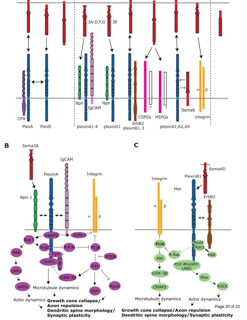
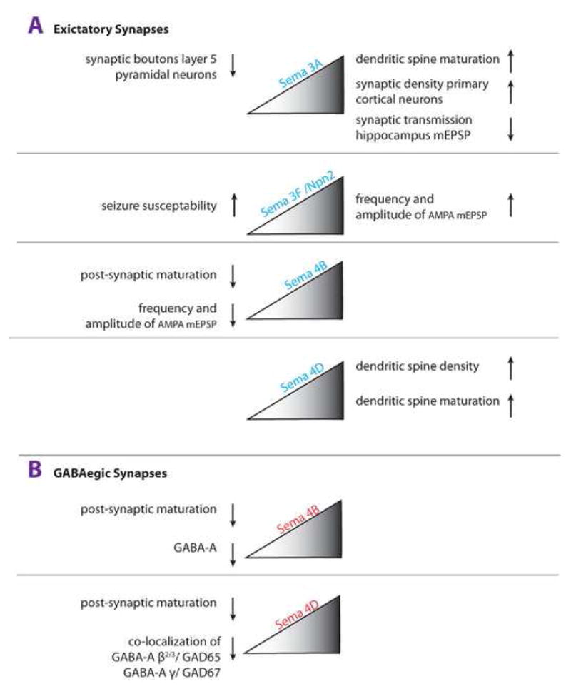
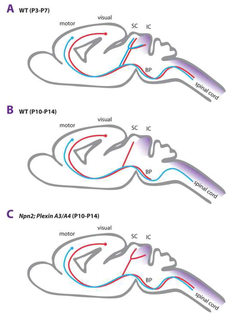
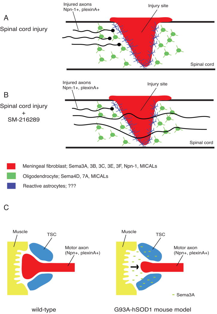
The semaphorins, originally discovered as evolutionarily conserved steering molecules for developing axons, also influence neuronal structure and function in the early postnatal and juvenile nervous systems through several refinement processes. Semaphorins control synaptogenesis, axon pruning, and the density and maturation of dendritic spines. In addition, semaphorins and their downstream signaling components regulate synaptic physiology and neuronal excitability in the mature hippocampus, and these proteins are also implicated in a number of developmental, psychiatric, and neurodegenerative disorders. Significant inroads have been made in defining the mechanisms by which semaphorins regulate dynamic changes in the neuronal cytoskeleton at the molecular and cellular levels during embryonic nervous system development. However, comparatively little is known about how semaphorins influence neuronal structure and synaptic plasticity during adult nervous system homeostasis or following injury and disease. A detailed understanding of how semaphorins function beyond initial phases of neural network assembly is revealing novel insights into key aspects of nervous system physiology and pathology.


Conflict of interest statement

9. Ethic in publishing: General statement

The authors declare no conflict of interest (RJP and RJG)

Figures

Figure 1
Figure 1. Semaphorins and their receptors and signaling pathways in the nervous system
(a) Semaphorins exist as secreted, membrane-spanning, or glycosylphosphatidylinositol (GPI)-anchored proteins many of which bind to plexin receptors. Invertebrate semaphorins in classes 1 and 2 (Sema1s and Sema2s) utilize PlexinA (PlexA) and PlexinB (PlexB) receptors, respectively. The co-receptor Off-track (OTK) functions with PlexA, and PlexA and PlexB might form heteromultimeric receptor complexes. In vertebrates, PlexinAs are receptors for Sema3s and Sema6s. In contrast to Sema3s, Sema6s do not require neuropilins (Npns) to direct plexinA binding. Ig-superfamily cell adhesion molecules (IgCAMs) function with Npns and plexinAs to mediate neuronal Sema3 functions. Unlike other Sema3s, Sema3E directly binds plexinD1. Sema3E-plexinD1-mediated axon attraction, but not repulsion, requires Npn-1. Sema4s associate with plexinBs and plexinB-ErbB2 interactions are required for certain Sema4 functions. HSPGs are required for Sema5A-mediated axon attraction and CSPGs switch Sema5A from an attractant to a repellent presumably by facilitating interactions with an unknown neuronal receptor. The GPI-linked semaphorin Sema7A binds neuronal integrin receptors. Recent evidence indicates that some membrane-spanning semaphorins might function both as ligands and receptors, and in addition might influence plexin receptors through cis interactions. (b) Sema3A binding to the Npn–plexinA complex promotes FARP2 dissociation from plexinA. Dissociated FARP2 activates Rac1, which facilitates Rnd1–plexinA associations and drives PIPKIγ661-mediated inhibition of integrin function. Active Rac1 also controls actin dynamics through a PAK-LIMK1-cofilin pathway. Rnd1–plexinA interactions stimulate plexinA RasGAP activity which suppresses R-Ras and inactivates PI3K-Akt signaling. Interestingly, Sema3s also regulate PI3K-Akt through PTEN. Downregulation of PI3K-Akt signaling leads to the inhibition of integrin-mediated adhesion, activation of myosin II (MyoII), and reduced phosphorylation of ERM and GSK-3β. Phosphorylation of CRMP2 by GSK-3β inactivates CRMP2 and relies on a Cdk5- and Fyn-dependent priming phopshorylation. CRMP2 regulates microtubule dynamics. (c) Sema4D–plexinB1 interactions promote phosphorylation of plexinB1 and ErbB2. Repulsive Sema4D–plexinB signaling involves four GTPases; Rnd1, R-Ras, Rho and Rac1. Sema4D–plexinB1 binding promotes Rnd1-dependent activation of the plexinB1 GAP domain and transient suppression of R-Ras activity. R-Ras inactivation promotes PI3K and Akt inactivation followed by GSK-3β activation and CRMP2 inactivation. In addition, plexinB1 associates with the RhoGEFs PDZ-RhoGEF and LARG to regulate Rho and ROCK activity and to influence cytoskeletal dynamics. Rac1–plexinB1 binding might sequester Rac1 away from PAK.
Figure 2
Figure 2. Class 3 and 4 semaphorins regulate synapse structure and physiology
A growing number of semaphorins have been implicated in the regulation of synaptogenesis, dendritic spine density, dendritic spine maturation, and synaptic transmission. Effects of decreased semaphorin signaling are shown on the left and effects in the presence of increased semaphorin signaling are shown on the right. (a) Excitatory Synapses: In glutamatergic neurons, Sema3A increases the density of clusters of synapsin I and postsynaptic density-95 in vitro. Loss of Sema3A results in a dendritic spine phenotype of layer 5 cortical projection neurons in vivo. Physiological studies showed that exogenously applied Sema3F to acute hippocampal slices modulates basal synaptic transmission by increasing the frequency and amplitude of mESPCs in dentate granule cells and CA1 pyramidal neurons. Sema3A and Sema3F appear to have opposite effects on synaptic transmission in the hippocampus, as in the presence of Sema3A CA1 neurons are more depressed. In primary hippocampal neurons, RNAi knock-down of Sema4B, but not Sema4D, attenuates postsynaptic maturation, as assessed by a decrease in the density of synapsin 1/PSD-95 colocalized puncta and a decrease in AMPA-receptor containing synapses. Exogenous application of Sema4D promotes dendritic spine density and spine maturation in vitro. In primary hippocampal neurons, RNAi knock-down of Sema4B (but not Sema4D) results in a decrease of the frequency and amplitude of AMPA-receptor mediated mESPCs. (b) GABAergic Synapses: In GABAergic neurons, RNAi knock-down of Sema4B or Sema4D attenuates postsynaptic maturation, as assessed by the density of GABA-A receptor puncta. In vivo, loss of Sema4D attenuates the development of GABAergic synapses in the hippocampus, as assessed by co-localization of GABA-A and GAD67 staining. In vivo, Sema3A and Sema3F influence GABAergic function indirectly by regulating tangential migration of cortical interneurons.
Figure 3
Figure 3. Semaphorins regulate stereotypic pruning
(a) In early mouse postnatal development (P3-P5), layer 5 projection neurons located in the visual cortex or motor cortex extend subcortical projections to similar target areas, including the superior (SC) and inferior colliculus (IC) in the diencephalon, and the dorsal spinal cord. Some of these targets are innervated by virtue of interstitial axon branching. Subsequent elimination (pruning) of specific subsets of axon branches is used to establish the mature innervation pattern. (b) In wild-type animals at P10-14, axon branches of upper motor neurons to the SC and IC and projections of visual cortical neurons to the IC and corticospinal tract (CST) are lost as a result of stereotypic pruning. As a consequence, visual cortical projections terminate in the SC and basilar pons (BP) and layer 5 motor projections give rise to the CST. (c) In Npn-2 null or PlexinA3/PlexinA4 double mutant mice pruning of visual cortical projections to the CST and IC is defective. Pruning of upper motorneuron axon collaterals to the SC and IC is normal and thus, independent of Npn-2 and PlexinA3/PlexinA4. The Npn-2 ligand Sema3F (purple) is expressed in the IC and dorsal spinal cord, and thus, a strong candidate for a ligand that signals pruning of exuberant visual cortical axon projections. The molecular mechanisms that regulate axon pruning of upper motorneuron projections to the SC and IC have not yet been defined.
Figure 4
Figure 4
Role for semaphorins in regeneration failure and amyotrophic lateral sclerosis (ALS)? (a, b) Schematic representation of the injured rodent spinal cord. Following injury to the central nervous system (CNS), meningeal fibroblasts migrate into the injury site and form the fibrotic core of the neural scar. This fibrotic scar is surrounded by a region of reactive astrocytes and, more distantly, oligodendrocytes. Meningeal fibroblasts and oligodendrocytes in CNS injury sites have been shown to express semaphorins, their receptors and signaling molecules which may act to inhibit regrowth of severed axons. (b) Application of the fungus-derived inhibitor SM-216289 into the lesion site of rats with spinal cord transection injuries enhances anatomical and functional regeneration, presumably by preventing interactions between Sema3A and axonal neuropilin-1 (Npn-1). (c) Schematic representation of the neuromuscular junction (NMJ) of wild-type and G93A-hSOD1 mice. G93A-hSOD1 mice, a mouse model for ALS, display a marked increase of Sema3A expression in terminal Schwann cells (TSCs) at the NMJ. Intriguingly, this increase is limited to TSCs of fast-fatigable type IIb and IIx muscle fibers which are the first muscle subtype to be lost in ALS. Increased expression of Sema3A in TSCs may lead to de-adhesion or repulsion of motor axons away from the NMJ, eventually resulting in axonal denervation and motor neuron degeneration.

Similar articles

Cited by

References

    1. Tran TS, Kolodkin AL, Bharadwaj R. Semaphorin regulation of cellular morphology. Annu Rev Cell Dev Biol. 2007;23:263–292. - PubMed
    1. Yuste R, Bonhoeffer T. Morphological changes in dendritic spines associated with long-term synaptic plasticity. Annu Rev Neurosci. 2001;24:1071–1089. - PubMed
    1. Yazdani U, Terman JR. The semaphorins. Genome Biol. 2006;7:211. - PMC - PubMed
    1. Franco M, Tamagnone L. Tyrosine phosphorylation in semaphorin signalling: shifting into overdrive. EMBO Rep. 2008;9:865–871. - PMC - PubMed
    1. Falk J, Bechara A, Fiore R, Nawabi H, Zhou H, Hoyo-Becerra C, Bozon M, Rougon G, Grumet M, Puschel AW, et al. Dual functional activity of semaphorin 3B is required for positioning the anterior commissure. Neuron. 2005;48:63–75. - PubMed

Publication types