. 2009 Jul 7;106(27):10917-21.
doi: 10.1073/pnas.0904792106. Epub 2009 Jun 18.

Non-blinking and photostable upconverted luminescence from single lanthanide-doped nanocrystals

Affiliations

Non-blinking and photostable upconverted luminescence from single lanthanide-doped nanocrystals

Shiwei Wu et al. Proc Natl Acad Sci U S A. .

Abstract

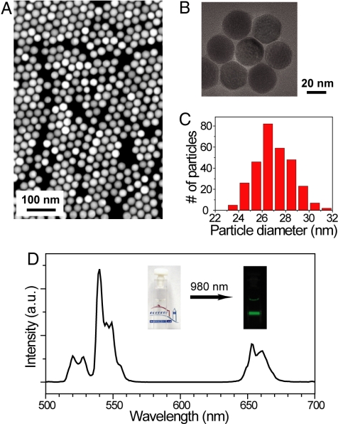
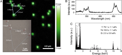
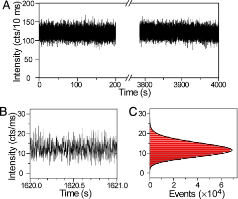
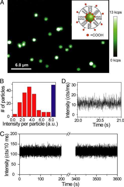
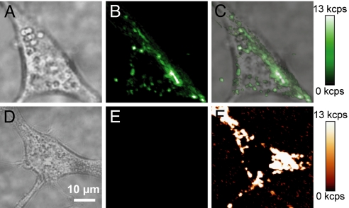
The development of probes for single-molecule imaging has dramatically facilitated the study of individual molecules in cells and other complex environments. Single-molecule probes ideally exhibit good brightness, uninterrupted emission, resistance to photobleaching, and minimal spectral overlap with cellular autofluorescence. However, most single-molecule probes are imperfect in several of these aspects, and none have been shown to possess all of these characteristics. Here we show that individual lanthanide-doped upconverting nanoparticles (UCNPs)--specifically, hexagonal phase NaYF(4) (beta-NaYF(4)) nanocrystals with multiple Yb(3+) and Er(3+) dopants--emit bright anti-Stokes visible upconverted luminescence with exceptional photostability when excited by a 980-nm continuous wave laser. Individual UCNPs exhibit no on/off emission behavior, or "blinking," down to the millisecond timescale, and no loss of intensity following an hour of continuous excitation. Amphiphilic polymer coatings permit the transfer of hydrophobic UCNPs into water, resulting in individual water-soluble nanoparticles with undiminished photophysical characteristics. These UCNPs are endocytosed by cells and show strong upconverted luminescence, with no measurable anti-Stokes background autofluorescence, suggesting that UCNPs are ideally suited for single-molecule imaging experiments.


Conflict of interest statement

The authors declare no conflict of interest.

Figures

Fig. 1.
Fig. 1.
Lanthanide-doped UCNPs (NaYF4: Yb3+/Er3+) showing near-infrared to visible upconverted luminescence. (A and B) TM-SEM and TEM images of the UCNPs. (C) A histogram of particle sizes obtained from TM-SEM images of approximately 300 nanoparticles (average particle size = 26.9 ± 1.7 nm). (D) Upconverted luminescence spectrum of the UCNPs (0.01 wt%) diluted in hexane when excited by a 980-nm CW diode laser at power density of approximately 8 W/cm2. Upconverted visible emission bands are centered at 540 nm and 655 nm. (Inset) Photographs of the transparent solution without laser illumination and the upconverted visible luminescence under laser illumination.
Fig. 2.
Fig. 2.
Individual UCNPs on a silicon nitride membrane. (A) Confocal upconverted luminescent image of individual UCNPs. Laser power density is approximately 3 × 10⁶ W/cm2, and dwell time per pixel is 10 ms. The TM-SEM image (Inset) taken at the upper left corner region of the optical image shows that the individual diffraction-limited luminescent spots are emitted from individual UCNPs. The 5 individual UCNPs are labeled as A–E; an impurity is labeled as F. (see Fig. S3 and Table S1, for their TM-SEM images and local chemical compositions.) (B) Upconverted luminescence spectrum of a single UCNP. (C) EDS of the UCNP labeled as C in A. From the EDS analysis of 20 individual UCNPs, the averaged ratios and their standard deviations of different elements (Y, Yb, and Er) are obtained and also noted in C.
Fig. 3.
Fig. 3.
Photostability and non-blinking behavior of a single UCNP. (A) The time trace of emission intensity from a single UCNP under continuous laser illumination for more than 1 h, suggesting the durable photostability of the UCNPs. The bin time for each data point is 10 ms. The laser power used on the sample is approximately 10 mW, corresponding to a power density of ≈5 × 10⁶ W/cm2. (B and C) The zoom-in time trace and histogram of emission intensity, showing no on/off behavior − non-blinking. Note that the bin time for each data point in (B and C) is reduced to 1 ms for higher time resolution. The histogram is fitted with a Poisson distribution function, where I is the emission intensity, A is the number of total events (6 × 10⁵), and λ is the fitting parameter (λ = 12.066 ± 0.009).
Fig. 4.
Fig. 4.
Upconverted luminescence of individual water-soluble UCNPs. (A) Confocal upconverted luminescent image of individual amphiphilic polymer-coated UCNPs (schematically shown in the Inset) sparsely dispersed on a clean coverglass. The laser power is approximately 10 mW, equivalent to approximately 5 × 10⁶ W/cm2. Some of the bright luminescent spots represent multiple UCNPs within the diffraction limited area, generating saturated “white” spots in the image. (B) A histogram of integrated emission intensity from over 200 upconverted luminescent spots, suggesting that most of the luminescent spots are from single polymer-coated UCNPs. The data were analyzed from confocal upconverted luminescent images over a 75 × 75 μm area, and the number of saturated “white” spots was shown in the histogram as a blue bar. Such single water-soluble UCNPs also exhibit exceptional photostability (C) and non-blinking behavior (D).
Fig. 5.
Fig. 5.
Live-cell imaging of UCNPs in NIH 3T3 murine fibroblasts. (A) Brightfield image of a cell with endocytosed UCNPs, (B) upconverted luminescence following 980-nm excitation, and (C) overlay. (D) Brightfield image of a cell without UCNPs, (E) upconverted luminescence following 980-nm excitation, and (F) cellular autofluorescence following 532-nm excitation. All confocal images are shown on the same intensity scale. (Scale bar, 10 μm.)

References

    1. Heer S, Kompe K, Gudel HU, Haase M. Highly efficient multicolour upconversion emission in transparent colloids of lanthanide-doped NaYF4 nanocrystals. Adv Mater. 2004;16:2102–2105.
    1. Mai H-X, Zhang Y-W, Sun L-D, Yan C-H. Highly efficient multicolor up-conversion emissions and their mechanisms of monodisperse NaYF 4:Yb,Er core and core/shell-structured nanocrystals. J Phys Chem C. 2007;111:13721–13729.
    1. Weiss S. Fluorescence spectroscopy of single biomolecules. Science. 1999;283:1676–1683. - PubMed
    1. Alivisatos AP, Gu W, Larabell C. Quantum dots as cellular probes. Annu Rev Biomed Eng. 2005;7:55–76. - PubMed
    1. Medintz IL, Uyeda HT, Goldman ER, Mattoussi H. Quantum dot bioconjugates for imaging, labeling, and sensing. Nat Mater. 2005;4:435–446. - PubMed

Publication types

LinkOut - more resources