. 2009 Jul 7;106(27):11085-9.
doi: 10.1073/pnas.0904716106. Epub 2009 Jun 18.

An icosahedral algal virus has a complex unique vertex decorated by a spike

Affiliations

An icosahedral algal virus has a complex unique vertex decorated by a spike

Mickaël V Cherrier et al. Proc Natl Acad Sci U S A. .

Abstract

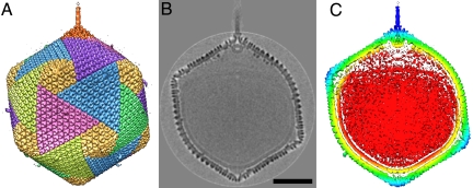
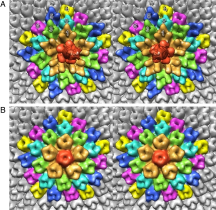
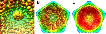
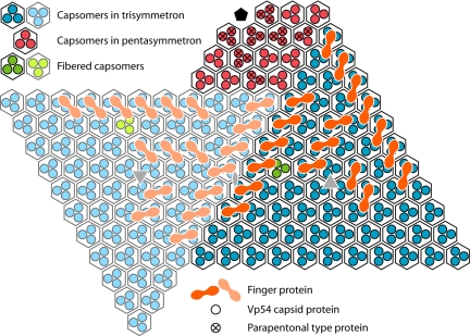
Paramecium bursaria Chlorella virus-1 is an icosahedrally shaped, 1,900-A-diameter virus that infects unicellular eukaryotic green algae. A 5-fold symmetric, 3D reconstruction using cryoelectron microscopy images has now shown that the quasiicosahedral virus has a unique vertex, with a pocket on the inside and a spike structure on the outside of the capsid. The pocket might contain enzymes for use in the initial stages of infection. The unique vertex consists of virally coded proteins, some of which have been identified. Comparison of shape, size, and location of the spike with similar features in bacteriophages T4 and P22 suggests that the spike might be a cell-puncturing device. Similar asymmetric features may have been missed in previous analyses of many other viruses that had been assumed to be perfectly icosahedral.


Conflict of interest statement

The authors declare no conflict of interest.

Figures

Fig. 1.
Fig. 1.
The 5-fold-averaged cryoEM structure of PBCV-1 viewed down a quasi-2-fold axis. (A) Hexagonal arrays of major capsomers form trisymmetrons and pentasymmetrons (yellow). The unique vertex with its spike structure is at the top. Capsomers in neighboring trisymmetrons are related by a 60° rotation, giving rise to the boundary between trisymmetrons. (B) Central cross-section of the cryoEM density. (Scale bar: 500 Å.) (C) The same view as in B but colored radially, with red density being within 680 Å, yellow between 680 and 745 Å, green between 745 and 810 Å, light blue between 810 and 880 Å, and dark blue greater than 880 Å. Note the typical lipid low-density gap surrounding the red nucleocapsid density.
Fig. 2.
Fig. 2.
Stereodiagrams of the pentasymmetrons at (A) the unique vertex and (B) the 5-fold axis opposite the unique vertex. Note the difference in the density distribution of the pentameric capsomer (red) in A and B. The top end of the narrow spike can also be seen in A. Although all of the major capsomers (Vp54 trimers) surrounding the pentasymmetrons and in the pentasymmetron shown in B have very similar density distributions, this is not the case for the pentasymmetron at the special vertex shown in A. The peripentonal capsomers around the unique vertex are shown in gold and have an extended amino acid insertion. The 10 capsomers surrounding the peripentonal capsomers are colored blue and green. Whereas the green capsomers are similar to the peripentonal capsomers, the blue capsomers are similar to the major capsomers. The capsomers in the outer pentasymmetron ring are colored yellow, dark blue, and pink. The yellow and pink capsomers are similar in structure to the major capsid protein, whereas the dark blue capsomers have a slightly different density distribution.
Fig. 3.
Fig. 3.
View of features inside the PBCV-1 capsid. All panels are colored as in Fig. 1C. (A) The portal within the capsid at the unique vertex. (B) View of the unique vertex from the inside of the virus. Note the finger proteins at a slightly smaller radius appear red. No finger proteins are associated with the pentasymmetron at the unique vertex. (C) View of the vertex opposite the unique vertex. Note the lack of finger protein features and the low-density gap corresponding to the lipid bilayer membrane roughly in the yellow density.
Fig. 4.
Fig. 4.
Diagrammatic organization of capsomers as seen from inside the virus viewed toward the unique vertex. Within each quasihexagonal capsomer, there are 3 Vp54 monomers, each represented by a dot (blue within one trisymmetron, light blue within the neighboring trisymmetron, and red within the pentasymmetron). Some of the capsomers are associated with minor capsid finger proteins, shown in orange in one trisymmetron and a lighter orange in the neighboring trisymmetron.

References

    1. Dunigan DD, Fitzgerald LA, Van Etten JL. Phycodnaviruses: A peek at genetic diversity. Virus Res. 2006;117:119–132. - PubMed
    1. Meints RH, Lee K, Burbank DE, Van Etten JL. Infection of a Chlorella-like alga with the virus, PBCV-1: Ultrastructural studies. Virology. 1984;138:341–346. - PubMed
    1. Van Etten JL, Lane LC, Meints RH. Viruses and viruslike particles of eukaryotic algae. Microbiol Rev. 1991;55:586–620. - PMC - PubMed
    1. Yan X, et al. Structure and assembly of large lipid-containing dsDNA viruses. Nat Struct Biol. 2000;7:101–103. - PMC - PubMed
    1. Meints RH, Burbank DE, Van Etten JL, Lamport DT. Properties of the Chlorella receptor for the virus PBCV-1. Virology. 1988;164:15–21. - PubMed

Publication types

MeSH terms

LinkOut - more resources