. 2009 Jun 30;106(26):10835-40.
doi: 10.1073/pnas.0903463106. Epub 2009 Jun 17.

Dendro-dendritic bundling and shared synapses between gonadotropin-releasing hormone neurons

Affiliations

Dendro-dendritic bundling and shared synapses between gonadotropin-releasing hormone neurons

Rebecca E Campbell et al. Proc Natl Acad Sci U S A. .

Abstract

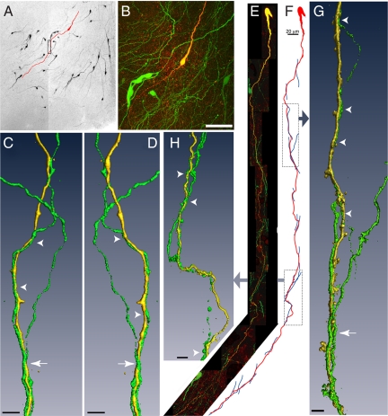
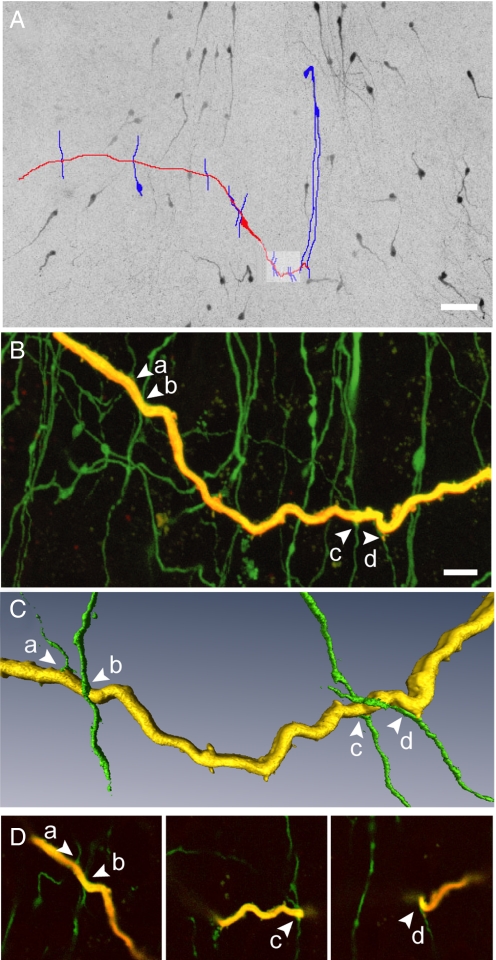
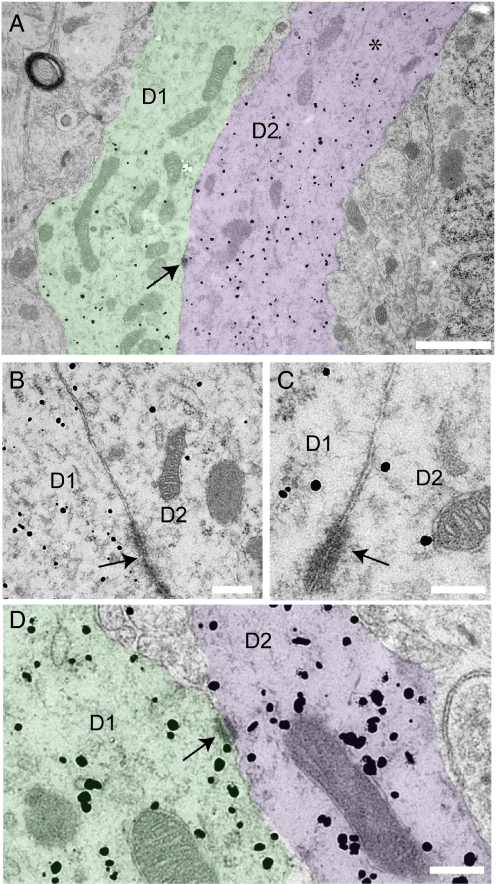
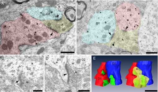
The pulsatile release of gonadotropin-releasing hormone (GnRH) is critical for mammalian fertility, but the mechanisms underlying the synchronization of GnRH neurons are unknown. In the present study, the full extent of the GnRH neuron dendritic tree was visualized by patching and filling individual GnRH neurons with biocytin in acute brain slices from adult GnRH-green fluorescent protein (GFP) transgenic mice. Confocal analysis of 42 filled GnRH neurons from male and female adult mice revealed that the dendrites of the great majority of GnRH neurons (86%) formed multiple close appositions with dendrites of other GnRH neurons. Two types of interactions were encountered; the predominant interaction was one of vertical dendritic bundling where dendrites were found to wrap around each other in the same axis. The other interaction was one in which a GnRH neuron dendrite intercepted other GnRH neuron dendrites in a perpendicular fashion. Electron microscopy using pre-embedded, silver-enhanced immunogold labeling for both GnRH and GFP peptides in GnRH-GFP transgenic mice, confirmed that GnRH neuron dendrites were often immediately juxtaposed. Membrane specializations, including punctae and zonula adherens, were found connecting adjacent dendritic elements of GnRH neurons. Remarkably, individual afferent axon terminals were found to synapse with multiple GnRH neuron dendrites at sites of bundling. Together, these data demonstrate that GnRH neurons are not isolated from one another but, rather, interconnected via their long dendritic extensions. The observation of shared synaptic input to bundled GnRH neuron dendrites suggests a mechanism of GnRH neuron synchronization.


Conflict of interest statement

The authors declare no conflict of interest.

Figures

Fig. 1.
Fig. 1.
Biocytin-filled GnRH neurons exhibiting dendro-dendritic bundling with other GnRH neurons in the verticle orientation. (A) Montage of low power confocal images through a thick slice of the rPOA shows GnRH-GFP neurons (black) and a single GnRH neuron that was filled with biocytin and subsequently labeled with a fluorescent marker (traced in red). (B) High power projection of confocal images of the filled neuron in A, showing endogenous GFP expression in multiple GnRH soma and in a plexus of processes (green) and fluorescently labeled, biocytin-filled GnRH neuron (yellow/red). (C and D) Three-dimensional isosurface rendering of GFP (green) and biocytin-filled (yellow) dendrites (rectangle in A) that bundle together viewed from 2 angles 180° apart. Arrowhead indicates 2 bundled dendrites; arrow shows the bundling of 3 dendrites. (E) Montage of high power confocal image stacks showing another filled GnRH neuron exhibiting dendritic contacts and bundling in the vertical orientation. (F) A schematic traced from confocal image data illustrates 15 apparent close associations between the filled dendrite (red) and other GnRH neuron dendrites (blue). Three-dimensional rendered images of the filled dendrite and apposing dendrites created from high power confocal images are shown for the highlighted regions. (G) Two juxtaposed GnRH neuron dendrites can be seen in the upper portion of the image (arrowheads), and 3 dendrites are found bundling together further down the length of the dendrite (arrow). (H) Points of contact are apparent between the filled dendrite and 2 dendrites that run in parallel with the filled dendrite (arrowheads). (Scale bars: B, 50 μm; C, D, G, and H, 5 μm.)
Fig. 2.
Fig. 2.
Dendro-dendritic associations in the horizontal orientation. (A) Montage of low power confocal images through a thick brain slice through the rPOA shows GnRH-GFP neurons (black) and a single GnRH neuron that was filled with biocytin and subsequently labeled with a fluorescent marker (traced in red). GnRH-GFP neuron dendrites in apparent close apposition with the dendrite of the filled GnRH neuron are traced in blue. (B) High power confocal projection of the area highlighted in panel A showing a portion of the filled dendrite (yellow) and other endogenously fluorescent GnRH processes. Arrows indicate 4 dendrites (a–d) found in close apposition with the filled dendrite. (C) Isosurface rendering of the dendrites in close apposition show the perpendicular nature of these interactions. (D) Individual optical sections (0.32 μm thick) showing the close apposition of dendritic processes. (Scale bars: A, 50 μm; B, 10 μm.)
Fig. 3.
Fig. 3.
Ultrastructural evidence of bundling GnRH dendrites. (A) Two distinct silver-enhanced immunogold labeled GnRH neuron dendrites (D1 and D2, pseudocolored for clarity) in parallel with membranes juxtaposed. Silver-enhanced immunogold label appears as scattered black dots. Note that both GnRH and GFP were labeled and that the latter does not associate with dense core vesicles. (A) Asterisk indicates an area of loosely arranged microtubules running longitudinally through the labeled dendrites. Arrow indicates a zonula adherens junction between the 2 dendrites. (B and C) Higher power view in adjacent serial sections through the juxtaposed dendrites shown in A, showing additional zonula adherens membrane specializations. (D) Another example of adjacent GnRH neuron dendrites (D1 and D2, pseudocolored for clarity). Arrow indicates punctae adherens junction (arrows) linking the 2 membranes. (Scale bars: A, 1 μm; B–D, 200 nm.
Fig. 4.
Fig. 4.
Double synapses are present on bundling GnRH dendrites. (A and B) Two examples from different animals of bundling GnRH neuron dendrites in cross section (D1 and D2) receiving shared afferent input (arrows) from single axon terminals (A). (B) Arrowhead indicates a single dense core vesicle amongst numerous clear vesicles in the terminal. Asterisks are positioned amongst typical microtubules, seen in cross section as small circles, in the labeled dendrites. (C–E) Serial ultra-thin images through the labeled, bundled GnRH dendrites shown in B were collected and a 3-dimensional model created (E). Postsynaptic densities (green) were present on the dendritic shaft (B) and on the neck of sessile spines (arrows, C and D). The 3-dimensional, reconstructed dendrites are pictured with and without the shared afferent terminal (yellow).

References

    1. Knobil E, Plant TM, Wildt L, Belchetz PE, Marshall G. Control of the rhesus monkey menstrual cycle: Permissive role of hypothalamic gonadotropin-releasing hormone. Science. 1980;4437:1371–1373. - PubMed
    1. Herbison AE. Physiology of the gonadotropin-releasing hormone neuronal network. In: Neill JD, editor. Knobil and Neil's Physiology of Reproduction. 3rd Ed. New York: Raven Press; 2006. pp. 1415–1482.
    1. Moenter SM, DeFazio AR, Pitts GR, Nunemaker CS. Mechanisms underlying episodic gonadotropin-releasing hormone secretion. Front Neuroendocrinol. 2003;24:79–93. - PubMed
    1. Wray S. Development of luteinizing hormone releasing hormone neurones. J Neuroendocrinol. 2001;13:3–11. - PubMed
    1. Purnelle G, Gerard A, Czajkowski V, Bourguignon JP. Pulsatile secretion of gonadotropin-releasing hormone by rat hypothalamic explants without cell bodies of GnRH neurons. Neuroendocrinology. 1997;66:305–312. - PubMed

Publication types

MeSH terms