. 2009 Sep;23(9):1466-78.
doi: 10.1210/me.2009-0119. Epub 2009 Jun 18.

Subcellular trafficking of the TRH receptor: effect of phosphorylation

Affiliations

Subcellular trafficking of the TRH receptor: effect of phosphorylation

Brian W Jones et al. Mol Endocrinol. 2009 Sep.

Abstract

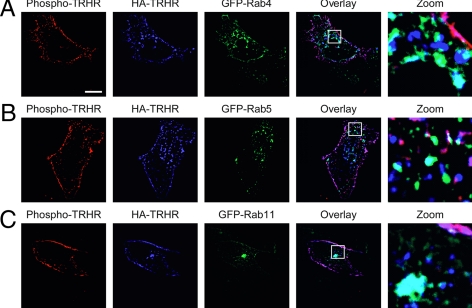
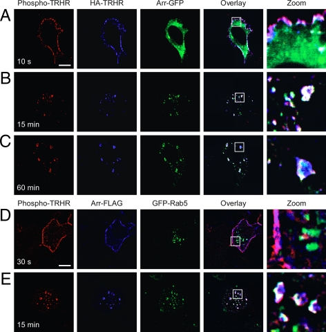
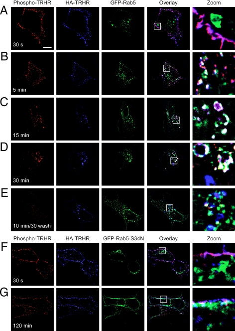
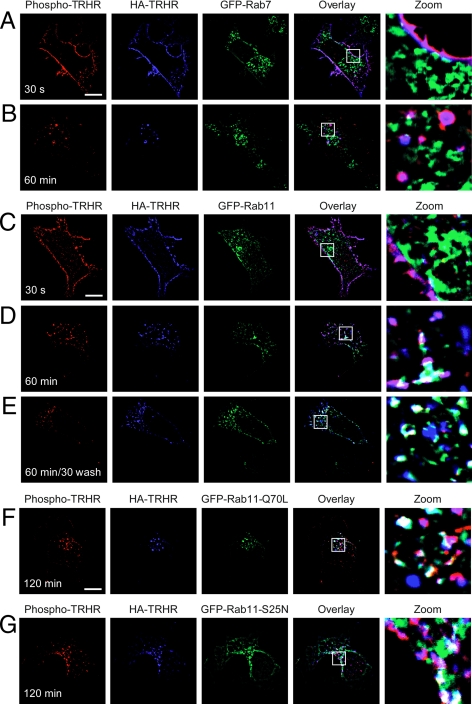
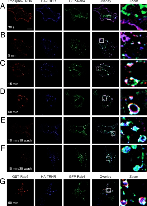
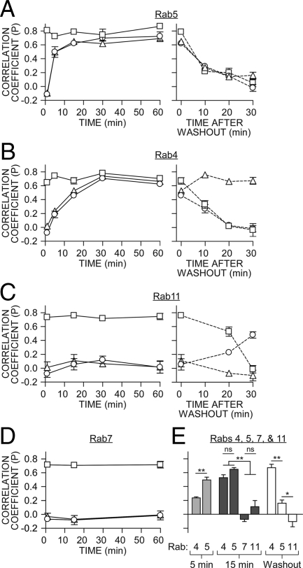
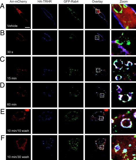
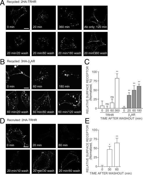
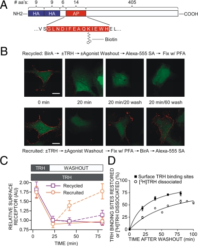
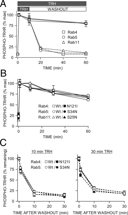
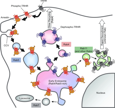
Activation of the G protein-coupled TRH receptor leads to its phosphorylation and internalization. These studies addressed the fundamental question of whether phosphorylation regulates receptor trafficking or endosomal localization regulates the phosphorylation state of the receptor. Trafficking of phosphorylated and dephosphorylated TRH receptors was characterized using phosphosite-specific antibody after labeling surface receptors with antibody to an extracellular epitope tag. Rab5 and phosphoreceptor did not colocalize at the plasma membrane immediately after TRH addition but overlapped extensively by 15 min. Dominant-negative Rab5-S34N inhibited receptor internalization. Later, phosphoreceptor was in endosomes containing Rab5 and Rab4. Dephosphorylated receptor colocalized with Rab4 but not with Rab5. Dominant-negative Rab4, -5, or -11 did not affect receptor phosphorylation or dephosphorylation, showing that phosphorylation determines localization in Rab4(+)/Rab5(-) vesicles and not vice versa. No receptor colocalized with Rab7; a small amount of phosphoreceptor colocalized with Rab11. To characterize recycling, surface receptors were tagged with antibody, or surface receptors containing an N-terminal biotin ligase acceptor sequence were labeled with biotin. Most recycling receptors did not return to the plasma membrane for more than 2 h after TRH was removed, whereas the total cell surface receptor density was largely restored in less than 1 h, indicating that recruited receptors contribute heavily to early repopulation of the plasma membrane.


Figures

Figure 1
Figure 1
TRH receptor in permeabilized cells. Cells expressing 2HA-AP-TRH receptor and GFP-Rab4, -5, or -11 were treated with 100 nm TRH for 30 sec, fixed, permeabilized, and stained with anti-HA and anti-phospho-TRH receptor antibodies. Overlap of blue HA-TRH receptor and green GFP-Rabs appears cyan in overlay. Bar, 10 μm. TRHR, TRH receptor.
Figure 2
Figure 2
Phospho-TRH receptor traffics with arrestin. A–C, Cells expressing 2HA-AP-TRH receptor and arrestin-GFP (green) were incubated with mouse anti-HA antibody to label surface receptors and then stimulated with 100 nm TRH for the indicated times. After fixation, cells were stained with antimouse Alexa Fluor 647 antibody to identify anti-HA antibody-labeled receptors (blue) or rabbit anti-phospho-TRH receptor antibody and antirabbit Alexa Fluor 555 antibody to identify phospho-TRH receptors (red). Pink shows overlay of red and blue channels; white shows overlay of all three channels. D and E, Cells expressing 2HA-AP-TRH receptor, arrestin-FLAG, and GFP-Rab5 were stimulated with 100 nm TRH for the indicated times and fixed and stained. Mouse anti-FLAG antibody followed by antimouse Alexa Fluor 647 antibody was used to label arrestin-FLAG (blue). Bar, 10 μm. Arr-, Arrestin; s, seconds; TRHR, TRH receptor.
Figure 3
Figure 3
TRH receptor internalizes via Rab5-positive endosomes. Cells expressing 2HA-AP-TRH receptor and (A–E) GFP-Rab5 or (F and G) GFP-Rab5-S34N were incubated with anti-HA antibody, stimulated with 100 nm TRH for the indicated times, and fixed and stained. Some cells were washed to remove TRH and allowed to recover for 30 min before fixation. The localization of GFP-Rab5-S34N was quite variable in different cells (e.g. panels F and G and supplemental Fig. 1). Bar, 10 μm. s, Seconds; TRHR, TRH receptor.
Figure 4
Figure 4
TRH receptor does not colocalize with Rab7 and minimally with Rab11. Cells expressing 2HA-AP-TRH receptor and either GFP-Rab7 (A and B) or GFP-Rab11 (C and D) were incubated with anti-HA antibody and stimulated with 100 nm TRH for the indicated times. Cells in panel E were washed to remove TRH and allowed to recover for 30 min before fixation and staining. Cells expressing 2HA-AP-TRH receptor and constitutively active GFP-Rab11-Q70L (panel F) or dominant-negative GFP-Rab11-S25N (panel G). Bar, 10 μm. s, Seconds; TRHR, TRH receptor.
Figure 5
Figure 5
Dephosphorylated TRH receptor traffics via Rab4. A–F, Cells expressing 2HA-AP-TRH receptor and GFP-Rab4 were incubated with anti-HA antibody, stimulated with 100 nm TRH for the indicated times, and fixed and stained. Some cells were washed to remove TRH and allowed to recover for the indicated times before fixation. G, Cells expressing 2HA-AP-TRH receptor, GFP-Rab4, and GST-Rab5 were treated as in panel D except that rabbit anti-GST antibody was used to label GST-Rab5. Bar, 10 μm. s, Seconds; TRHR, TRH receptor.
Figure 6
Figure 6
Colocalization of Rabs with TRH receptor. Pearson’s correlation coefficients for: 2HA-AP-TRH receptor with phospho-TRH receptor (squares), 2HA-AP-TRH receptor with GFP-Rabs (triangles) or phospho-TRH receptor with GFP-Rabs (circles) are shown as mean ± se (n = 3–12). A–D: Left panel, values from cells stimulated with TRH; A–C, right panel, values after agonist washout. E, Comparison of selected coefficients from panels A–D for colocalization of 2HA-AP-TRH receptor and particular Rab GTPases 5 or 15 min after stimulation with TRH or 30 min after agonist washout. **, P < 0.001; *, P < 0.05; nonsignificant (ns), P > 0.05 by one-way ANOVA.
Figure 7
Figure 7
Arrestin-receptor complex colocalizes with Rab4. Cells expressing 2HA-AP-TRH receptor, GFP-Rab4, and arrestin-mCherry were incubated with anti-HA antibody, stimulated with 100 nm TRH for the indicated times, fixed, and stained. Some cells were washed to remove TRH and allowed to recover for the indicated times before fixation. Bar, 10 μm. Arr-, Arrestin; s, seconds.
Figure 8
Figure 8
TRH and β2-adrenergic receptor trafficking. To measure recycling, cells expressing (A) 2HA-AP-TRH receptor or (B) HA-β 2-adrenergic receptor were incubated with anti-HA antibody, stimulated with 100 nm TRH or 1 μm isoproterenol, respectively, for the indicated times, fixed, and stained. Some cells were washed to remove agonist and allowed to recover for the indicated times before fixation; 1 μm propranolol was included in the recovery buffer for cells expressing HA-β2-adrenergic receptor. C, Quantification by line scan of relative surface receptor from cells treated as in panels A and B. D, To measure recruitment, cells expressing 2HA-AP-TRH receptor were treated as indicated; anti-HA antibody was then added to intact cells to identify surface receptors selectively. mCherry was coexpressed to allow positive identification of transfected cells (data not shown). E, Quantification by line scan of relative surface receptor from cells treated as in panel D. Shown are mean ± se of three to five cells from independent experiments, except the 60-min washout in panel E, which is the mean ± range from two determinations. **, P < 0.01; *, P < 0.05; nonsignificant (ns), P > 0.05 by one-way ANOVA compared with 0 min after washout. Bar, 10 μm. β2-AR, β2-adrenergic receptor; TRHR, TRH receptor.
Figure 9
Figure 9
Recycling of biotinylated TRH receptor. A, Model of 2HA-AP-TRH receptor. B and C, Cells expressing 2HA-AP-TRH receptor were incubated with or without 100 nm TRH with or without washout, as shown in the middle of panel B. Cells were fixed with paraformaldehyde (PFA), biotinylated with BirA, and labeled with Alexa Fluor 555 streptavidin in the order shown above (recycled) or below (recruited) the micrographs. Control experiments established that staining of naïve cells was comparable when fixation preceded incubation with BirA or when the order was reversed. Bar, 10 μm. C, Quantification by line scan of relative surface receptor. Shown are mean ± se of four to seven cells from four independent experiments. Solid lines show results of cells exposed to TRH throughout, and dashed lines show cells after TRH washout. D, To measure restoration of TRH binding sites after internalization, cells stably expressing 2HA-TRH receptors were incubated with or without 100 nm TRH for 20 min to cause receptor internalization, washed, and incubated for various times. Dishes were placed on ice, and the number of TRH-binding sites on the cell surface was titrated by incubating cells with 10 nm [3H]MeTRH on ice as described in Materials and Methods. To measure receptor cycling, cells were incubated with [3H]TRH for 20 min, washed, and incubated for different times before determination of the fraction of [3H]TRH released as described in Materials and Methods. Points show the mean ± se from five to nine experiments, each performed in duplicate. Error bars were within symbol size where not visible. aa, Amino acids.
Figure 10
Figure 10
Effect of Rab GTPase overexpression on TRH receptor phosphorylation and dephosphorylation. Cells expressing 2HA-AP-TRH receptor and (A–C) wild-type or (B and C) dominant-negative GFP-Rab4, -5, or -11 were stimulated with 100 nm TRH for the indicated times. Some cells were washed to remove TRH and incubated for the indicated times. Phosphorylated receptor was quantified by ELISA; shown are mean ± se of pooled replicates from at least three independent experiments. Solid lines show results of cells exposed to TRH throughout, and dashed lines show cells after TRH washout. GFP fluorescence was examined in all experiments. All of the GFP-rabs were well expressed and detectable in essentially all cells expressing receptors. Differences were not significant by one-way ANOVA. TRHR, TRH receptor; Wt, wild type.
Figure 11
Figure 11
TRH receptor trafficking. Primary routes of receptor trafficking are marked with thick arrows; minor or unused pathways have thin arrows. TRH receptors on the cell surface (gray) are rapidly phosphorylated (red) in response to TRH and recruit arrestin to the plasma membrane. Receptors can be dephosphorylated while still on the plasma membrane after agonist is removed. Arrestin escorts phospho-TRH receptor to clathrin-coated vesicles (CCV), where the arrestin-receptor complex is internalized on Rab5-positive endosomes (shaded blue). The arrestin-receptor complex is delivered to early endosomes rich in Rab5 and Rab4. Some phosphoreceptor bound to arrestin moves into Rab11-positive endosomes (shaded green). Once dephosphorylated, TRH receptor (blue) moves rapidly to exclusively Rab4-positive vesicles (shaded pink) and is slowly recycled to the plasma membrane. Before treatment with TRH, an intracellular pool of reserve TRH receptors (gray) colocalizes in part with Rab11 in a perinuclear compartment. TRHR, TRH receptor.

Similar articles

Cited by

References

    1. Jones BW, Song GJ, Greuber EK, Hinkle PM 2007 Phosphorylation of the endogenous thyrotropin-releasing hormone receptor in pituitary GH3 cells and pituitary tissue revealed by phosphosite-specific antibodies. J Biol Chem 282:12893–12906 - PubMed
    1. Groarke DA, Wilson S, Krasel C, Milligan G 1999 Visualization of agonist-induced association and trafficking of green fluorescent protein-tagged forms of both β-arrestin-1 and the thyrotropin-releasing hormone receptor-1. J Biol Chem 274:23263–23269 - PubMed
    1. Vrecl M, Anderson L, Hanyaloglu A, McGregor AM, Groarke AD, Milligan G, Taylor PL, Eidne KA 1998 Agonist-induced endocytosis and recycling of the gonadotropin-releasing hormone receptor: effect of β-arrestin on internalization kinetics. Mol Endocrinol 12:1818–1829 - PubMed
    1. Yu R, Hinkle PM 1998 Signal transduction, desensitization, and recovery of responses to thyrotropin-releasing hormone after inhibition of receptor internalization. Mol Endocrinol 12:737–749 - PubMed
    1. Moore CA, Milano SK, Benovic JL 2007 Regulation of receptor trafficking by GRKs and arrestins. Annu Rev Physiol 69:451–482 - PubMed

Publication types

Substances