. 2009 Aug;15(8):1528-42.
doi: 10.1261/rna.1578409. Epub 2009 Jun 18.

IGF2BP1 enhances HCV IRES-mediated translation initiation via the 3'UTR

Affiliations

IGF2BP1 enhances HCV IRES-mediated translation initiation via the 3'UTR

Susan Weinlich et al. RNA. 2009 Aug.

Abstract

The positive-strand RNA genome of the Hepatitis C virus (HCV) contains an internal ribosome entry site (IRES) in the 5'untranslated region (5'UTR) and structured sequence elements within the 3'UTR, but no poly(A) tail. Employing a limited set of initiation factors, the HCV IRES coordinates the 5'cap-independent assembly of the 43S pre-initiation complex at an internal initiation codon located in the IRES sequence. We have established a Huh7 cell-derived in vitro translation system that shows a 3'UTR-dependent enhancement of 43S pre-initiation complex formation at the HCV IRES. Through the use of tobramycin (Tob)-aptamer affinity chromatography, we identified the Insulin-like growth factor-II mRNA-binding protein 1 (IGF2BP1) as a factor that interacts with both, the HCV 5'UTR and 3'UTR. We report that IGF2BP1 specifically enhances translation at the HCV IRES, but it does not affect 5'cap-dependent translation. RNA interference against IGF2BP1 in HCV replicon RNA-containing Huh7 cells reduces HCV IRES-mediated translation, whereas replication remains unaffected. Interestingly, we found that endogenous IGF2BP1 specifically co-immunoprecipitates with HCV replicon RNA, the ribosomal 40S subunit, and eIF3. Furthermore eIF3 comigrates with IGF2BP1 in 80S ribosomal complexes when a reporter mRNA bearing both the HCV 5'UTR and HCV 3'UTR is translated. Our data suggest that IGF2BP1, by binding to the HCV 5'UTR and/or HCV 3'UTR, recruits eIF3 and enhances HCV IRES-mediated translation.


Figures
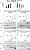
FIGURE 1.
FIGURE 1.
HCV IRES-mediated translation initiation is enhanced by the HCV 3′UTR in vitro. (A,B, upper panel) Schematic representation of HCV 5′UTR and HCV 3′UTR bearing Luc reporter mRNAs. (Lower panel) Cytoplasmic Huh7 cell extract was used in cell-free translation reactions programmed with the [32P] trace-labeled mRNAs HCV Luc or HCVΔ3′UTR Luc for the times indicated. Luc activity is expressed as Luc units. Error bars denote the standard deviation from the mean of three different experiments. [32P] trace-labeled mRNAs were extracted from the translation reaction at the time points indicated. [32P] trace-labeled 5′cap sORF mRNA was used as an extraction control. Extracted mRNAs were analyzed by agarose gel electrophoresis. The incorporation of [32P] was determined by phosphorimaging (Storm 860 Phosphor Imager; Molecular Dynamics, Image Quant software) and expressed as a percentage of signal intensity. Error bars denote the standard deviation from the mean of three different experiments. (C) [32P]-labeled HCV sORF mRNA (left panel) or HCVΔ3′UTR sORF mRNA (right panel) was incubated in Huh7 cell extract in the presence of 2 mM cycloheximide for 1 and 15 min, as indicated. (D) [32P]-labeled HCV sORF mRNA (left panel) or HCVΔ3′UTR sORF mRNA (right panel) was incubated in Huh 7 cell extract in the presence of 2 mM cycloheximide and 5 mM GMP-PNP for 1 and 15 min, as indicated. (C,D) Translation initiation complexes were allowed to assemble for the time indicated and subsequently resolved by centrifugation on 5%–25% linear sucrose gradients. After fractionation from the bottom to the top of the gradient, the radioactivity was monitored, expressed as a percentage of incorporation, and plotted against the fraction number. RNA extracted from 22 fractions was analyzed on agarose gels and visualized by ethidium bromide staining. The positions of 18S and 28S rRNA are indicated. The distribution of rpS3 and eIF6 in the sucrose gradient fractions, determined by Western blotting, indicates the position of 80S ribosomes and ribosomal 60S and 40S subunits, respectively.
FIGURE 2.
FIGURE 2.
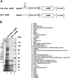
Tob-aptamer affinity purification of HCV 5′UTR and HCV 3′UTR interacting proteins from cytoplasmic Huh7 cell extract. (A) Schematic representation of the transcripts applied in the RNA affinity purification. (B, left panel) Silver stain of affinity-purified proteins (SDS-PAGE): (lane 1) purification with Tob-HCV sORF; (lane 2) control purification with HCV sORF lacking the Tob-aptamer; (lane 3) protein standard. Numbers at the left indicate the position of gel slices used for the identification of proteins shown at the right.
FIGURE 3.
FIGURE 3.
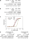
IGF2BP1 binds to the HCV 5′UTR and 3′UTR in vitro. (A) UV-cross-linking of [32P]-labeled 5′UTR and recombinant IGF2BP1 (lane 1) in the presence of 10- or 100-fold molar excess of unlabeled competitor RNAs: (lanes 2,3) 5′UTR; (lanes 4,5) 3′UTR; and (lanes 6,7) β-globin 5′-leader. (B) UV-cross-linking of [32P]-labeled 3′UTR and recombinant IGF2BP1 (lane 1) in the presence of 10- or 100-fold molar excess of unlabeled competitor RNAs: (lanes 2,3) 5′UTR; (lanes 4,5) 3′UTR; and (lanes 6,7) β-globin 5′leader. (C) Filter binding assay performed with [32P]-labeled (black) HCV 5′UTR; (red) HCV 3′UTR; or (yellow) β-globin 5′leader and increasing amounts of purified recombinant IGF2BP1. (Inset) KD values. (D, left panel) UV-cross-linking of [32P]-labeled 5′UTR and recombinant IGF2BP1 (lane 1) in the presence of 100-fold molar excess of unlabeled competitor RNAs: (lane 2) complete 5′UTR (nucleotides 1–375); (lane 3) fragment (1) (nucleotides 1–128); (lane 4) fragment (2) (nucleotides 128–315); (lane 5) fragment (3) (nucleotides 315–375); or (lane 6) β-globin 5′leader. (Right panel) UV-cross-linking of [32P]-labeled 3′UTR and recombinant IGF2BP1 (lane 1) in the presence of 100-fold molar excess of unlabeled competitor RNAs: (lane 2) complete 3′UTR (nucleotides 1–234); (lane 3) fragment (1) (nucleotides 1–151); (lane 4) fragment (2) (nucleotides 151–234); or (lane 5) β-globin 5′leader.
FIGURE 4.
FIGURE 4.
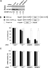
IGF2BP1 is required for HCV IRES-mediated translation, but not for 5′cap-dependent translation in vivo. (A) Huh7 cells were transfected with (lane 1) no siRNA; (lane 2) control siRNA; or (lanes 3,4) two independent siRNAs against IGF2BP1. The RNAi effect on protein expression was analyzed using antibodies against IGF2BP1 and vinculin to monitor RNAi specificity. (B, upper panel) Schematic representation of HCV Luc, HCVΔ3′UTR Luc, and 5′cap Luc mRNAs. (Lower panel) To quantify the influence of reduced IGFBP1 expression on HCV IRES-mediated or 5′cap-dependent translation, the cells were transfected with the reporter mRNAs indicated. Luc activity was measured and expressed as percentage (average of three experiments with standard deviations). (C) Total RNA was isolated, and the integrity of Luc RNA levels was analyzed by qRT-PCR. Relative RNA amounts were determined by the ΔΔCt method using endogenous cyclophilin A mRNA for normalization.
FIGURE 5.
FIGURE 5.
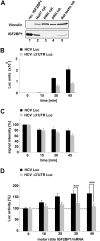
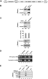
IGF2BP1 enhances HCV IRES-mediated translation via the 3′UTR in rat primary hepatocyte extract. (A) Detection of IGF2BP1 and vinculin (control) in Western blot assays: (lane 1) recombinant IGF2BP1; (lane 2) cytoplasmic Huh 7 cell extract; (lane 3) cytoplasmic rat primary hepatocyte extract; (lane 4) total rat primary hepatocyte extract; and (lane 5) total extract of rat testis (antibody control). (B) Cytoplasmic extract of rat primary hepatocytes was used in cell-free translation reactions, programmed with [32P] trace-labeled reporter mRNAs HCV Luc or HCVΔ3′UTR Luc for the times indicated. Luciferase activity is expressed as relative Luc units. Error bars denote the standard deviation from the mean of three different experiments. (C) [32P] trace-labeled mRNAs were extracted from the translation reaction at the time points indicated. [32P] trace-labeled 5′cap sORF mRNA was used as an extraction control. Extracted mRNAs were analyzed by agarose gel electrophoresis. The incorporation of [32P] was determined by phosphorimaging (Storm 860 PhosphorImager; Molecular Dynamics Image Quant software) and expressed as a percentage of signal intensity. Error bars denote the standard deviation from the mean of three different experiments. (D) Reporter mRNAs HCV Luc or HCVΔ3′UTR Luc were incubated in cytoplasmic extracts of rat primary hepatocytes in the presence of increasing amounts of recombinant IGF2BP1. Luciferase activity is expressed as relative Luc units. Error bars denote the standard deviation from the mean of three different experiments. Translation was set to 100% in the absence of recombinant IGF2BP1. In the presence of recombinant protein, data were analyzed with the t-test (two-tailed), (***) p < 0.0005.
FIGURE 6.
FIGURE 6.
Reduced expression of IGF2BP1 in HCV RNA-replicon containing Huh 7 cells does affect HCV IRES-mediated translation, but not HCV RNA replication. (A) Schematic representation of the bi-cistronic replicon RNA. (B) Detection of IGF2BP1 and vinculin (control) in (lane 1) cytoplasmic extracts of Huh7 and (lane 2) Huh7 replicon cells in Western blot assays. (C) Huh7 replicon cells were transfected with (ctrl.) control or (IGF2BP1/1) IGF2BP1 siRNAs. IGF2BP1, vinculin, neomycin phosphotransferase II (NPT2), and NS5A were detected by specific antibodies. HCV RNA replication was measured by qRT-PCR. (D, lane 3) IGF2BP1 was immunoprecipitated with a specific IGF2BP1 antibody from cytoplasmic extracts of Huh7 replicon cells. Control immunoprecipitations were performed with (lane 5) a control antibody (Fyn) or (lane 7) beads alone. Coimmunoprecipitated HCV RNA was detected by RT-PCR. Cyclophilin A mRNA served as specificity control. (E) IGF2BP1 was immunoprecipitated with a specific IGF2BP1 antibody from cytoplasmic extracts of Huh7 replicon cells in the (lane 4) absence and (lane 5) presence of RNase A. Control immunoprecipitations were performed with (lanes 2,3) beads alone or (lanes 6,7) a control antibody (Fyn). Immunoprecipitated proteins were resolved on SDS-PAGE and analyzed with antibodies against IGF2BP1, eIF3c, rpS3, or eIF6.
FIGURE 7.
FIGURE 7.
eIF3 and IGF2BP1 comigrate with 80S ribosomes assembled on HCV 5′UTR and 3′UTR bearing mRNA in Huh7 extract. [32P]-labeled (left panel) HCV sORF mRNA or (right panel) 5′cap sORF mRNA was incubated in micrococcal nuclease treated Huh7 cell extract in the presence of 2 mM cycloheximide for 15 min. (A,B) Translation initiation complexes were allowed to assemble and subsequently resolved by centrifugation on 5%–25% linear sucrose gradients. After fractionation from the bottom to the top of the gradient, the radioactivity was monitored, expressed as the percentage of incorporation, and plotted against the fraction number. The distribution of rpS3, eIF6, IGF2BP1, or eIF3c in the sucrose gradient fractions was determined using specific antibodies.

Similar articles

Cited by

References

    1. Ali N, Siddiqui A. Interaction of polypyrimidine tract-binding protein with the 5′ noncoding region of the Hepatitis C virus RNA genome and its functional requirement in internal initiation of translation. J Virol. 1995;69:6367–6375. - PMC - PubMed
    1. Amrani N, Ghosh S, Magnus DA, Jacobson A. Translation factors promote the formation of two states of the closed-loop mRNP. Nature. 2008;453:1276–1280. - PMC - PubMed
    1. Anthony D, Merrick WC. Analysis of 40 S and 80 S complexes with mRNA as measured by sucrose density gradients and primer extension inhibition. J Biol Chem. 1992;267:1554–1562. - PubMed
    1. Atlas R, Behar L, Sapoznik S, Ginzburg I. Dynamic association with polysomes during P19 neuronal differentiation and an untranslated-region-dependent translation regulation of the τ mRNA by the τ mRNA-associated proteins IMP1, HuD, and G3BP1. J Neurosci Res. 2007;85:173–183. - PubMed
    1. Barton DJ, Flanegan JB. Coupled translation and replication of poliovirus RNA in vitro: Synthesis of functional 3D polymerase and infectious virus. J Virol. 1993;67:822–831. - PMC - PubMed

Publication types

MeSH terms