

Cerebellar lesions in multiple system atrophy: postmortem MR imaging-pathologic correlations
- PMID: 19541777
- PMCID: PMC7051491
- DOI: 10.3174/ajnr.A1662
Cerebellar lesions in multiple system atrophy: postmortem MR imaging-pathologic correlations
Abstract
Background and purpose: Cerebellar atrophy and white matter T2-hyperintensities have been characterized as cerebellar lesions of multiple system atrophy (MSA). The aim of the study was to correlate MR images with histologic findings in cerebellar lesions of MSA.
Materials and methods: Postmortem T2-weighted images using 1.5T were compared with histologic findings in 7 postmortem-proved cases with MSA. The MR imaging findings in the cerebellar cortices and deep white matter dentate nucleus regions were compared with their histologic findings in each case.
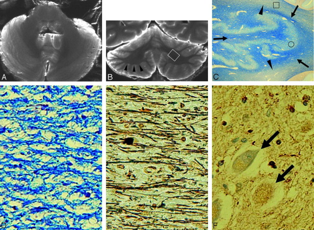
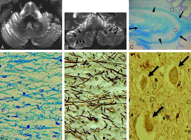
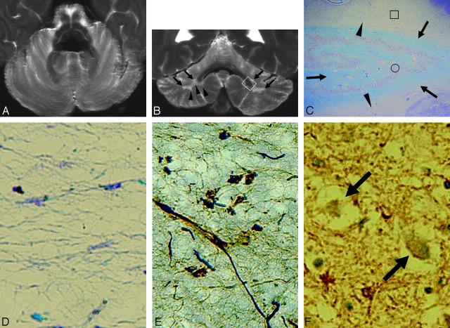
Results: We detected 3 types of cerebellar changes: type 1, no apparent atrophy or signal-intensity changes; type 2, cerebellar atrophy and inhomogeneous (patchy and/or confluent) cerebellar white matter hyperintensities; and type 3, cerebellar atrophy and diffuse white matter hyperintensities. Hypointensities were seen in the dentate nucleus regions. Atrophy of the cerebellar white matter was more severe than that of cerebellar cortices, and this anatomy was well depicted on coronal images. Histologically, degeneration was more severe in the cerebellar white matter than in the cerebellar cortices. Hyperintensities in the cerebellar white matter showed loss of myelinated fibers and gliosis. Hypointensities in the dentate nucleus regions revealed diffuse ferritin deposition in preserved dentate nuclei and white matter both around and within the nuclei.
Conclusions: Hyperintensities in the cerebellar white matter reflect degenerated white matter associated with loss of myelinated fibers and gliosis, whereas hypointensities in the dentate nucleus regions reflect diffuse ferritin deposition in preserved dentate nuclei and white matter around and within the nuclei. Degeneration is more severe in the cerebellar white matter than in the cerebellar cortices.
Figures
Similar articles
-
Putaminal lesion in multiple system atrophy: postmortem MR-pathological correlations.Neuroradiology. 2008 Jul;50(7):559-67. doi: 10.1007/s00234-008-0381-y. Epub 2008 May 8. Neuroradiology. 2008. PMID: 18463858
-
In vivo voxel-based morphometry in multiple system atrophy of the cerebellar type.Arch Neurol. 2003 Oct;60(10):1431-5. doi: 10.1001/archneur.60.10.1431. Arch Neurol. 2003. PMID: 14568814 Clinical Trial.
-
Prominent White Matter Involvement in Multiple System Atrophy of Cerebellar Type.Mov Disord. 2020 May;35(5):816-824. doi: 10.1002/mds.27987. Epub 2020 Jan 29. Mov Disord. 2020. PMID: 31994808
-
Structural and Functional Magnetic Resonance Imaging of the Cerebellum: Considerations for Assessing Cerebellar Ataxias.Cerebellum. 2016 Feb;15(1):21-25. doi: 10.1007/s12311-015-0738-9. Cerebellum. 2016. PMID: 26521073 Review.
-
Cerebellar involvement in metabolic disorders: a pattern-recognition approach.Neuroradiology. 1998 Jun;40(6):347-54. doi: 10.1007/s002340050597. Neuroradiology. 1998. PMID: 9689620 Review.
Cited by
-
Human alpha-synuclein overexpressing MBP29 mice mimic functional and structural hallmarks of the cerebellar subtype of multiple system atrophy.Acta Neuropathol Commun. 2021 Apr 14;9(1):68. doi: 10.1186/s40478-021-01166-x. Acta Neuropathol Commun. 2021. PMID: 33853667 Free PMC article.
-
Detecting ferroptosis and immune infiltration profiles in multiple system atrophy using postmortem brain tissue.Front Neurosci. 2023 Dec 29;17:1269996. doi: 10.3389/fnins.2023.1269996. eCollection 2023. Front Neurosci. 2023. PMID: 38222105 Free PMC article.
-
Multiple System Atrophy: An Oligodendroglioneural Synucleinopathy1.J Alzheimers Dis. 2018;62(3):1141-1179. doi: 10.3233/JAD-170397. J Alzheimers Dis. 2018. PMID: 28984582 Free PMC article. Review.
-
Hot cross bun sign in a case with multisystem atrophy.Iran J Neurol. 2014 Apr 3;13(2):110-1. Iran J Neurol. 2014. PMID: 25295157 Free PMC article. No abstract available.
-
Altered Brain Volume, Microstructure Metrics and Functional Connectivity Features in Multiple System Atrophy.Front Aging Neurosci. 2022 May 19;14:799251. doi: 10.3389/fnagi.2022.799251. eCollection 2022. Front Aging Neurosci. 2022. PMID: 35663568 Free PMC article.
References
-
- Papp MI, Kahn JE, Lantos PL, et al. Glial cytoplasmic inclusions in the CNS of patients with multiple system atrophy (striatonigral degeneration, olivopontocerebellar atrophy and Shy-Drager syndrome). J Neurol Sci 1989;94:79–100 - PubMed
-
- Spillantini MG, Crowther RA, Jakes R, et al. Filamentous alpha-synuclein inclusions link multiple system atrophy with Parkinson's disease and dementia with Lewy bodies. Neurosci Lett 1998;251:205–08 - PubMed
-
- Galvin JE, Lee VMY, Trojanowski JQ. Synucleinopathies: clinical and pathological implications. Arch Neurol 2001;58:186–90 - PubMed
-
- Wenning GK, Ben Shlomo Y, Magalhaes M, et al. Clinical features and natural history of multiple system atrophy: an analysis of 100 cases. Brain 1994;117:835–45 - PubMed
MeSH terms
LinkOut - more resources
Full Text Sources
Medical