. 2010 May;42(5):552-63.
doi: 10.1165/rcmb.2009-0068OC. Epub 2009 Jun 18.

Conditional overexpression of connective tissue growth factor disrupts postnatal lung development

Affiliations

Conditional overexpression of connective tissue growth factor disrupts postnatal lung development

Shu Wu et al. Am J Respir Cell Mol Biol. 2010 May.

Abstract

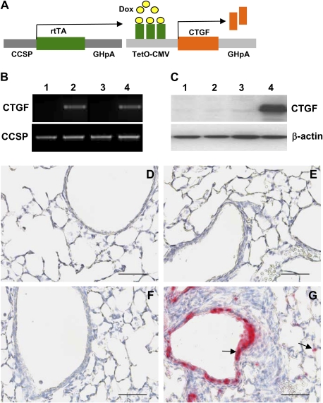
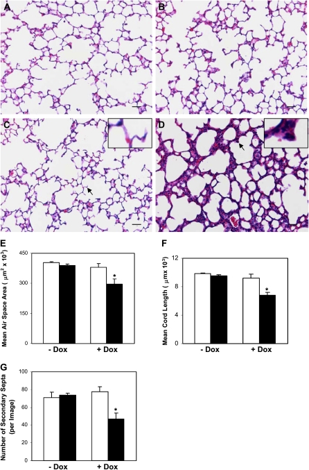
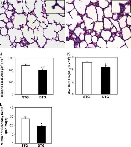
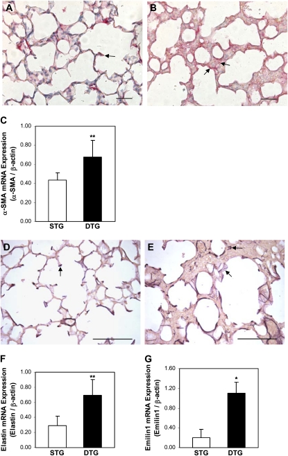
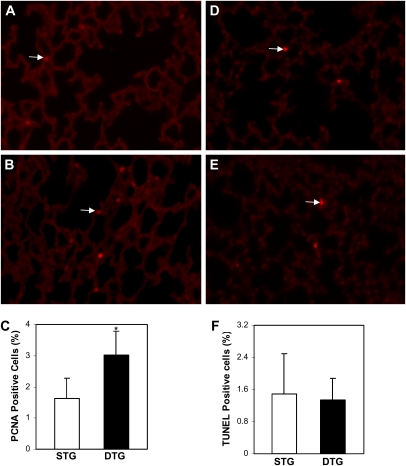
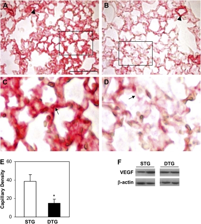
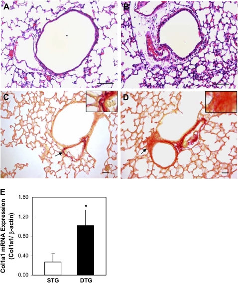
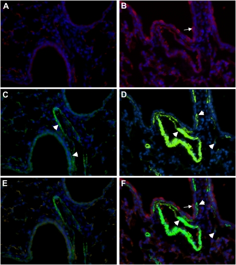
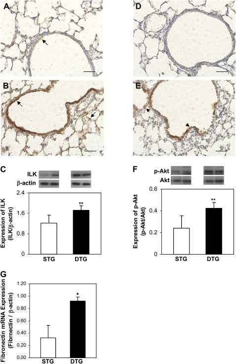
Connective tissue growth factor (CTGF) is a member of an emerging family of immediate-early gene products that coordinates complex biological processes during development, differentiation, and tissue repair. Overexpression of CTGF is associated with mechanical ventilation with high tidal volume and oxygen exposure in newborn lungs. However, the role of CTGF in postnatal lung development and remodeling is not well understood. In the present study, a double-transgenic mouse model was generated with doxycycline-inducible overexpression of CTGF in respiratory epithelial cells. Overexpression of CTGF from Postnatal Days 1-14 resulted in thicker alveolar septa and decreased secondary septal formation. This is correlated with increased myofibroblast differentiation and disorganized elastic fiber deposition in alveolar septa. Overexpression of CTGF also decreased alveolar capillary network formation. There were increased alpha-smooth muscle actin expression and collagen deposition, and dramatic thickening in the peribronchial/peribronchiolar and perivascular regions in the double-transgenic lungs. Furthermore, overexpression of CTGF increased integrin-linked kinase expression, activated its downstream signaling target, Akt, as well as increased mRNA expression of fibronectin. These data demonstrate that overexpression of CTGF disrupts alveologenesis and capillary formation, and induces fibrosis during the critical period of alveolar development. These histologic changes are similar to those observed in lungs of infants with bronchopulmonary dysplasia.


Figures

Figure 1.
Figure 1.
Generation of double-transgenic mouse model with conditional overexpression of connective tissue growth factor (CTGF) in respiratory epithelium. (A) Mating homozygous Clara cell secretory protein (CCSP)–reverse tetracycline-responsive transactivator (rtTA) mice to heterozygous tetracycline operator (TetO)–CTGF mice produced double-transgenic pups containing CCSP-rtTA and TetO-CTGF transgenes. Administration of doxycycline (Dox) to the dams from Postnatal Days 1–14 induced overexpression of CTGF in airway epithelium. (B) PCR analysis of tail DNA with CCSP-rtTA and TetO-CTGF primers identified CCSP-rtTA single-transgenic (STG) mice (lanes 1 and 3) and CCSP-rtTA/TetO-CTGF double-transgenic (DTG) mice (lanes 2 and 4) without (lanes 1 and 2) and with (lanes 3 and 4) Dox administration. (C) Western blot analysis demonstrated high level of CTGF protein expression in DTG lungs exposed to Dox (lane 4) compared with extremely low levels of CTGF protein expression in STG lungs without (lane 1) and with (lane 3) Dox treatment and in DTG transgenic lungs without Dox treatment (lane 2). (D) STG lungs without Dox administration. (E) DTG lungs without Dox exposure. (F) STG lungs treated with Dox. (G) Immunohistochemistry revealed extensive CTGF expression in bronchiolar and alveolar epithelium only in Dox-treated DTG lungs (arrows). Magnification, 40×. Scale bars, 50 μm.
Figure 2.
Figure 2.
CTGF disrupted alveolarization. Histological examination on hematoxylin and eosin (H&E)–stained lung tissue sections at Postnatal Day 14 demonstrated normal alveolar morphogenesis with thin septa and well formed secondary septa in STG lungs without (A) and with (C) Dox administration, and in DTG lungs without (B) Dox exposure. Administration of Dox disrupted alveolarization with hypercellular septa and poorly formed secondary septa in DTG lungs (D). Mean alveolar airspace area (E), mean chord length (MCL) (F), and number of secondary septa (G) were significantly decreased in Dox-treated DTG lungs at Postnatal Day 14. Similar changes in lung histology (STG [H]; DTG [I]), mean alveolar airspace area (J), MCL (K), and number of secondary septa (L) were detected as early as Postnatal Day 6 after Dox treatment. Insets are higher magnification views of secondary septa indicated by the arrows. n = 3 in groups without Dox administration; n = 4 in groups with Dox administration at Postnatal Day 14; n = 4 in STG group; and n = 5 in DTG group at Postnatal Day 6. Open bars, STG; solid bars, DTG; * P < 0.001, ** P < 0.005. Magnification, 20×. Scale bars, 50 μm.
Figure 2.
Figure 2.
CTGF disrupted alveolarization. Histological examination on hematoxylin and eosin (H&E)–stained lung tissue sections at Postnatal Day 14 demonstrated normal alveolar morphogenesis with thin septa and well formed secondary septa in STG lungs without (A) and with (C) Dox administration, and in DTG lungs without (B) Dox exposure. Administration of Dox disrupted alveolarization with hypercellular septa and poorly formed secondary septa in DTG lungs (D). Mean alveolar airspace area (E), mean chord length (MCL) (F), and number of secondary septa (G) were significantly decreased in Dox-treated DTG lungs at Postnatal Day 14. Similar changes in lung histology (STG [H]; DTG [I]), mean alveolar airspace area (J), MCL (K), and number of secondary septa (L) were detected as early as Postnatal Day 6 after Dox treatment. Insets are higher magnification views of secondary septa indicated by the arrows. n = 3 in groups without Dox administration; n = 4 in groups with Dox administration at Postnatal Day 14; n = 4 in STG group; and n = 5 in DTG group at Postnatal Day 6. Open bars, STG; solid bars, DTG; * P < 0.001, ** P < 0.005. Magnification, 20×. Scale bars, 50 μm.
Figure 3.
Figure 3.
CTGF increased myofibroblast differentiation and caused abnormal elastic fiber deposition in alveolar septa. Immunohistochemistry detected α–smooth muscle actin (α-SMA) expression in cells at the tips of secondary septa in Dox-exposed STG lungs (arrow [A]). In Dox-exposed DTG lungs, α-SMA was detected irregularly and with increased abundance in alveolar septa and on the surface of alveolar septa (arrows [B]). (C) Quantitative real-time RT-PCR revealed significant increase in α-SMA mRNA expression in Dox-treated DTG lungs. Hart's staining detected organized elastin fibers at the tips of secondary septa and along parts of alveolar septa in Dox-treated STG lungs (arrow [D]). In Dox-treated DTG lungs, elastin fibers were localized along alveolar septa, at poorly formed secondary septa that are fragmented and irregular, and in thick alveolar septa (arrows [E]). Quantitative real-time RT-PCR analysis demonstrated increased mRNA expressions of elastin (F) and emilin1 (G) in Dox-exposed DTG lungs. n = 4/group; *P < 0.001, **P < 0.05. Magnification, 40×. Scale bars, 50 μm (A and B), 100 μm (D and E).
Figure 4.
Figure 4.
Effects of CTGF on cell proliferation and apoptosis. Immunofluorescence staining with an anti–proliferating cell nuclear antigen (PCNA) antibody detected proliferating cells (arrows) in Dox-administrated STG (A) and DTG (C) lungs. (D) Quantification of proliferating cell index demonstrated increased proliferating cells in Dox-treated DTG lungs. Apoptotic cells (arrows) were detected by a TUNEL assay in Dox administrated STG (B) and DTG (D) lungs. (F) Quantification of apoptotic index revealed no difference in these lungs. n = 4/group; *P < 0.05. Magnification, 40×.
Figure 5.
Figure 5.
CTGF disrupted capillary development. Immunohistochemistry using an anti–platelet endothelial cell adhesion molecule (PECAM)–1 antibody detected intense signal in intra-acinous vessels (arrowhead) and capillary networks (arrow) in Dox-treated STG lungs (A and C). PECAM-1 was detected with intense signal in intra-acinous vessels (arrowhead) but with dramatically decreased intensity and continuity in capillary networks (arrow) in Dox treated DTG lungs (B and D). (E) Quantification of capillary density determined by the percentage of PECAM-1–stained area versus alveolar tissue area. (F) Western blot analysis of vascular endothelial growth factor (VEGF) protein expression. n = 4/group; *P < 0.01. Magnification, 40×. Scale bars, 100 μm.
Figure 6.
Figure 6.
CTGF induced fibrosis. Histologic examination on H&E-stained lung tissue sections revealed dramatic thickening of peribronchial/peribronchiolar and perivascular regions in Dox-administrated DTG lungs (B) compared with STG lungs (A). Picro Sirius Red staining demonstrated increased collagen deposition in these regions in DTG lungs (D) compared with STG lungs (C). Insets are higher magnification views of the area indicated by the arrows. Quantitative real-time RT-PCR demonstrated significantly increased mRNA expression of collagen type I, α1 (col1a1) (E) in DTG lungs. n = 4/group; *P < 0.01. Magnification, 40× (A and B), 20× (C and D). Scale bars, 50 μm.
Figure 7.
Figure 7.
CTGF increased α-SMA expression in mesenchymal tissues surrounding conducting airways and blood vessels. Dual immunofluorescence staining was performed on lung tissue sections from Dox-administrated STG and DTG lungs with an anti-CTGF antibody (red signal) and an anti–α-SMA antibody (green signal) and DAPI nuclear staining (blue signal). CTGF (A) was undetectable and α-SMA (arrowheads [C]) was detected in bronchial/bronchiolar walls and vessels in STG lungs. In DTG lungs, CTGF (arrow [B]) was detected in airway epithelium and α-SMA (arrowheads [D]) was detected with increased intensity in bronchial/bronchiolar walls adjacent to CTGF-expressing cells, and in vessels and surrounding mesenchymal tissues. E represents a merged image of A and C; and F represents a merged image of B and D. Magnification, 40×.
Figure 8.
Figure 8.
CTGF activated integrin-linked kinase (ILK)–Akt signaling. Immunohistochemistry was performed with anti-ILK and anti–p-Akt antibodies on lung tissue sections from Dox-treated STG and DTG mice. In STG lungs, ILK was detected with low intensity in bronchiolar epithelial cells (arrow [A]), and p-Akt was undetectable (D). In DTG lungs, ILK (arrows [B]) and p-Akt (arrowheads [E]) were detected with strong intensity in bronchiolar epithelial cells, and weakly in surrounding mesenchymal tissues and alveolar septa. Western blot demonstrated that overexpression of CTGF increased ILK expression (C) and activated Akt (F) in DTG lungs. Quantitative real-time RT-PCR demonstrated increased fibronectin mRNA expression in DTG lungs (G). n = 4/group; *P < 0.001, **P < 0.05. Magnification, 40×. Scale bars, 50 μm.

Similar articles

Cited by

References

    1. Jobe AH, Bancalari E. Bronchopulmonary dysplasia. Am J Respir Crit Care Med 2001;163:1723–1729. - PubMed
    1. Husain AN, Siddiqui NH, Stocker JT. Pathology of arrested acinar development in post surfactant bronchopulmonary dysplasia. Hum Pathol 1998;29:710–717. - PubMed
    1. Coalson JJ, Winter VT, Delemos RA. Decreased alveolarization in baboon survivors with bronchopulmonary dysplasia. Am J Respir Crit Care Med 1995;152:640–646. - PubMed
    1. Coalson JJ. Pathology of bronchopulmonary dysplasia. Semin Perinatol 2006;30:179–184. - PubMed
    1. Jobe AJ. The new BPD: an arrest of lung development. Pediatr Res 1999;46:641–643. - PubMed

Publication types

MeSH terms

Substances