. 2009 Jul;175(1):376-91.
doi: 10.2353/ajpath.2009.081160. Epub 2009 Jun 18.

Modulation of the E2F1-driven cancer cell fate by the DNA damage response machinery and potential novel E2F1 targets in osteosarcomas

Affiliations

Modulation of the E2F1-driven cancer cell fate by the DNA damage response machinery and potential novel E2F1 targets in osteosarcomas

Michalis Liontos et al. Am J Pathol. 2009 Jul.

Abstract

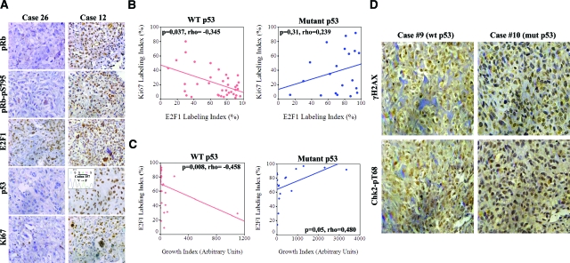
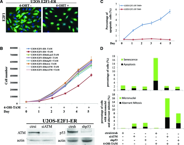
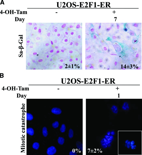
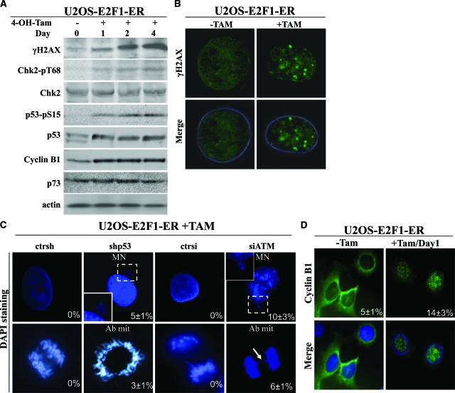
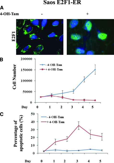
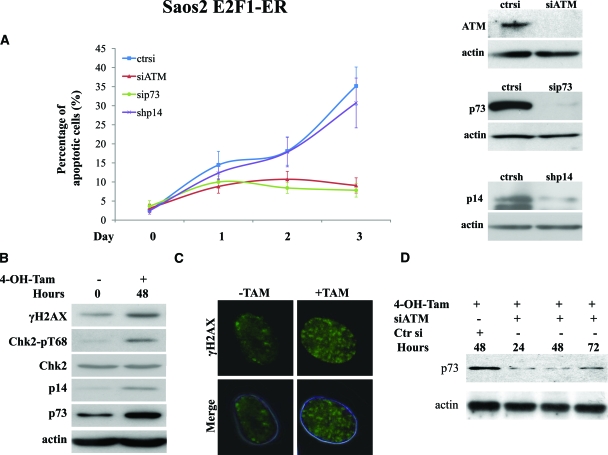
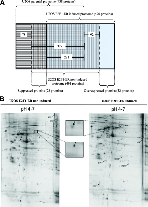
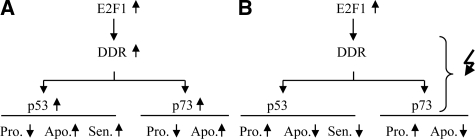
Osteosarcoma is the most common primary bone cancer. Mutations of the RB gene represent the most frequent molecular defect in this malignancy. A major consequence of this alteration is that the activity of the key cell cycle regulator E2F1 is unleashed from the inhibitory effects of pRb. Studies in animal models and in human cancers have shown that deregulated E2F1 overexpression possesses either "oncogenic" or "oncosuppressor" properties, depending on the cellular context. To address this issue in osteosarcomas, we examined the status of E2F1 relative to cell proliferation and apoptosis in a clinical setting of human primary osteosarcomas and in E2F1-inducible osteosarcoma cell line models that are wild-type and deficient for p53. Collectively, our data demonstrated that high E2F1 levels exerted a growth-suppressing effect that relied on the integrity of the DNA damage response network. Surprisingly, induction of p73, an established E2F1 target, was also DNA damage response-dependent. Furthermore, a global proteome analysis associated with bioinformatics revealed novel E2F1-regulated genes and potential E2F1-driven signaling networks that could provide useful targets in challenging this aggressive neoplasm by innovative therapies.


Figures

Figure 1
Figure 1
p53 status determines the association of E2F1 expression in relation to proliferation and apoptosis in osteosarcomas. A: Immunohistochemical analysis (IHC) of pRb, pRb-pS795, E2F1, p53, and Ki-67 in two representative osteosarcoma cases with different p53 status. Inset: confirmation of p53 mutation by DNA sequencing. B: Scatter plots depicting the correlation of E2F1 and Ki-67 expression according to p53 status. Note the statistically significant inverse relationship between E2F1 IHC expression and proliferation index (P = 0.037, Spearman test) in cases retaining wild-type p53. C: Scatter plots showing the relation of E2F1 expression with Growth Index in cases with intact and mutant p53 (P = 0.008 and P = 0.05 respectively, Spearman test). D: IHC analysis for γH2AX and Chk2-pT68 in representative osteosarcomas with wild-type (wt) and mutant (mut) p53, respectively.
Figure 2
Figure 2
E2F1 overexpression in U2OS cells raises anti-tumor barriers. A: Addition of 400 nmol/L 4-OH-Tamoxifen (4-OH-Tam) leads to nuclear translocation of E2F1. B: Growth curves of untransfected U2OS E2F1-ER cells and cells transfected with control siRNA, siATM, or shp53 before and after the addition of 4-OH-Tamoxifen. The lines are the means of three independent experiments. Immunoblots for p53 and ATM, confirming gene silencing. C: Percentage of apoptotic cells as determined by fluorescence-activated cell sorting analysis (the lines are the means of three independent experiments) in untreated and 4-OH-Tamoxifen administered U2OS E2F1-ER cells. D: Histograms depicting the activation of the anti-tumor barriers (apoptosis and senescence), the presence of micronuclei and the aberrant mitoses in the U2OS E2F1-ERshp53 and U2OS E2F1-ERsiATM cells, and their corresponding controls, before and after the addition of 4-OH-Tamoxifen.
Figure 3
Figure 3
A: Sa-β-Gal staining after 7 days of 4-OH-Tamoxifen addition in U2OS E2F1-ER cells (percentages presented as an inset). B: Cells with morphological characteristics of “mitotic catastrophe” after the addition of 4-OH-Tamoxifen (percentages presented as an inset).
Figure 4
Figure 4
The effect of E2F1 overexpression in U2OS cells is mediated by DDR activation. A: Western blot analysis for γH2AX, Chk2-pT68, Chk2, p53-pS15, p53, Cyclin B1, and p73. Actin serves as loading control. B: Immunofluorescence analysis for γH2AX in U2OS E2F1-ER cells showing foci after addition of 4-OH-Tamoxifen. C: U2OS E2F1-ERshp53 and U2OS E2F1-ERsiATM cells, as well as their corresponding controls, were stained with 4,6-diamidino-2-phenylindole and analyzed for morphological indications of genomic instability (MN: micronuclei and Ab mit: aberrant mitosis). The arrow indicates a nucleoplasmic bridge. D: Immunofluorescence analysis showing Cyclin B1 nuclear translocation in the U2OS E2F1-ER cells after 4-OH-Tamoxifen induction of E2F1.
Figure 5
Figure 5
E2F1 overexpression in Saos2 cells raises anti-tumor barriers. A: Addition of 400 nmol/L 4-OH-Tamoxifen leads to nuclear translocation of E2F1. B: Growth curves of Saos2 E2F1-ER cells with and without the addition of 4-OH-Tamoxifen (4-OH-Tam). The lines are the means of three independent experiments. C: Percentage of apoptotic cells as determined by fluorescence-activated cell sorting analysis. The lines are the means of three independent experiments.
Figure 6
Figure 6
The outcome of E2F1 overexpression in Saos2 cells is mediated by DDR activation. A: Apoptotic rate in the Saos2 E2F1-ERsiATM, Saos2 E2F1-ERsip73, and Saos2 E2F1-ERshp14ARF cells before and after the addition of 4-OH-Tamoxifen (4-OH-Tam), respectively. The lines are the means of three independent experiments. Immunoblots for ATM, p73, and p14ARF confirmed corresponding gene silencing. B: Western blot analysis of γH2AX, Chk2-pT68, Chk2, and p73. Actin serves as loading control. C: Immunofluorescence analysis in Saos E2F1-ER cells showing the presence of H2AX foci before and after addition of 4-OH-Tamoxifen. D: Western blot analysis for p73 expression in Saos2 E2F1-ER cells treated with 4-OH-Tamoxifen and silenced for ATM expression.
Figure 7
Figure 7
A: The subtractive algorithm used for the identification of overexpressed and suppressed proteins after E2F1 induction in U2OS E2F1-ER cells. B: Representative two-dimensional gel analysis of proteins from U2OS E2F1-ER cells either untreated (U2OS E2F1-ER non-induced) or 48 hours after 4-OH-Tamoxifen treatment (U2OS E2F1-ER induced). Proteins were extracted and separated on a non-linear immobilized pH-gradient strip pH 4 to 7, followed by a 12% SDS-polyacrylamide gel separation. The gel was stained with Coomassie blue. Identification of protein spots was performed by peptide mass fingerprint and/or postsource decay MS. Proteins found to be suppressed or overexpressed are annotated on the gels. Names and descriptions of each annotated protein are presented in supplemental Tables 3A and 3B (see http://ajp.amjpatho.org). Insets are magnified photos indicating suppression of K2C1 protein.
Figure 8
Figure 8
Proposed model summarizing the DDR-dependent modulation of E2F1 activity in human osteosarcoma cells (A) with intact DDR response machinery, and (B) with aberrations in the DDR response machinery. DDR, DNA damage response; Pro, proliferation; Apo, apoptosis; Sen, senescence. Symbols: increase (upward pointing arrow), decrease (downward pointing arrow), aberration(s) (zigzag arrow).

References

    1. Meyers PA, Gorlick R. Osteosarcoma. Pediatr Clin North Am. 1997;44:973–989. - PubMed
    1. Wadayama B, Toguchida J, Shimizu T, Ishizaki K, Sasaki MS, Kotoura Y, Yamamuro T. Mutation spectrum of the retinoblastoma gene in osteosarcomas. Cancer Res. 1994;54:3042–3048. - PubMed
    1. Tsantoulis PK, Gorgoulis VG. Involvement of E2F transcription factor family in cancer. Eur J Cancer. 2005;41:2403–2414. - PubMed
    1. Iaquinta PJ, Lees JA. Life and death decisions by the E2F transcription factors. Curr Opin Cell Biol. 2007;19:649–657. - PMC - PubMed
    1. Bell LA, Ryan KM. Life and death decisions by E2F-1. Cell Death Differ. 2004;11:137–142. - PubMed

Publication types

MeSH terms