

doi: 10.1126/science.1173155.
Epub 2009 Jun 18.
Functional amyloids as natural storage of peptide hormones in pituitary secretory granules
Affiliations
- PMID: 19541956
- PMCID: PMC2865899
- DOI: 10.1126/science.1173155
Item in Clipboard
Functional amyloids as natural storage of peptide hormones in pituitary secretory granules
Science.
.
Abstract
Amyloids are highly organized cross-beta-sheet-rich protein or peptide aggregates that are associated with pathological conditions including Alzheimer's disease and type II diabetes. However, amyloids may also have a normal biological function, as demonstrated by fungal prions, which are involved in prion replication, and the amyloid protein Pmel17, which is involved in mammalian skin pigmentation. We found that peptide and protein hormones in secretory granules of the endocrine system are stored in an amyloid-like cross-beta-sheet-rich conformation. Thus, functional amyloids in the pituitary and other organs can contribute to normal cell and tissue physiology.
Figures
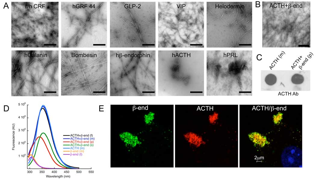
Amyloid-like aggregation and coaggregation of hormones. (A) EM of ten hormones incubated for 30 days indicate the formation of amyloid fibrils. In fig. S3 the entire set of 42 hormones studied are shown. The aggregations of the hormones were followed at 37° C at a concentration of 2 mg/ml in the presence of 0.4 mM LMW heparin in 5% D-mannitol (pH 5.5) under slight agitation. The human prolactin (hPRL) was fibrillized in presence of 400 µM chondroitin sulfate A. Transmission electron microscopy of negative stained samples was performed. Scale bars are 500 nm. (B-E) Coaggregation of ACTH with β-endorphin measured by (B) EM, (C) dot blot, (D) Trp and Tyr fluorescence, and (E) colocalization in AtT20 cells. (B) EM of ACTH – β-endorphin mixture at 37° C at hormone concentrations of 1 mg/ml each in the presence of 0.4 mM LMW heparin in 5% D-mannitol (pH 5.5) incubated under slight agitation for 14 days. (C) The presence of ACTH in these amyloid fibrils of the ACTH – β-endorphin mixture was identified by dot blot staining with ACTH antibody of the aggregates harvested by centrifugation (p). For a positive control of the antibody staining, ACTH monomer (m) was used. (D) Trp and Tyr fluorescence was measured for an aggregated sample of ACTH – β-endorphin mixture labeled with (f) and displayed in black. High speed centrifugation at 120,000 g for 1 h was performed with this sample and the Trp and Tyr fluorescence was measured both for the corresponding pellet (p) displayed in red, as well as the supernatant (s) displayed in green. Furthermore, a fresh solution of a mixture of ACTH – β-endorphin (m) displayed in blue, a fresh solution of ACTH (m) displayed in blue, a fresh solution of β-endorphin (m) displayed in yellow, and an aggregated β-endorphin fibrillar sample (f) displayed in violet were measured accordingly. The Trp fluorescence signal in pellet suggests that ACTH is present in fibrils since only ACTH has a Trp. The λmax of the Trp signal is blue shifted by 15 nm when compared to monomeric/non-fibrillated ACTH suggested that the Trp is less solvent exposed in the fibril structure than in its monomeric state. Furthermore, the fluorescence study suggested the aggregation of about half of the ACTH, because the Trp fluorescence intensities of the pellet containing fibrils and the corresponding supernatant containing ACTH monomer were similar. Since the Tyr fluorescence of monomeric β-endorphin, the β-endorphin fibrils harvested by centrifugation, as well as the harvested ACTH – β-endorphin fibrils were similar in intensity and no Tyr fluorescence signal was observed in the corresponding supernatant, the entire β-endorphin population appeared to have aggregated into amyloid fibrils. (E) Colocalization of the two hormones in the tumor cell line AtT20 by double immunohistochemistry with mouse ACTH (red) and rabbit endorphin antibodies (green). The nuclear marker DAPI is shown in blue.
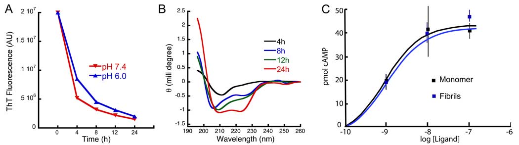
Release of monomeric, α-helical and functional CRF from its amyloid fibrils. CRF amyloid fibrils were dialyzed against buffer with a 10 kDa cut-off membrane. (A) Time-dependent decrease of Thio T fluorescence inside the membrane at two pH’s as labeled. The decrease of Thio T indicates loss of amyloid fibrils due to dialysis. (B) Time-resolved CD spectroscopy outside the membrane measuring released CRF. The time-dependent increase of the signal indicates release of CRF from the amyloid. The released CRF is likely to be monomeric because of the 10 kDa cut-off of the dialysis membrane. The CD spectrum of the released CRF is of helical structure, which corresponds to the active conformation of CRF. (C) Functional studies of monomeric and amyloid fibrillar sample of CRF by measuring in a hormone concentration-dependent manner the activation of intracellular cyclic AMP in CHO cells stably expressing CRF-R1. Both samples show similar potencies.
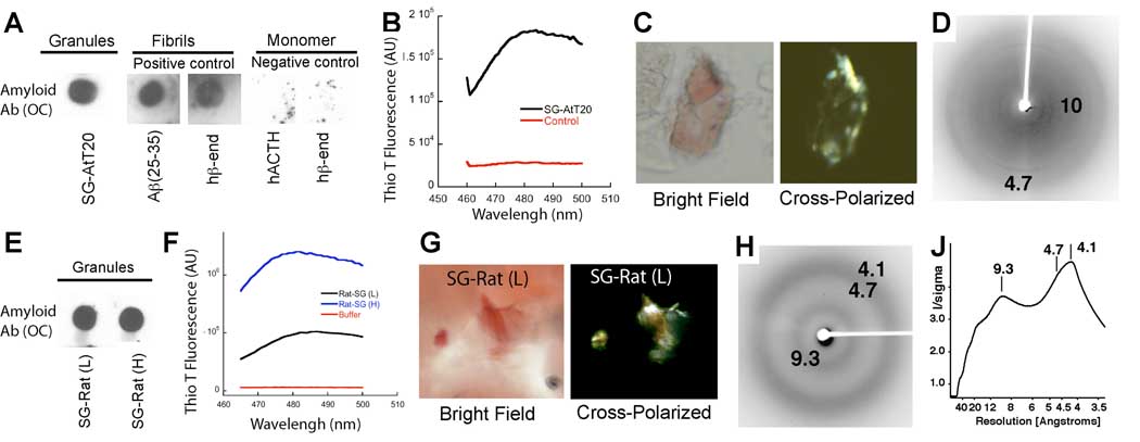
Purified secretory granules from the AtT20 cell line and from rat pituitary are composed of an amyloid-like structure as determined by an amyloid-specific antibody OC (A, E), the amyloid-specific dies Thio-T (B, F), CR (C-G), and x-ray fiber diffraction (D,H,J). (A) Dot blot staining of purified secretory granules from AtT20 cells against the amyloid-specific antibody OC. As positive controls, staining of amyloid fibrils of Aβ(25–35) fibrils and β-endorphin fibrils (in presence of LMW heparin) are shown. As negative controls, staining of monomeric human ACTH and β-endorphin are presented. (B) Thio T fluorescence between 460–500 nm is shown for purified secretory granules (black) and buffer-only (red). Thio T was excited at 450 nm. The strong binding of Thio T to secretory granules suggests that they are composed of amyloid-like structure. (C) Congo Red birefringence of purified secretory granules. CR birefringence is shown that requests the presence of ordered (amyloid) aggregates. The picture represents the bright field microscope image with 10X resolutions. The same section is shown under cross-polarized light with 10X magnification, respectively. (D) X-ray fiber diffraction of purified membrane-depleted secretory granules that were treated with 1% Lubrol PX. The two reflections at 4.7 Å and ~10 Å consistent with cross-β-sheet structure are labeled. (E–J) Purified light (L) and heavy (H) secretory granules from rat pituitary are composed of amyloid-like structure using an amyloid-specific antibody OC (E), the amyloid-specific dies Thio-T (F), CR birefringence (G), and x-ray fiber diffraction (H, J). The same measurement parameters as in (A-D) were used. In (J) the X-ray fiber diffraction measurements of purified membrane-less secretory granules are shown as a full image radial profile. The two reflections at ~4.7 Å and ~9.3 Å consistent with cross-β-sheet structure are labeled. In addition, a strong 4.1 Å is present attributed to the remaining lipid content of the granules under study.
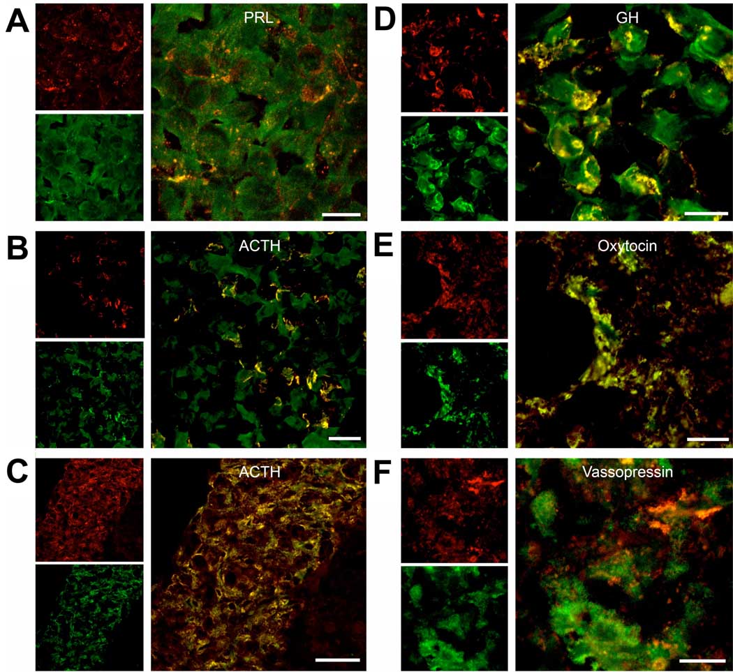
Immunohistochemical staining of the mouse pituitary with Thio S (green) and with polyclonal antibodies (red) to (A) prolactin, (B) and (C) ACTH, (D) growth hormone (GH), (E) oxytocin, and (F) vasopressin. Regions selected of the pituitary are (A), (B), and (D) anterior lobe, (C) intermediate lobe, and (E) and (F) posterior lobe. Scale bars are 20 µm.
References
Publication types
MeSH terms
Substances
Grants and funding
LinkOut - more resources
Full Text Sources
Other Literature Sources
