. 2009 Jun 19;324(5934):1580-2.
doi: 10.1126/science.1172890.

Merkel cells are essential for light-touch responses

Affiliations

Merkel cells are essential for light-touch responses

Stephen M Maricich et al. Science. .

Abstract

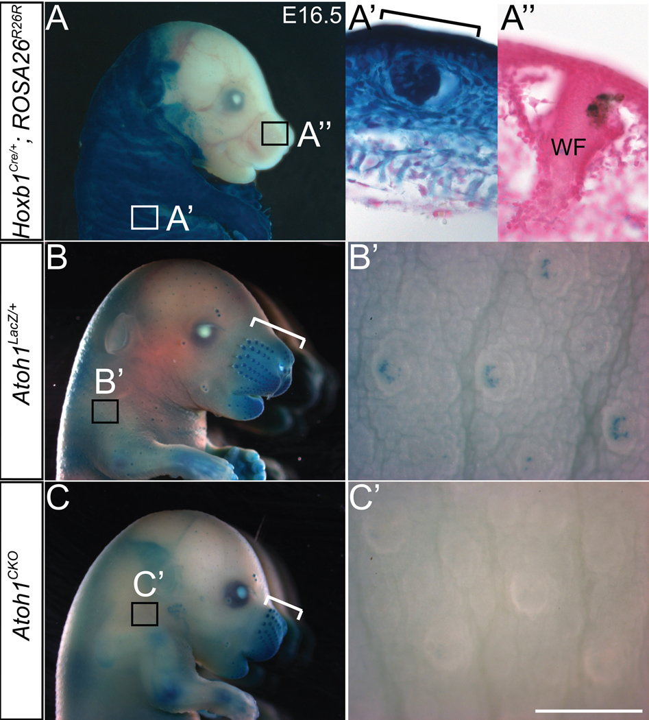
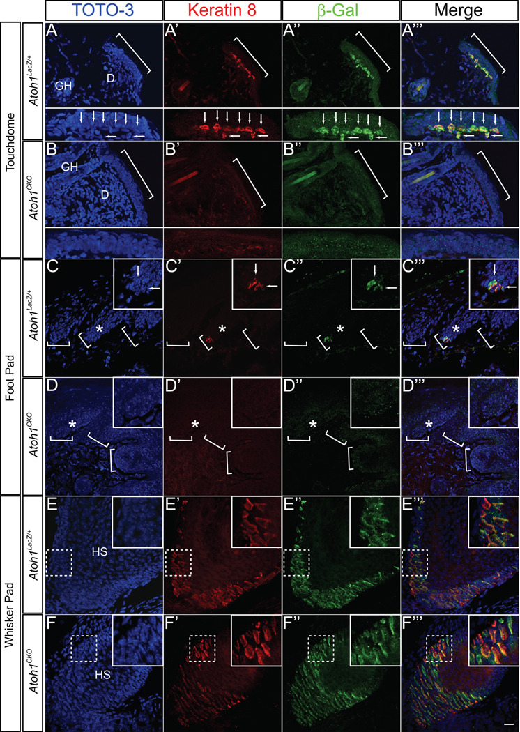
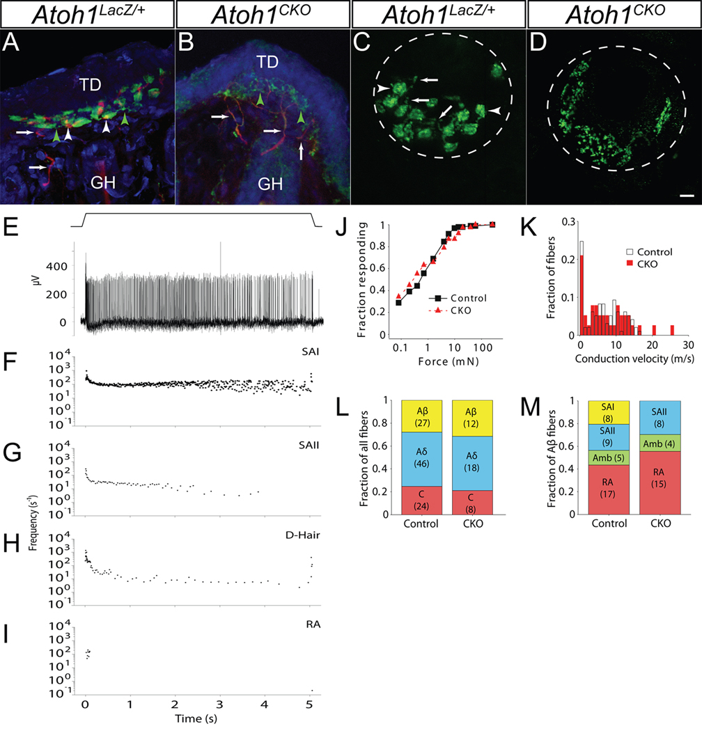
The peripheral nervous system detects different somatosensory stimuli, including pain, temperature, and touch. Merkel cell-neurite complexes are touch receptors composed of sensory afferents and Merkel cells. The role that Merkel cells play in light-touch responses has been the center of controversy for over 100 years. We used Cre-loxP technology to conditionally delete the transcription factor Atoh1 from the body skin and foot pads of mice. Merkel cells are absent from these areas in Atoh1(CKO) animals. Ex vivo skin/nerve preparations from Atoh1(CKO) animals demonstrate complete loss of the characteristic neurophysiologic responses normally mediated by Merkel cell-neurite complexes. Merkel cells are, therefore, required for the proper encoding of Merkel receptor responses, suggesting that these cells form an indispensible part of the somatosensory system.


Figures

Fig. 1
Fig. 1. Hoxb1Cre abolishes Atoh1flox expression in the body but not the face
Wholemount X-gal staining of E16.5 embryos revealed β-galactosidase expression driven from the ROSA26R26R (A-A”) or Atoh1LacZ (B–C’) loci. A) Hoxb1Cre/+; ROSA26R26R embryo. Hoxb1Cre expression is absent from the majority of the head. Boxes denote regions shown in (A’) and (A”). A’) and A”) Body skin and whisker pad, respectively, from a Hoxb1Cre/+; ROSA26R26R embryo counterstained with nuclear fast red. Staining is present throughout the epidermis and dermis of the body skin, while virtually no staining is seen in the whisker pad. Bracket in (A’) denotes a developing touchdome. WF – whisker follicle. B) Atoh1LacZ/+ embryo showing β-galactosidase expression in the whisker pad (bracket) and touch domes of the skin. Box denotes region shown in (B’). B’) Hoxb1+/+; Atoh1LacZ/flox embryo body skin showing staining in individual touch domes. C) Atoh1CKO embryo showing β-galactosidase expression driven from the Atoh1 locus in the whisker pad (bracket), but absent from touch domes of the skin (box). C’) Atoh1CKO embryo body skin. Touch domes are present but lack the wildtype staining pattern. Scale bar: 5 mm (A, B, C); 400 µm (B’, C’); 100µm (A’, A”).
Fig. 2
Fig. 2. Merkel cells are absent from the body skin and foot pads of Atoh1CKO animals
All tissue is from P22 animals, and all images are z-stack projections of confocal images. A–B”’) Skin from Atoh1LacZ/+ (A-A”’) and Atoh1CKO (B-B”’) animals. Brackets denote a single touch dome. Subpanels in A-A”’ show Merkel cells (arrows) from the wildtype touch dome denoted by the bracket, while none are seen in the Atoh1CKO animal (subpanels B-B”’). D – dermis, GH – guard hair. C–D”’) Hind foot pad from Atoh1LacZ/+ (C-C”’) and Atoh1CKO (D-D”’) animals. Brackets denote epidermal rete ridges, asterisks denote areas shown in insets, and arrows in (C-C”’) mark individual Merkel cells. Cells positive for Keratin 8 and β-galactosidase are absent from Atoh1CKO body skin and foot pad. E–F”’) Whisker pad from Atoh1LacZ/+ (E-E”’) and Atoh1CKO (F-F”’) animals. There is no difference in the immunostaining patterns between the two genotypes. Dotted boxes outline areas shown in insets. HS – whisker hair shaft. All Keratin 8-positive cells were also β-galactosidase-positive and vice versa in all tissues of both genotypes. Scale bar: 25µm in all panels, 12.5 µm in subpanels and insets.
Fig. 3
Fig. 3. Touch dome afferents are present but SAI responses are absent in Atoh1CKO mice
A, B) Touch dome sections from P22 Atoh1LacZ/+ (A) and Atoh1CKO (B) animals immunostained for NF200 (red) and VGLUT2 (green). Arrows mark nerve branches innervating the touch dome, white arrowheads mark nerve terminal branches contacting individual Merkel cells, and green arrowheads mark VGLUT2-positive, NF200-negative nerve terminal branches. Counterstain (blue) is TOTO3. Note the lack of cellular staining but increased nerve branching pattern in (B). GH – guard hair, TD – touch dome. C, D) In vivo FM 1–43 dye labeling of adult Atoh1LacZ/+ (C) and Atoh1CKO (D) touch domes. Dotted lines delineate touch dome boundaries. Arrows (C) show sensory nerve branches; arrowheads mark Merkel cells. Note the excessive sensory nerve branching but absence of Merkel cells in (D). Scale bar: 12.5µm. E) Semi-intact recordings from touch-sensitive afferents innervating hairy skin of wildtype mice. Top: Displacement trace showing a 5-s touch to the skin surface. Bottom: slowly-adapting type I (SAI) response to the 5-s mechanical stimulus shown above. F–I) Representative plots of instantaneous firing frequency vs. time for 5-s mechanical stimuli of wildtype afferent fibers. F) Aβ SAI fiber. G) Aβ slowly-adapting type II (SAII) fiber. H) Aδ down hair fiber (D-Hair). I) Aβ rapidly-adapting (RA) fiber. J, K) Atoh1CKO fiber populations did not differ from wildtype littermates in mechanical sensitivity (by von Frey threshold, J) or conduction velocity (K) (Atoh1CKO n=38, wildtype n=97; p>0.10, Mann-Whitney U-test). L) A survey of all mechanosensitive afferents revealed that fiber type proportions (Aβ, Aδ and C) were not significantly different in Atoh1CKO mice compared to wildtype littermate control animals (p>0.10, Fisher exact test). M) Directed survey of Aβ-subtypes in Atoh1CKO and control mice. No SAI responses were found among Aβ fibers in Atoh1CKO mice (p<0.02, Fisher exact test). Number of fibers in (L) and (M) are shown in parentheses.

Similar articles

Cited by

References

    1. Johnson KO. Curr Opin Neurobiol. 2001 Aug;11:455. - PubMed
    1. Johnson KO, Lamb GD. J Physiol. 1981 Jan;310:117. - PMC - PubMed
    1. Johnson KO, Yoshioka T, Vega-Bermudez F. J Clin Neurophysiol. 2000 Nov;17:539. - PubMed
    1. Iggo A, Muir AR. J Physiol. 1969 Feb;200:763. - PMC - PubMed
    1. Woodbury CJ, Koerber HR. J Comp Neurol. 2007 Dec 10;505:547. - PubMed

Publication types

Substances