. 2009 Jul 17;105(2):158-66.
doi: 10.1161/CIRCRESAHA.109.197517. Epub 2009 Jun 18.

MicroRNA-145, a novel smooth muscle cell phenotypic marker and modulator, controls vascular neointimal lesion formation

Affiliations

MicroRNA-145, a novel smooth muscle cell phenotypic marker and modulator, controls vascular neointimal lesion formation

Yunhui Cheng et al. Circ Res. .

Abstract

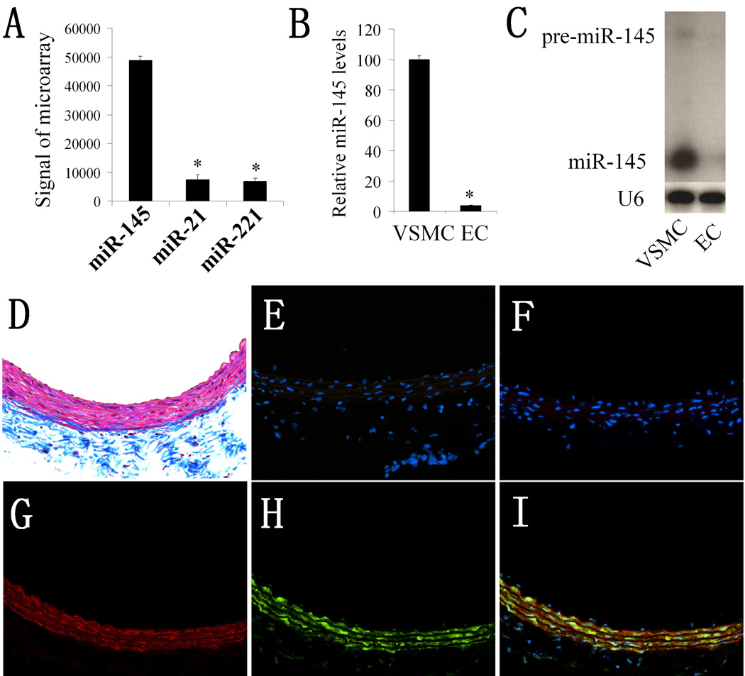
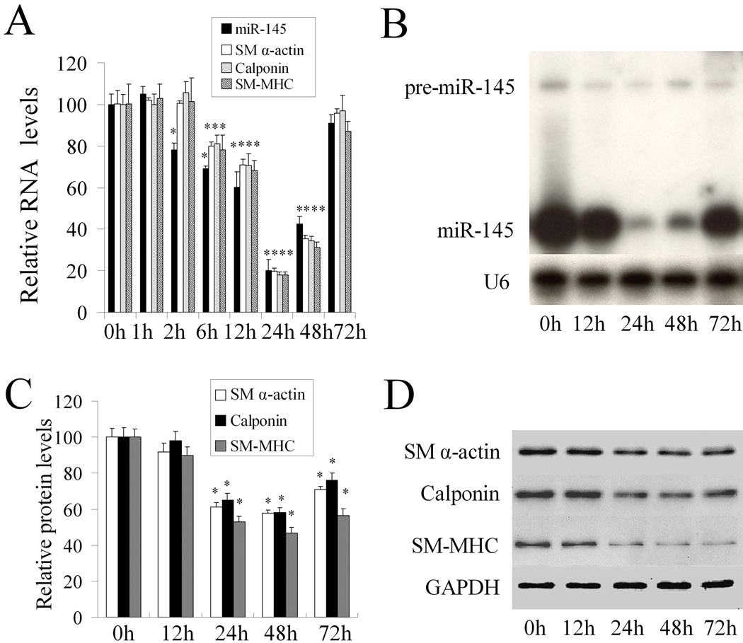
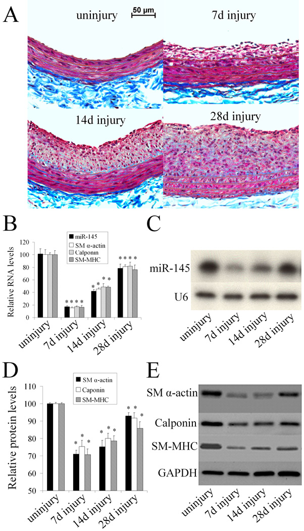
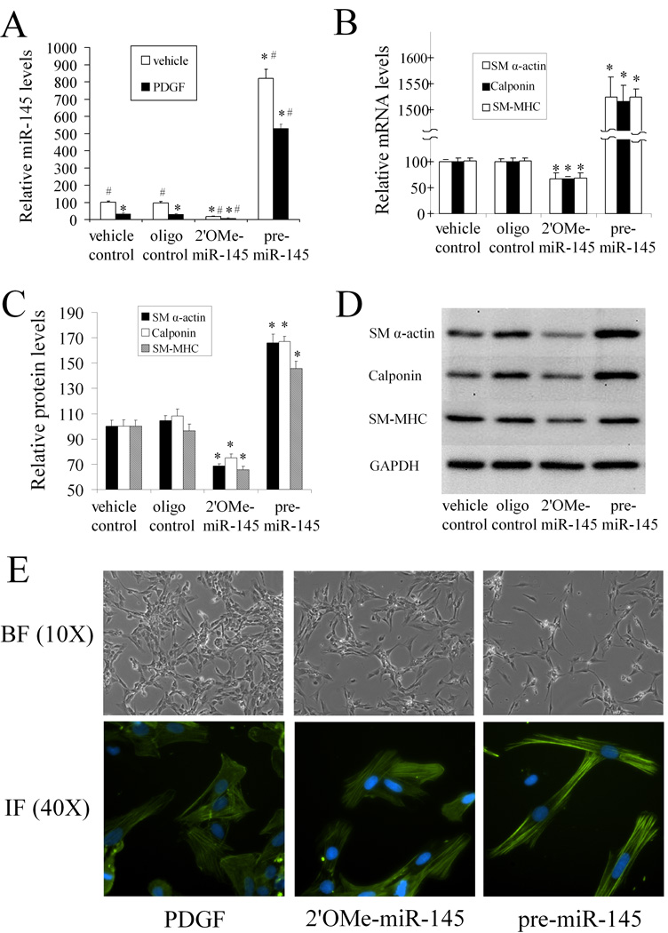
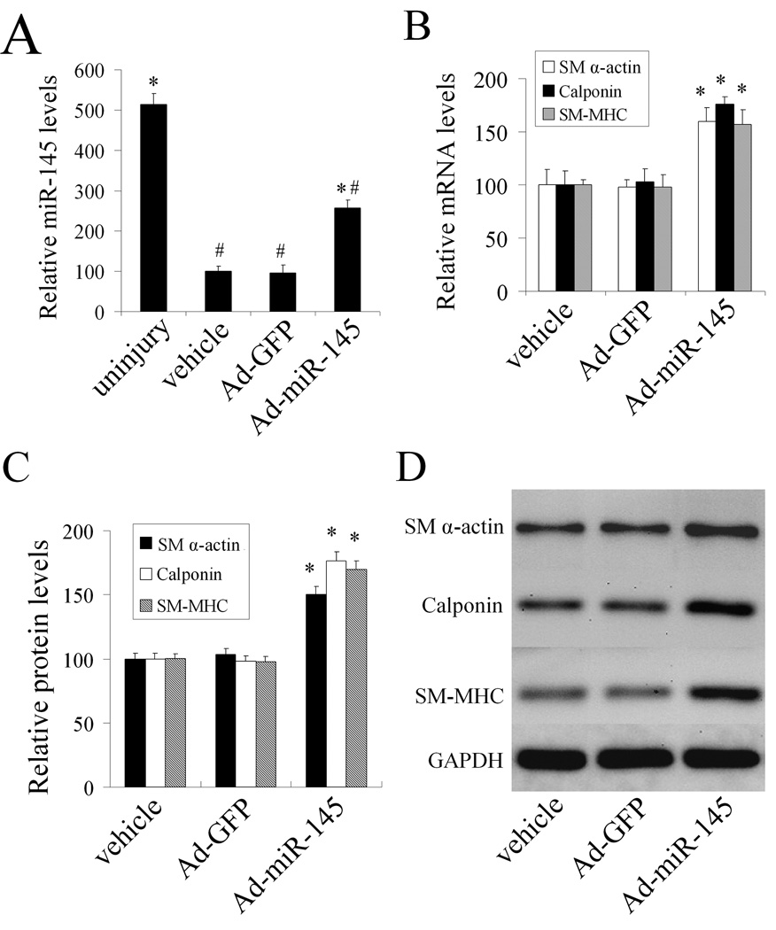
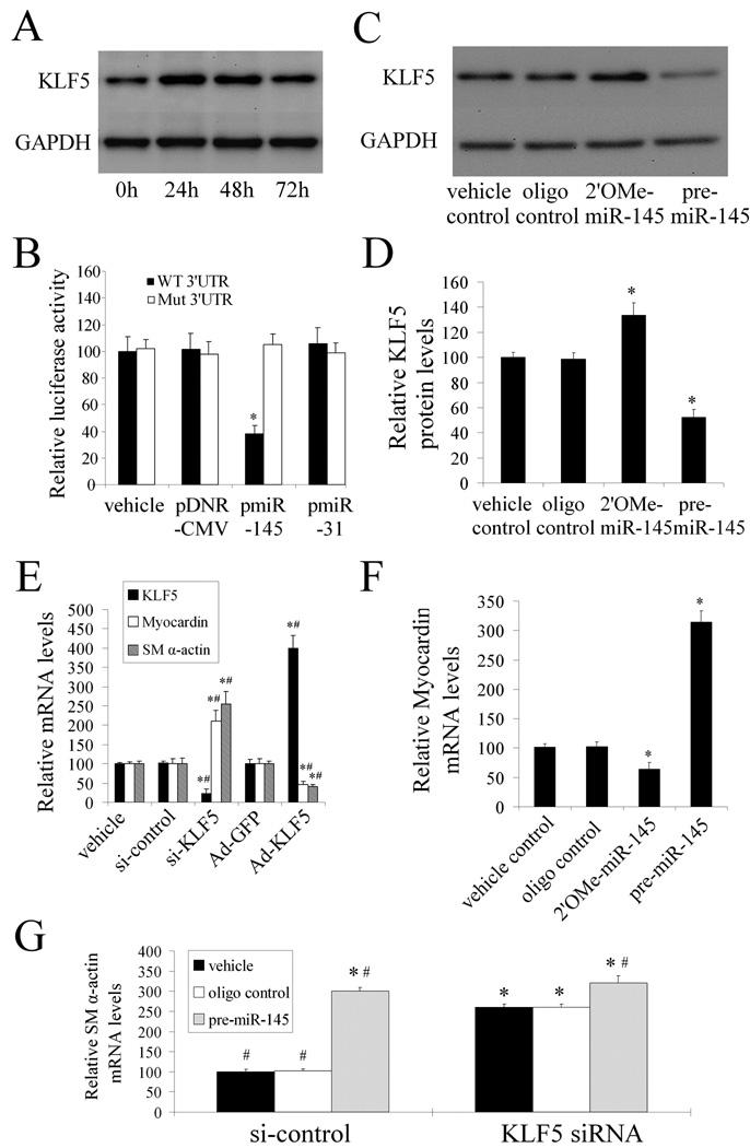
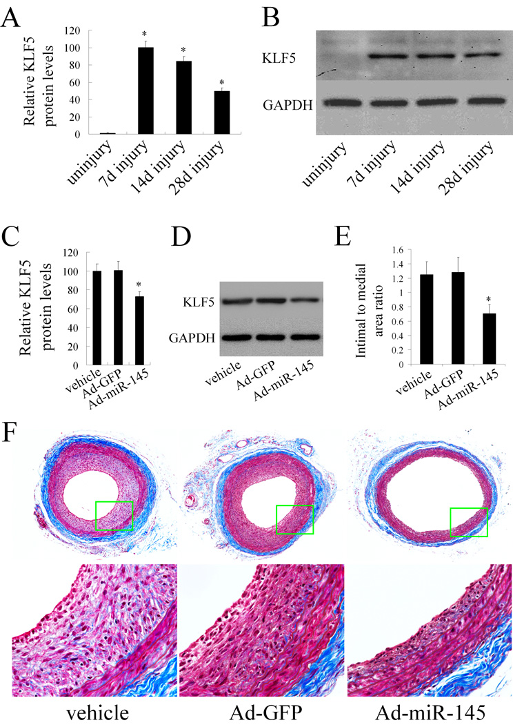
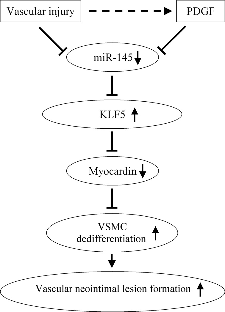
Phenotypic modulation of vascular smooth muscle cells (VSMCs) plays a critical role in the pathogenesis of a variety of proliferative vascular diseases. Recently, we have found that microRNA (miRNA) miR-145 is the most abundant miRNA in normal vascular walls and in freshly isolated VSMCs; however, the role of miR-145 in VSMC phenotypic modulation and vascular diseases is currently unknown. Here we find that miR-145 is selectively expressed in VSMCs of the vascular wall and its expression is significantly downregulated in the vascular walls with neointimal lesion formation and in cultured dedifferentiated VSMCs. More importantly, both in cultured rat VSMCs in vitro and in balloon-injured rat carotid arteries in vivo, we demonstrate that the noncoding RNA miR-145 is a novel phenotypic marker and a novel phenotypic modulator of VSMCs. VSMC differentiation marker genes such as SM alpha-actin, calponin, and SM-MHC are upregulated by premiR-145 or adenovirus expressing miR-145 (Ad-miR-145) but are downregulated by the miR-145 inhibitor 2'OMe-miR-145. We have further identified that miR-145-mediated phenotypic modulation of VSMCs is through its target gene KLF5 and its downstream signaling molecule, myocardin. Finally, restoration of miR-145 in balloon-injured arteries via Ad-miR-145 inhibits neointimal growth. We conclude that miR-145 is a novel VSMC phenotypic marker and modulator that is able of controlling vascular neointimal lesion formation. These novel findings may have extensive implications for the diagnosis and therapy of a variety of proliferative vascular diseases.


Figures

Figure 1
Figure 1. miR-145 is selectively expressed in VSMCs of the vascular wall
(A). The expression levels of miR-145, miR-21, and miR-221 in normal rat carotid arteries. Note: n=6; *P<0.05 compared with miR-145. (B). The relative expression of miR-145 in rat VSMCs and ECs determined by qRT-PCR. Note: n=6; *P<0.05 compared with that in VSMCs. (C). The expression of miR-145 and pre-miR-145 in rat VSMCs and ECs determined by northern blot analysis. (D). Masson's trichrome staining of rat carotid artery. (E). Negative control of In situ hybridization (no miRNA probe). (F). Scrambled probe control. (G). Immunofluorescence with the smooth muscle marker SM α-actin (red color). (H). In situ hybridization of miR-145 (green color). (I). Merged images of (G) and (H). Note: blue color is the cell nuclear staining by DAPI.
Figure 2
Figure 2. miR-145 is a phenotypic marker for VSMCs in vitro in cultured cells
(A). PDGF-BB (20 ng/ml) caused a time-dependent suppression of miR-145 and VSMC differentiation marker genes such as SM α-actin, calponin, and SM-MHC as determined by qRT-PCR. (B). miR-145 and pre-miR-145 in PDGF-BB (20 ng/ml)-treated VSMCs as determined by northern blot analysis. (C). Quantitative analysis of VSMC differentiation marker genes by western blot. (D). Representative western blots in VSMCs treated with PDGF. Note: n=6; *P<0.05 compared with 0 h group.
Figure 3
Figure 3. miR-145 is a phenotypic marker for VSMCs in the vascular wall in vivo
(A). Representative Masson's trichrome staining in uninjured and injured rat carotid arteries at 7, 14, and 28 days after angioplasty. (B). The expression of miR-145, VSMC differentiation marker genes, S M α-actin, calponin, and SM-MHC in uninjured and injured rat carotid arteries determined by qRT-PCR. (C). miR-145 expression in the vascular walls determined by northern blot analysis. (D). Quantitative analysis of VSMC differentiation marker genes by western blot. (E). Representative western blots of VSMC differentiation marker genes. Note: n=6; *P<0.05 compared with uninjured control group.
Figure 4
Figure 4. miR-145 modulates VSMC phenotype in vitro in cultured cells
The VSMCs were pre-treated with vehicle, control oligo, 2'OMe-miR-145, or pre-miR-145 for 4h followed by PDGF or vehicle for 24 h. (A). Modulation of miR-145 expression by 2'OMe-miR-145 (100 nM) and pre-miR-145 (100 nM) in VSMCs with or without PDGF (20 ng/ml). n=6; *P<0.05 compared with oligo control group treated with vehicle. # p<0.05 compared with oligo control group treated with PDGF. (B). 2'OMe-miR-145 strengthened, whereas pre-miR-145 inhibited PDGF-mediated effects on VSMC maker genes as determined by qRT-PCR. n=6; *P<0.05 compared with oligo control. (C). 2'OMe-miR-145 strengthened, whereas pre-miR-145 inhibited PDGF-mediated effects on VSMC maker genes as determined by western blot. n=6; *P<0.05 compared with oligo control. (D). Representative western blots of VSMC differentiation marker genes. (E). Up panel: Representative morphological changes of primary cultured VSMCs treated with PDGF (20 ng/ml), 2'OMe-miR-145 (100 nM), or pre-miR-145 (100 nM) for 48 hours. Bottom panel: Representative immunofluorescence images of the VSMCs via anti-SM α-actin antibody (green color). Note: blue color is the cell nuclear staining by DAPI.
Figure 5
Figure 5. miR-145 modulates the VSMC phenotype in vivo in balloon injured rat carotid arteries
(A). miR-145 expression in uninjured arteries, and in balloon-injured arteries at 7 days after angioplasty, which were treated with vehicle, Ad-GFP, or Ad-miR-145. (B). Ad-miR-145 increased the VSMC differentiation marker genes in injured arteries as determined by qRT-PCR. (C). Ad-miR-145 increased the VSMC differentiation marker genes in injured arteries as determined by western blot. (D). Representative western blots of VSMC differentiation marker genes. Note: n=6; *P<0.05 compared with Ad-GFP control. #p<0.05 compared with uninjured control group.
Figure 6
Figure 6. KLF5 is the critical target gene of miR-145 that is responsible for miR-145-mediated effect on VSMC phenotypic modulation
(A). KLF5 expression was increased in VSMCs after PDGF (20 ng/ml)-stimulation. (B). pmiR-145, but not pDNR-CMV, or unrelated pmiR-31 inhibited luciferase activity. In the mutated control group, the inhibitory effect of pmiR-145 on luciferase activity was abrogated. n=5; *P<0.05 compared with vehicle control. (C). Representative western blots of KLF5 in miR-145 modulated VSMCs. (D). 2'OMe-miR-145 increased, whereas pre-miR-145 decreased KLF5 expression in cultured VSMCs. n=6; *P<0.05 compared with oligo control. (E).The effects of KLF5 on the expression of myocardin and SM α–actin. n=5; * P<0.05 compared with siRNA control (si-control) and, # P<0.05 compared with Ad-GFP. (F). The effect of miR-145 on the expression of myocardin. n=6; *P<0.05 compared with oligo control. (G). The effects of pre-miR-145 on the expression of SM α–actin in VSMCs with or without KLF5 depletion. Note: n=6; *P<0.05 compared with oligo control in siRNA control (si-control)-treated groups. #P<0.05 compared with oligo control in KLF5 siRNA-treated groups.
Figure 7
Figure 7. miR-145 modulates KLF5 expression and vascular neointimal lesion formation in vivo in balloon-injured rat carotid arteries
(A). The expression of KLF5 in uninjured and balloon-injured rat carotid arteries. n=6; *P<0.05 compared with uninjured control. (B). Representative western blots of KLF5 in uninjured and balloon-injured rat carotid arteries. (C).The effect of Ad-miR-145 on the expression of KLF5 in balloon-injured arteries. n=5; *P<0.05 compared with Ad-GFP. (D). Representative western blots of KLF5 in balloon-injured rat carotid arteries treated with vehicle, Ad-GFP or Ad-miR-145. (E). The effect of Ad-miR-145 on vascular neointimal lesion formation in rat carotid arteries at 14 days after angioplasty. n=9; *P<0.05 compared with Ad-GFP. (F). Representative Masson's trichrome stained photomicrographs of rat carotid arteries treated with vehicle, Ad-GFP or Ad-miR145.
Figure 8
Figure 8. Molecular mechanisms of miR-145-mediated effects on VSMC phenotypic modulation and vascular neointimal lesion formation

References

    1. Owens GK, Kumar MS, Wamhoff BR. Molecular regulation of vascular smooth muscle cell differentiation in development and disease. Physiol Rev. 2004;84:767–801. - PubMed
    1. Rzucidlo EM, Martin KA, Powell RJ. Regulation of vascular smooth muscle cell differentiation. J Vasc Surg. 2007;45:A25–A32. - PubMed
    1. Ambros V. The functions of animal microRNAs. Nature. 2004;431:350–355. - PubMed
    1. Chen K, Rajewsky N. The evolution of gene regulation by transcription factors and microRNAs. Nat Rev Genet. 2007;8:93–103. - PubMed
    1. Lewis BP, Burge CB, Bartel DP. Conserved seed pairing, often flanked by adenosines, indicates that thousands of human genes are microRNA targets. Cell. 2005;120:15–20. - PubMed

Publication types

MeSH terms