

Robust docosahexaenoic acid-mediated neuroprotection in a rat model of transient, focal cerebral ischemia
- PMID: 19542051
- PMCID: PMC2745047
- DOI: 10.1161/STROKEAHA.109.555979
Robust docosahexaenoic acid-mediated neuroprotection in a rat model of transient, focal cerebral ischemia
Abstract
Background and purpose: Docosahexaenoic acid (DHA; 22:6n-3), an omega-3 essential fatty acid family member, is the precursor of neuroprotectin D1, which downregulates apoptosis and, in turn, promotes cell survival. This study was conducted to assess whether DHA would show neuroprotective efficacy when systemically administered in different doses after middle cerebral artery occlusion (MCAo) in rats.
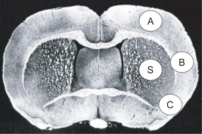
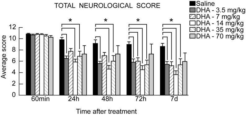
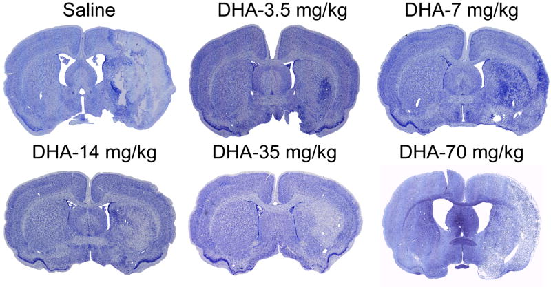
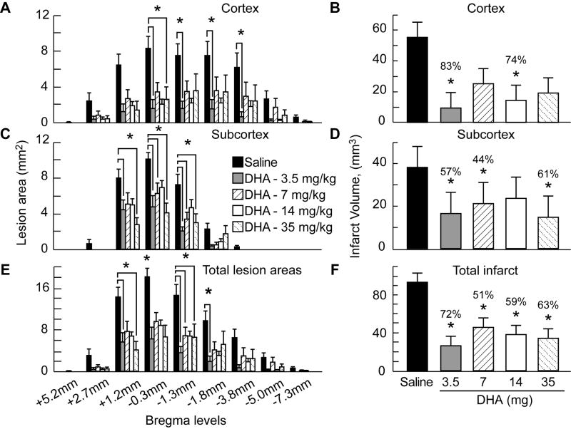
Methods: Sprague-Dawley rats were anesthetized with isoflurane and subjected to 2 hour of MCAo. Animals were treated with either DHA (low doses=3.5 or 7 mg/kg; medium doses=16 or 35 mg/kg; and high dose=70 mg/kg) or an equivalent volume of saline intravenously 3 hours after MCAo onset. Neurologic status was evaluated during occlusion (60 minutes) and on days 1, 2, 3, and 7 after MCAo. Seven days after MCAo, brains were perfusion-fixed, and infarct areas and volumes were determined.
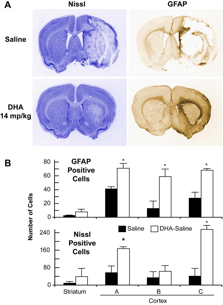
Results: Only the low and medium doses of DHA significantly improved the neurologic score compared with vehicle-treated rats at 24 hours, 48 hours, 72 hours, and 7 days. DHA markedly reduced total corrected infarct volume in all treated groups compared with vehicle-treated rats (3.5 mg/kg, 26+/-9 mm(3); 7 mg/kg, 46+/-12 mm(3); 16 mg/kg, 37+/-5 mm(3); and 35 mg/kg, 34+/-15 mm(3) vs vehicle, 94+/-12 mm(3)). Cortical and striatal infarct volumes were also significantly reduced by treatment with DHA. No neuroprotective effects were observed with 70 mg/kg DHA.
Conclusions: We conclude that DHA experimental therapy at low and medium doses improves neurologic and histologic outcomes after focal cerebral ischemia and might provide benefits in patients after ischemic stroke.
Conflict of interest statement
Figures
Similar articles
-
Human albumin therapy of acute ischemic stroke: marked neuroprotective efficacy at moderate doses and with a broad therapeutic window.Stroke. 2001 Feb;32(2):553-60. doi: 10.1161/01.str.32.2.553. Stroke. 2001. PMID: 11157196
-
Docosahexaenoic acid signaling modulates cell survival in experimental ischemic stroke penumbra and initiates long-term repair in young and aged rats.PLoS One. 2012;7(10):e46151. doi: 10.1371/journal.pone.0046151. Epub 2012 Oct 30. PLoS One. 2012. PMID: 23118851 Free PMC article.
-
Docosahexaenoic acid complexed to albumin provides neuroprotection after experimental stroke in aged rats.Neurobiol Dis. 2014 Feb;62:1-7. doi: 10.1016/j.nbd.2013.09.008. Epub 2013 Sep 21. Neurobiol Dis. 2014. PMID: 24063996 Free PMC article.
-
Acute treatment with docosahexaenoic acid complexed to albumin reduces injury after a permanent focal cerebral ischemia in rats.PLoS One. 2013 Oct 23;8(10):e77237. doi: 10.1371/journal.pone.0077237. eCollection 2013. PLoS One. 2013. PMID: 24194876 Free PMC article.
-
Novel aspirin-triggered neuroprotectin D1 attenuates cerebral ischemic injury after experimental stroke.Exp Neurol. 2012 Jul;236(1):122-30. doi: 10.1016/j.expneurol.2012.04.007. Epub 2012 Apr 19. Exp Neurol. 2012. PMID: 22542947 Free PMC article.
Cited by
-
Dietary Omega-3 Fatty Acids Do Not Change Resistance of Rat Brain or Liver Mitochondria to Ca(2+) and/or Prooxidants.J Lipids. 2012;2012:797105. doi: 10.1155/2012/797105. Epub 2012 Aug 27. J Lipids. 2012. PMID: 22970378 Free PMC article.
-
Partial rescue of Rett syndrome by ω-3 polyunsaturated fatty acids (PUFAs) oil.Genes Nutr. 2012 Jul;7(3):447-58. doi: 10.1007/s12263-012-0285-7. Epub 2012 Mar 8. Genes Nutr. 2012. PMID: 22399313 Free PMC article.
-
Unsaturated fatty acids supplementation reduces blood lead level in rats.Biomed Res Int. 2015;2015:189190. doi: 10.1155/2015/189190. Epub 2015 May 14. Biomed Res Int. 2015. PMID: 26075218 Free PMC article.
-
Lettuce glycoside B ameliorates cerebral ischemia reperfusion injury by increasing nerve growth factor and neurotrophin-3 expression of cerebral cortex in rats.Indian J Pharmacol. 2014 Jan-Feb;46(1):63-8. doi: 10.4103/0253-7613.125171. Indian J Pharmacol. 2014. PMID: 24550587 Free PMC article.
-
Preventative strategies for early-onset bipolar disorder: towards a clinical staging model.CNS Drugs. 2010 Dec;24(12):983-96. doi: 10.2165/11539700-000000000-00000. CNS Drugs. 2010. PMID: 21090835 Review.
References
-
- Bazan NG. Synaptic lipid signaling: significance of polyunsaturated fatty acids and platelet-activating factor. J Lipid Res. 2003;44:2221–2233. - PubMed
-
- Gamoh S, Hashimoto M, Sugioka K, Shahdat Hossain M, Hata N, Misawa Y, Masumura S. Chronic administration of docosahexaenoic acid improves reference memory-related learning ability in young rats. Neuroscience. 1999;93:237–241. - PubMed
-
- McGahon BM, Martin DS, Horrobin DF, Lynch MA. Age-related changes in synaptic function: analysis of the effect of dietary supplementation with omega-3 fatty acids. Neuroscience. 1999;94:305–314. - PubMed
-
- Rodriguez de Turco EB, Belayev L, Liu Y, Busto R, Parkins N, Bazan NG, Ginsberg MD. Systemic fatty acid responses to transient focal cerebral ischemia: influence of neuroprotectant therapy with human albumin. J Neurochem. 2002;83:515–524. - PubMed
-
- Belayev L, Marcheselli VL, Khoutorova L, Rodriguez de Turco EB, Busto R, Ginsberg MD, Bazan NG. Docosahexaenoic acid complexed to albumin elicits high-grade ischemic neuroprotection. Stroke. 2005;36:118–123. - PubMed
Publication types
MeSH terms
Substances
Grants and funding
LinkOut - more resources
Full Text Sources
Other Literature Sources
