. 2009 Sep 4;284(36):24622-33.
doi: 10.1074/jbc.M109.011858. Epub 2009 Jun 19.

Antigen-specific proteolysis by hybrid antibodies containing promiscuous proteolytic light chains paired with an antigen-binding heavy chain

Affiliations

Antigen-specific proteolysis by hybrid antibodies containing promiscuous proteolytic light chains paired with an antigen-binding heavy chain

Gopal Sapparapu et al. J Biol Chem. .

Abstract

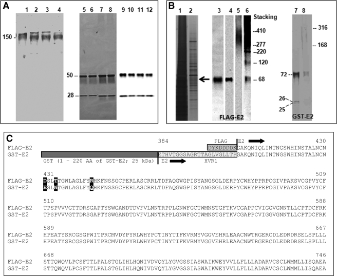
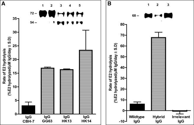
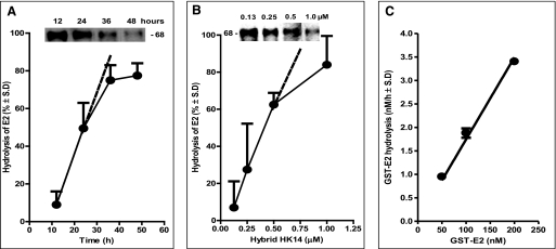
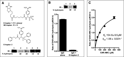
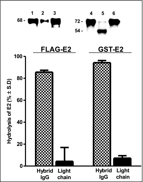
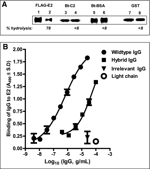
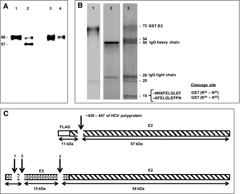
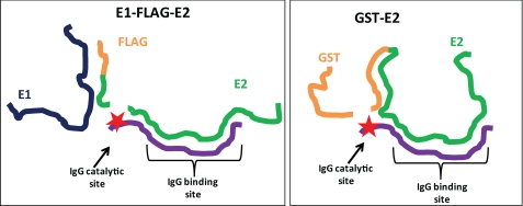
The antigen recognition site of antibodies consists of the heavy and light chain variable domains (V(L) and V(H) domains). V(L) domains catalyze peptide bond hydrolysis independent of V(H) domains (Mei, S., Mody, B., Eklund, S. H., and Paul, S. (1991) J. Biol. Chem. 266, 15571-15574). V(H) domains bind antigens noncovalently independent of V(L) domains (Ward, E. S., Güssow, D., Griffiths, A. D., Jones, P. T., and Winter, G. (1989) Nature 341, 544-546). We describe specific hydrolysis of fusion proteins of the hepatitis C virus E2 protein with glutathione S-transferase (GST-E2) or FLAG peptide (FLAG-E2) by antibodies containing the V(H) domain of an anti-E2 IgG paired with promiscuously catalytic V(L) domains. The hybrid IgG hydrolyzed the E2 fusion proteins more rapidly than the unpaired light chain. An active site-directed inhibitor of serine proteases inhibited the proteolytic activity of the hybrid IgG, indicating a serine protease mechanism. The hybrid IgG displayed noncovalent E2 binding in enzyme-linked immunosorbent assay tests. Immunoblotting studies suggested hydrolysis of FLAG-E2 at a bond within E2 located approximately 11 kDa from the N terminus. GST-E2 was hydrolyzed by the hybrid IgG at bonds in the GST tag. The differing cleavage pattern of FLAG-E2 and GST-E2 can be explained by the split-site model of catalysis, in which conformational differences in the E2 fusion protein substrates position alternate peptide bonds in register with the antibody catalytic subsite despite a common noncovalent binding mechanism. These studies provide proof-of-principle that the catalytic activity of a light chain can be rendered antigen-specific by pairing with a noncovalently binding heavy chain subunit.


Figures

FIGURE 1.
FIGURE 1.
Hybrid Abs and HCV E2 fusion protein substrates. A, expression and purification of full-length hybrid IgGs. Coomassie Blue-stained nonreducing SDS-electrophoresis gels of wild type IgG CBH-7, hybrid IgG HK13, hybrid IgG HK14, and hybrid IgG GG63 (respectively, lanes 1–4); silver-stained reducing SDS gels of these IgGs (respectively, lanes 5–8) and immunoblots of reducing SDS gels of these IgGs stained with a mixture of anti-heavy chain and anti-light chain Abs (respectively, lanes 9–12). IgGs were purified from culture supernatants by affinity chromatography on Protein G-agarose. The 150-kDa band corresponds to intact tetrameric IgGs. The 50- and 28-kDa bands are the heavy and light chains, respectively. B, E2 fusion protein substrates. Shown are reducing SDS gels of the crude lysate from stably transfected Chinese hamster ovary cells coexpressing FLAG-E2 and E1 (lane 1) and the lysate after fractionation on immobilized anti-FLAG Ab stained with silver (lane 2), an anti-E2 Ab (lane 3), or anti-FLAG Ab under reducing (lanes 4) or non-reducing conditions (lane 5 and its overexposed version, lane 6). Lanes 7 and 8 are, respectively, reducing and non-reducing SDS gels of GST-E2 stained with Coomassie Blue. C, FLAG-E2 and GST-E2 primary structure. The E2 fusion proteins have identical sequences except: (a) GST (amino acid residues 1–220 of GST-E2) and FLAG tags (residues 1–8 of FLAG-E2); (b) the hypervariable 1 region is deleted in FLAG-E2; and (c) mismatches at positions 431, 434, and 444 (bold, numbering corresponding to HCV H77 polyprotein).
FIGURE 2.
FIGURE 2.
Hydrolysis of E2 fusion proteins by hybrid IgG Abs. A, GST-E2 hydrolysis. Inset, reducing SDS gels stained with anti-GST Ab showing GST-E2 (130 nm) incubated for 5 days with diluent (lane 1), wild type IgG CBH-7 (lane 2), hybrid IgG GG63 (lane 3), hybrid IgG HK13 (lane 4), or hybrid IgG HK14 (lane 5; all IgG samples tested at 1 μm). The bar plot shows GST-E2 hydrolysis rates computed by densitometry of the intact GST-E2 band. A reaction product at 54 kDa is evident. B, FLAG-E2 hydrolysis. Inset, reducing SDS gels stained with polyclonal anti-E2 Abs showing FLAG-E2 (1 nm) incubated for 2 days with wild type IgG CBH-7 (lane 1), hybrid IgG HK14 (lane 2), or irrelevant anti-gp120 IgG GL2 (lane 3; IgG samples tested at 0.5 μm). Bars show hydrolysis of FLAG-E2 determined as cleavage of the intact 68-kDa protein band as in panel A. Data in panels A and B are mean ± S.D. of three independent reactions.
FIGURE 3.
FIGURE 3.
Characteristics of E2 fusion protein hydrolysis by hybrid HK14. A, time dependence of FLAG-E2 hydrolysis. Hydrolysis of FLAG-E2 (1 nm) by the hybrid IgG (0.5 μm) was measured as described in the legend to Fig. 2B. Mean ± S.D. of two reactions is shown. Inset, reducing SDS gels stained with polyclonal anti-E2 Abs showing cleavage of the 68-kDa FLAG-E2 band after incubation with hybrid IgG for 12, 24, 36, and 48 h. B, hybrid IgG concentration dependence. FLAG-E2 (1 nm) hydrolysis determined after incubation for 3 days with increasing hybrid IgG concentrations. Mean ± S.D. of two reactions is shown. Inset, reducing SDS gels stained with polyclonal anti-E2 Abs showing cleavage of the 68-kDa FLAG-E2 band after incubation with 0.13, 0.25, 0.5, and 1.0 μm hybrid IgG HK14. C, substrate concentration dependence. Increasing GST-E2 concentrations were incubated for 48 h with the hybrid IgG (0.5 μm). Hydrolysis was determined from cleavage of the intact 72-kDa protein band stainable with polyclonal anti-E2 Abs. Mean ± S.D. of two reactions is shown. The linear increase in rate confirms that substrate is present at concentrations that do not saturate the IgG, permitting measurement of the effect of noncovalent substrate recognition on the reaction rate.
FIGURE 4.
FIGURE 4.
Serine protease inhibitor effect and kinetics of Glu-Ala-Arg-AMC hydrolysis. A, phosphonate diester hapten inhibition of FLAG-E2 hydrolysis. Reducing SDS gels showing FLAG-E2 (1 nm) incubated with diluent (lane 1) or hybrid IgG HK14 (0.5 μm) in the presence of 2% dimethyl sulfoxide (lane 2), the non-electrophilic compound NE-hapten (lane 3), E-hapten 1 (lane 4), or E-hapten 2 (lane 5). Inhibitor concentrations: E-hapten 1 and NE-hapten, 500 μm; E-hapten 2, 200 μm. Reaction time was 48 h. Staining was with polyclonal anti-E2 Abs. Numbers below the lanes indicate % hydrolysis compared with diluent control (lane 1). Top, structures of NE-hapten, E-hapten 1, and E-hapten 2. B, phosphonate diester inhibition of Glu-Ala-Arg-AMC hydrolysis. Shown are the hydrolysis rates of the substrate (400 μm) incubated with hybrid IgG (0.1 μm) for 24 h in the presence or absence of E-hapten 2 (200 μm). Mean ± S.D. of three reactions is shown. Inset, inhibition of FLAG-E2 hydrolysis by the alternate substrate Glu-Ala-Arg-AMC. Shown are reducing SDS gels showing FLAG-E2 (1 nm) incubated with the hybrid IgG (0.5 μm) in the absence (lane 2) or presence (lane 3) of Glu-Ala-Arg-MCA (200 μm) for 48 h. Lane 1, diluent control without IgG. Staining with anti-E2 Ab. Numbers below the lanes indicate % hydrolysis compared with diluent control (lane 1). Mean ± S.D. of two reactions is shown. C, kinetic parameters for hybrid IgG catalyzed Glu-Ala-Arg-AMC hydrolysis. IgG was 0.1 μm. Initial velocities (V) fitted to the Michaelis-Menten equation by nonlinear regression (r2 = 0.98). Data are means of three reactions computed as slopes of plots of rate versus time at each substrate concentration.
FIGURE 5.
FIGURE 5.
Hydrolytic specificity of hybrid IgG: failure of the light chain alone to hydrolyze E2 proteins. Hydrolysis of FLAG-E2 (left) or GST-E2 (right) by hybrid IgG HK14 (1 μm, stippled bars) or light chain HK14 alone (2 μm, solid bars) was measured as described in the legend to Fig. 2 (24 and 96 h incubation, respectively, for FLAG-E2 and GST-E2). Data are mean ± S.D. of two reactions. Insets, reducing SDS gels showing FLAG-E2 incubated with wild type IgG CBH-7 (lane 1), hybrid IgG HK14 (lane 2), or light chain HK14 (lane 3) and GST-E2 incubated with wild type IgG CBH-7 (lane 4), hybrid IgG HK14 (lane 5), or light chain HK14 (lane 6).
FIGURE 6.
FIGURE 6.
Hydrolytic specificity of hybrid IgG: failure to hydrolyze irrelevant proteins and noncovalent E2 binding activity. A, substrate specificity. Reducing SDS gels showing reaction mixtures of hybrid IgG HK14 (0.5 μm) or diluent and FLAG-E2 (respectively, lanes 1 and 2), biotinylated Factor VIII C2 domain (respectively, lanes 3 and 4), biotinylated bovine serum albumin (respectively, lanes 5 and 6), or GST devoid of the E2 polypeptide (respectively, lanes 7 and 8). FLAG-E2 was 1 nm. Other substrates were 10 μm; reaction time was 48 h. FLAG-E2 staining was with anti-FLAG Ab, biotinylated protein staining with peroxidase-conjugated streptavidin, and GST staining with anti-GST Ab. Numbers below the lanes indicate % hydrolysis compared with diluent control. B, FLAG-E2 binding. Binding of hybrid IgG HK14, wild type IgG CBH-7, irrelevant anti-gp120 IgG GL2, and light chain HK14 to FLAG-E2 (4 ng/well) was measured by ELISA. Shown are A490 values versus Ab concentration curves fitted to A490 = bottom + (top − bottom)/(1 + 10((log EC50X) × Hill slope)) (r2 > 0.97). Mean ± S.D. of two wells each.
FIGURE 7.
FIGURE 7.
E2 fusion protein cleavage regions. A, reducing SDS gel showing reaction mixtures of FLAG-E2 (1 nm) incubated with wild type IgG CBH-7 or hybrid IgG HK14 (0.5 μm) for 48 h and stained with anti-E2 monoclonal Ab (respectively, lanes 1 and 2) or anti-FLAG Ab (respectively, lanes 3 and 4). Note the 57-kDa anti-E2 stainable product band generated by the hybrid IgG (lane 2) that was not stained by anti-FLAG Ab (lane 4). B, reducing SDS gel lanes of GST-E2 alone (lane 1), the hybrid IgG alone (lane 2), and the reaction mixture of GST-E2 and hybrid IgG HK14 (lane 3) stained with Coomassie Blue. GST-E2 was 3 μm and hybrid IgG was 1 μm with incubation for 5 days. The N-terminal 10 residues of the 15-kDa band determined by Edman's degradation and the deduced scissile bonds are indicated. Yields of the phenylthiohydantoin-derivitized amino acids in individual sequencing cycles were 0.53–1.61 pmol. C, schematic representation of FLAG-E2 (top) and GST-E2 (bottom) showing the cleavage sites deduced from the mass and immunoreactivity of the FLAG-E2 57-kDa product, and from amino acid sequencing and mass of the GST-E2 15-kDa product (see “Results”).
FIGURE 8.
FIGURE 8.
Split-site model for shared noncovalent E2 binding specificity and divergent hydrolytic specificity of hybrid IgG. The noncovalent antigen binding pocket of the hybrid IgG is occupied by the E2 epitope. The hydrolytic subsite of the IgG (★) is located distant from the binding site. If the conformations of the substrates (FLAG-E2, GST-E2) are divergent, different peptide bonds can be placed in register with the hydrolytic subsite despite noncovalent IgG binding to the same epitope in the substrates.

Similar articles

Cited by

References

    1. Sun M., Li L., Gao Q. S., Paul S. (1994) J. Biol. Chem. 269, 734–738 - PubMed
    1. Edelman G. M., Olins D. E., Gally J. A., Zinder N. D. (1963) Proc. Natl. Acad. Sci. U.S.A. 50, 753–761 - PMC - PubMed
    1. Roholt O., Onoue K., Pressman D. (1964) Proc. Natl. Acad. Sci. U.S.A. 51, 173–178 - PMC - PubMed
    1. Ward E. S., Güssow D., Griffiths A. D., Jones P. T., Winter G. (1989) Nature 341, 544–546 - PubMed
    1. Wilson I. A., Stanfield R. L., Rini J. M., Arevalo J. H., Schulze-Gahmen U., Fremont D. H., Stura E. A. (1991) CIBA Found. Symp. 159, 13–28; 28–39 - PubMed

Publication types

MeSH terms