. 2009 Aug 21;284(34):22916-25.
doi: 10.1074/jbc.M109.035519. Epub 2009 Jun 19.

Mirk regulates the exit of colon cancer cells from quiescence

Affiliations

Mirk regulates the exit of colon cancer cells from quiescence

Kideok Jin et al. J Biol Chem. .

Abstract

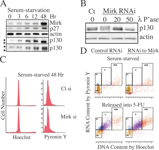
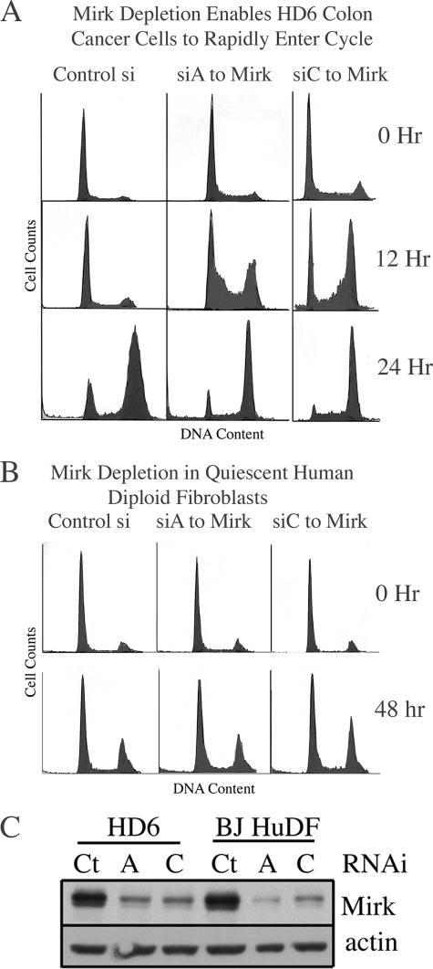
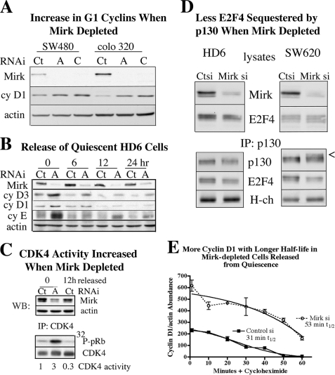
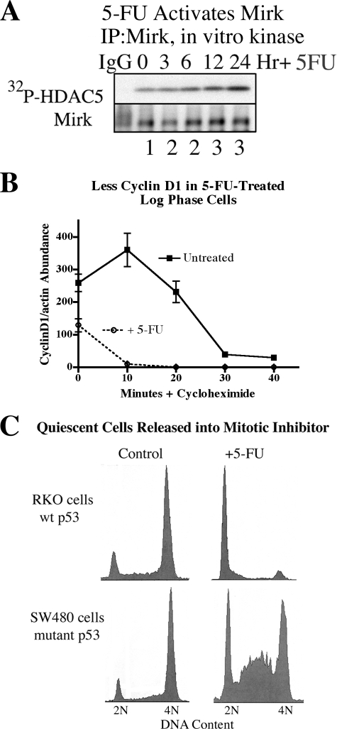
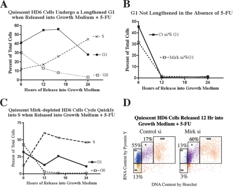
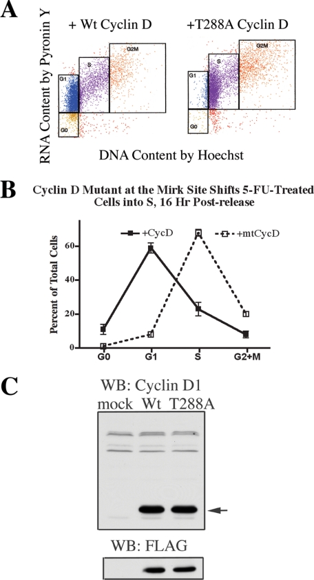
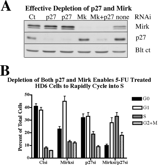
Mirk/Dyrk1B is a serine/threonine kinase widely expressed in colon cancers. Serum starvation induced HD6 colon carcinoma cells to enter a quiescent G0 state, characterized by a 2N DNA content and a lower RNA content than G1 cells. Compared with cycling cells, quiescent cells exhibited 16-fold higher levels of the retinoblastoma protein p130/Rb2, which sequesters E2F4 to block entry into G1, 10-fold elevated levels of the CDK inhibitor p27kip1, and 10-fold higher levels of Mirk. However, depletion of Mirk did not prevent entry into G0, but enabled quiescent HD6, SW480, and colo320 colon carcinoma cells to acquire some biochemical characteristics of G1 cells, including increased levels of cyclin D1 and cyclin D3 because of slower turnover, increased activity of their CDK4/cyclin D complexes, and increased phosphorylation and decreased E2F4 sequestering ability of the CDK4 target, p130/Rb2. As a result, depletion of Mirk allowed some cells to escape quiescence and enabled cells released from quiescence to traverse G1 more quickly. The kinase activity of Mirk was increased by the chemotherapeutic drug 5-fluorouracil (5-FU). Treatment of p53 mutant colon cancer cells with 5-FU led to an elongated G1 in a Mirk-dependent manner, as G1 was shortened by ectopic overexpression of cyclin D1 mutated at the Mirk phosphorylation site (T288A), but not by wild-type cyclin D1. Mirk, through regulating cyclin D turnover, and the CDK inhibitor p27, as shown by depletion studies, functioned independently and additively to regulate the exit of tumor cells from quiescence.


Figures

FIGURE 1.
FIGURE 1.
Characterization of quiescent HD6 colon carcinoma cells. A, HD6 colon carcinoma cells were cultured in serum-free medium for 0–48 h and examined for abundance of Mirk, p130/Rb2, p27kip1, and actin by Western blotting. The top four panels were scanned from a 10% acrylamide gel, while the lowest panel of p130/Rb2 was scanned from a 6% acrylamide gel. The dots mark the different positions of the more phosphorylated, slowly migrating p130 band, and the less phosphorylated, faster migrating p130 band. Similar data were obtained in a second experiment. B, E2F4 sequestering protein p130/Rb2HD6 is more phosphorylated in quiescent cells when Mirk is depleted. HD6 colon carcinoma cells were depleted of Mirk by transient transfection with RNAi duplex siA (Mirk si) or mock-depleted by transfection with GC-matched control RNAi (Ct si), cultured in serum-free medium for 48 h, then released into serum-containing growth medium. Lysates were treated with 20 or 50 μl of lambda phosphatase before Western analysis for p130/Rb2 and actin. Similar data obtained in two other experiments (not shown). C, HD6 colon carcinoma cells were depleted of Mirk or mock-depleted and then made quiescent as in B. 10,000 cells from each culture were analyzed for DNA content by staining with Hoechst 33258, then stained for RNA content (primarily polyribosomes) by Pyronin Y. Similar data showing accumulation of serum-starved cells with a 2N DNA content found in 3 two-parameter flow cytometric analyses using Hoechst/Pyronin Y staining and in 5 one-parameter flow cytometric analysis using propidium iodide to analyze DNA content (Table 1). D, serum-starved HD6 cells arrest predominantly in G0, but retain the capacity to enter cycle when released by addition of FBS. Data from C plotted together in the upper panel. In the lower panel, parallel cultures were released for 24 h by a change to growth medium containing 7% FBS, the toxic concentration of 1 μm 5-fluoruracil to damage DNA and block cycling cells in S phase by engagement of checkpoints, and 50 ng/ml nocodazole to block any remaining cycling cells in G2+M so that only one cell cycle was allowed. 10,000 cells from each culture were analyzed for DNA content by staining with Hoechst 33258, then stained for RNA content with Pyronin Y. The analyses of the serum-starved cultures and the released cultures were performed at the same time using the same gating. Similar data were seen in two additional experiments. The fraction of cells in each cell cycle position in the serum-starved culture control-depleted were 86% G0 (gold), 2% G1 (blue), 2% S (purple), and 5% G2+M (red) and Mirk-depleted were 71% G0, 1% G1, 1% S, and 7% G2+M. The serum-starved mock-depleted cultures released into 5-FU were 22% G0, 14% G1, 34% S, and 12% G2+M. The Mirk-depleted cultures released into 5-FU were 14% G0, 3% G1, 49% S, and 27% G2+M. The values do not equal 100% because of the presence of sub-G0 cells (not shown) and some cells with >2N DNA but lower RNA content than normal S phase cells.
FIGURE 2.
FIGURE 2.
Mirk depletion enables colon cancer cells to rapidly traverse G1 when released from quiescence, but does not detectably affect cycling of human diploid fibroblasts. A, HD6 colon carcinoma cells were depleted of Mirk by RNAi duplexes siA or siC or mock-depleted and then made quiescent as in Fig. 1. Cells were then released by change to medium containing 7% FBS and the mitotic inhibitor vinblastine at 2 nm so only one cell cycle would be analyzed by flow cytometry after propidium iodide staining. Data shown are representative of five replicate experiments, with the quantitation shown in Table 1. B, Mirk depletion does not detectably affect cell cycle progression of human diploid fibroblasts. Strain BJ cells were depleted of Mirk by transient transfection with RNAi duplexes siA and siC or mock-depleted with control RNAi by a 48-h transfection in growth medium with Lipofectamine 2000, then accumulated in G0/G1 by 24 h culture in serum-free medium. Cells were then released by a change to medium containing 7% FBS and 2 nm vinblastine to arrest cycling cells in G2+M and were analyzed for cell cycle distribution by flow cytometry 0 and 48 h following release. The number of cells analyzed per panel was 3680 ± 1218 (S.D.) (n = 6), with similar results in three similar experiments. C, parallel Western blots from the experiments shown in A and B, showing Mirk and actin levels in HD6 colon carcinoma cells and in BJ human diploid fibroblasts (BJ HuDF).
FIGURE 3.
FIGURE 3.
Mirk destabilizes cyclin D in colon cancer cells. A, SW480 and Colo 320 colon carcinoma cells were depleted of Mirk or mock-depleted as in Fig. 1. Lysates were examined for levels of Mirk, cyclin D1, and actin. Mirk depletion reduced cyclin D1 levels 2-fold in SW480 cells and 4-fold in Colo 320 cells. B, Mirk was depleted in HD6 cells by siA and made quiescent as in Fig. 1, then released into growth medium containing 2 nm vinblastine to collect cycling cells in G2+M. Parallel cultures were examined at 0, 6, 12, and 24 h after release for Mirk, cyclin D1, cyclin D3, cyclin E, or actin by Western blotting. Data shown are representative of two experiments. C, Mirk was depleted in HD6 cells by siA and made quiescent as in Fig. 1, then released into growth medium containing 2 nm vinblastine to collect cycling cells in G2+M. Lysates were Western blotted for the abundance of Mirk and actin (upper) and immunoprecipitated with anti-CDK4 antibody conjugated to agarose. An in vitro kinase reaction was carried out on recombinant pRb (lower). The ratio of phospho-pRb to immunoprecipitated CDK4 is shown below the respective lanes and is representative of two experiments. D, Mirk was depleted in HD6 cells and in SW620 cells by siD and made quiescent for 3 days as in Fig. 1. The E2F4 sequestering protein p130/Rb2 was immunoprecipitated and the amounts of p130 and co-immunoprecipitated E2F4 were determined by Western blotting, with immunoglobulin heavy chain (H-ch) as control. Cell lysates were examined for total Mirk and E2F4 levels by Western blotting. E, Mirk was depleted in HD6 cells by siA and made quiescent as in Fig. 1, then released into growth medium containing 20 μg/ml cycloheximide for the times indicated before lysates were analyzed by Western blotting. Sigmoidal curve-fitting (r2 = 0.9782) yielded a half-life of 31 min for cyclin D1 in the control-depleted cells and 53 min for the Mirk-depleted cells (n = 2, mean ± S.D. shown).
FIGURE 4.
FIGURE 4.
Activation of Mirk kinase activity by the chemotherapeutic drug 5-FU correlates with a lengthened G1 phase in colon cancer cells with mutant p53. A, HD6 (p53 mutant) colon carcinoma cells were exposed to 10 μm 5-fluorouracil for 0–24 h. Mirk was immunoprecipitated and an immune complex kinase assay was performed using the 1–283 amino acid fragment of histone deacetylase 5 (HDAC5) as the substrate (Mirk site at Ser-279). The amount of Mirk in the immunoprecipitates was determined by Western blotting. The ratio of Mirk kinase activity to total Mirk protein is given below each lane. B, Mirk was depleted in HD6 cells by siA and made quiescent as in Fig. 1, then released into growth medium containing 1 μm 5-FU for 24 h. Cells were then treated with 20 μg/ml cycloheximide for the times indicated before lysates were analyzed by Western blotting (n = 2, mean ± S.D. shown). C, RKO (p53 wild-type) and SW480 (p53 mutant) colon carcinoma cells were made quiescent by culture in DMEM+0.2% FBS for 2 days, then released for 24 h into DMEM + 7% FBS plus the mitotic inhibitor nocodazole (50 ng/ml) to block cycling cells in G2+M, and either 1 μm 5-FU or no additives. Cell cycle position was determined by one-parameter flow cytometry for DNA content after propidium iodide staining.
FIGURE 5.
FIGURE 5.
When quiescent HD6 colon carcinoma cells were released into fresh growth medium containing 5-fluorouracil, they were maintained in G1 for several hours, while Mirk-depleted cells rapidly traversed G1 and entered S phase. HD6 cells were depleted of Mirk or mock-depleted made quiescent as in Fig. 1, then released by change to growth medium containing 1 μm 5-FU. 50 ng/ml of the mitotic inhibitor nocodazole was added to arrest any cycling cells in G2+M so that only one cell cycle was allowed, and cells were collected 6, 12, 16, and 24 h following release. A parallel experiment was performed with depletion of Mirk by RNAi duplex siD directed to another sequence in the Mirk mRNA, cells were collected 24 h after release, and data were averaged together with the data from the experiment utilizing siA. Similar results were seen in three experiments. A, time course showing cell cycle distribution of mock-depleted HD6 cells 6–24 h after release from quiescence in the presence of 1 μm 5-FU. B, time course showing percent of cells in G1 of either mock-depleted or Mirk-depleted HD6 cells 6–24 h after release from quiescence, no 5-FU added. C, identical time course to A, with Mirk-depleted HD6 cells. D, HD6 cells were depleted of Mirk or mock-depleted, made quiescent as in Fig. 1 and then released for 12 h into growth medium containing 1 μm 5-FU. 50 ng/ml of the mitotic inhibitor nocodazole was added to arrest any cycling cells in G2+M so that only one cell cycle was allowed. Cell cycle phases were color-coded: G0 (gold), G1 (blue), S (purple), G2+M (red). Note that at this time of release the majority of mock-depleted cells were in G1 while the majority of Mirk-depleted cells were in S.
FIGURE 6.
FIGURE 6.
Expression of cyclin D1 mutated at the Mirk phosphorylation site conserved in all cyclin D isoforms gives a phenocopy of Mirk-depletion: cells can move into cycle even when challenged with 5-FU. HD6 cells were transfected with expression plasmids (5 μg) for either wild-type FLAG-cyclin D1 or FLAG-cyclin D1-T288A, or an empty vector, for 48 h in growth medium together with siA to deplete Mirk (data not shown) or with GC-matched control RNAi duplexes to mock deplete Mirk, made quiescent as in Fig. 1, then released for 16 h into growth medium containing 1 μm 5-FU. The cells which remained cycling under these conditions were arrested in G2+M by 50 ng/ml nocodazole. 95% of cells either depleted of Mirk or mock-depleted were collected at G2+M by nocodazole in control cultures not transfected with cyclin D constructs (not shown). Cells were analyzed by two-parameter flow cytometry after staining with Hoechst 33258 for DNA followed by Pyronin Y for RNA. 6341 ± 649 cells were analyzed per sample. A, two-parameter flow cytometry showing the effect of overexpression of wild-type cyclin D1 or mutant cyclin D1-T288A on cell cycle distribution in mock-depleted cells released from quiescence in the presence of 1 μm 5-FU for 16 h. B, plot of the data from A. C, lysates from HD6 cells transfected with either wild-type FLAG-cyclin D1, FLAG-cyclin D1-T288A, mutated at the Mirk phosphorylation site, or mock-transfected for 48 h in growth medium. Lysates analyzed for expression of plasmids by Western blotting for cyclin D1 (arrow) or for the FLAG tag. The upper bands in the cyclin D1 blot are cross-reacting bands showing equal loading. The endogenous cyclin D1 is seen as a low abundant band migrating just before the exogenous cyclin D constructs.
FIGURE 7.
FIGURE 7.
Depletion of both the CDK inhibitor p27 and Mirk enables 5-FU treated HD6 cells to rapidly cycle into S phase. HD6 cells were depleted of p27, Mirk or both by transient transfection with RNAi duplexes for 6 h, not the 48 h used in all prior experiments, made quiescent as in Fig. 1, and then released for 16 h into growth medium containing 1 μm 5-FU. The cells which remained cycling under these conditions were arrested in G2+M by 50 ng/ml nocodazole. A, Western blots of parallel cultures showing depletion of Mirk (Mk) and p27 with RNAi duplexes. Blt ct, blot control of cross-reacting band. B, cells were analyzed by two-parameter flow cytometry after staining with Hoechst 33258 for DNA followed by Pyronin Y for RNA. 7014 ± 2526 cells were analyzed per sample. Mean ± S.D. is shown. Values for untreated proliferating cells not in graph were 19% in G0, 29% in G1, 22% in S, and 29% in G2+M.

Similar articles

Cited by

References

    1. Coller H., Sang L., Roberts J. M. (2006) PLOS Biol. 4,329–349 - PMC - PubMed
    1. Chen C., Liu Y., Liu R., Ikenoue T., Guan K. L., Liu Y., Zheng P. (2008) J. Exp. Med. 205,2397–2408 - PMC - PubMed
    1. Deng X., Ewton D. Z., Friedman E. (2009) Cancer Research 69,3317–3324 - PMC - PubMed
    1. Deng X., Ewton D. Z., Pawlikowski B., Maimone M., Friedman E. (2003) J. Biol. Chem. 278,41347–41354 - PubMed
    1. Zou Y., Ewton D. Z., Deng X., Mercer S. E., Friedman E. (2004) J. Biol. Chem. 279,27790–27798 - PubMed

Publication types

MeSH terms

LinkOut - more resources