. 2009 Aug 21;284(34):22815-24.
doi: 10.1074/jbc.M109.032490. Epub 2009 Jun 19.

Lactogenic hormonal induction of long distance interactions between beta-casein gene regulatory elements

Affiliations

Lactogenic hormonal induction of long distance interactions between beta-casein gene regulatory elements

Elena B Kabotyanski et al. J Biol Chem. .

Abstract

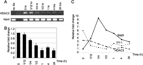
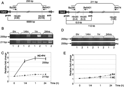
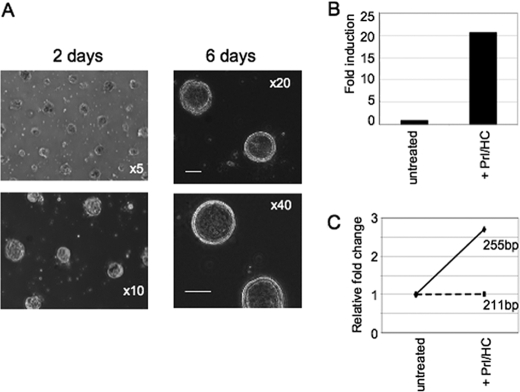
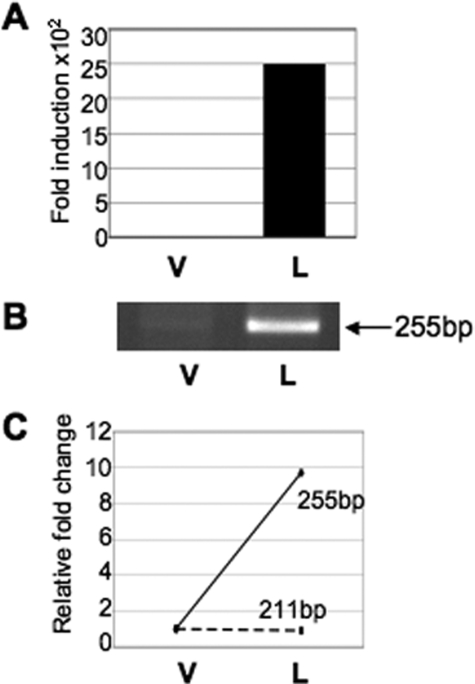
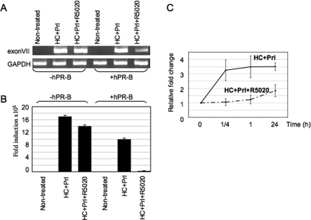
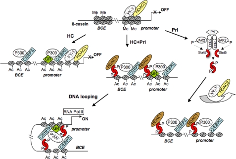
Lactogenic hormone regulation of beta-casein gene expression in mammary epithelial cells provides an excellent model in which to study the mechanisms by which steroid and peptide hormone signaling control gene expression. Prolactin- and glucocorticoid-mediated induction of beta-casein gene expression involves two principal regulatory regions, a proximal promoter and a distal enhancer located in the mouse approximately -6 kb upstream of the transcription start site. Using a chromosome conformation capture assay and quantitative real time PCR, we demonstrate that a chromatin loop is created in conjunction with the recruitment of specific transcription factors and p300 in HC11 mammary epithelial cells. Stimulation with both prolactin and hydrocortisone is required for the induction of these long range interactions between the promoter and enhancer, and no DNA looping was observed in nontreated cells or cells treated with each of the hormones separately. The lactogenic hormone-induced interaction between the proximal promoter and distal enhancer was confirmed in hormone-treated primary three-dimensional mammary acini cultures. In addition, the developmental regulation of DNA looping between the beta-casein regulatory regions was observed in lactating but not in virgin mouse mammary glands. Furthermore, beta-casein mRNA induction and long range interactions between these regulatory regions were inhibited in a progestin-dependent manner following stimulation with prolactin and hydrocortisone in HC11 cells expressing human PR-B. Collectively, these data suggest that the communication between these regulatory regions with intervening DNA looping is a crucial step required to both create and maintain active chromatin domains and regulate transcription.


Figures

FIGURE 1.
FIGURE 1.
Reciprocal regulation of histone deacetylase HDAC3 and YY-1 as compared with Stat5 binding at the β-casein proximal promoter in HC11 cells stimulated with lactogenic hormones. A, ChIP assays were performed in untreated cells and cells treated with HC and Prl for different periods of time using antibodies (Ab) to HDAC3. PCR was performed using primers specific for β-casein promoter followed by gel electrophoresis. B, presence of HDAC3 at different time points was measured by quantitative real time PCR. Immunoprecipitation data were normalized to input DNA, and the amounts were expressed as the fold-change relative to untreated cells. C, reciprocal dynamics of Stat5 and HDAC3:YY-1 binding to the proximal promoter in cells stimulated with hormones for different periods of time.
FIGURE 2.
FIGURE 2.
3C analysis indicates that long range interactions between the β-casein gene proximal promoter and distal enhancer are observed only following hormonal stimulation of HC11 cells with both prolactin and glucocorticoids but not in cells treated separately with these hormones. A, schematic representation of β-casein regulatory regions, primers, and amplicons used in PCR analysis. Cross-linked chromatin from nontreated and hormone-stimulated HC11 cells was digested with HindIII. The ligated DNA was PCR-amplified with primers as indicated. Two different primer sets, Bp290_Be5401 and Bp290_Be474, were designed to compare cross-linking frequency between restriction fragments in the promoter (prom) and enhancer (enh) region (255-bp PCR product) and between the promoter and a distal regions (112 kb from promoter; 211-bp PCR product). The sequences of the primers are given under “Experimental Procedures.” B, 3C assay was performed using HindIII-digested chromatin from untreated HC11 cells and cells treated with HC and Prl for 15 min, 1 h, and 24 h. Agarose gel electrophoresis of the PCR products obtained with primer set Bp290_Be5401 indicates interactions between the promoter and enhancer (upper panel), whereas the primers Bp290_Be474 represent the random ligation efficiency (lower panel). Lanes 1 and 2 show PCR products obtained from the diluted 1:10 and undiluted 3C templates, respectively. C, interaction frequency at different time points was measured by quantitative real time PCR. PCR products obtained with the 3C templates for both primer sets were normalized to control BAC template, and the amounts were expressed as the fold-change relative to untreated cells. The results shown are averages of at least three different amplifications with five different dilutions in each linear range with the standard deviations. D, 3C assay was performed using HindIII-digested chromatin from untreated HC11 cells and cells treated with HC (upper panel) or Prl (lower panel) for 15 min, 1 h, and 24 h. The PCR products (255 bp) using the Bp290_Be5401 primer set were run on agarose gels. Lanes 1 and 2 showed PCR products obtained from the diluted 1:10 and undiluted 3C templates, respectively. E, interaction frequency at different time points was determined as described above.
FIGURE 3.
FIGURE 3.
3C analysis detects hormonally regulated long range interactions between the β-casein proximal promoter and distal enhancer in three-dimensional acini cultures. A, light microscope images of three-dimensional structures grown on Matrigel (bar, 20 μm). MECs were isolated from mid-late pregnant mice (pooled from 12 to 14 mice), plated, and grown on Matrigel for 6 days. Cultures were incubated for 48 h in medium without serum and EGF followed by hormonal stimulation for an additional 48 h. B, total RNA prepared from three-dimensional acini in the absence (untreated) and presence of hormones (+Prl/HC) was reverse-transcribed and amplified using exon VII primers specific to the β-casein gene. The accumulation of transcripts was measured by quantitative real time PCR. Each value was corrected by GAPDH and expressed as a relative fold induction. C, 3C assay was performed using HindIII-digested chromatin in both untreated three-dimensional acini cultures and in cultures treated with lactogenic hormones. The interaction frequency was measured by quantitative real time PCR as described above. PCR products obtained with the 3C templates for both primer sets were normalized to the control BAC template, and the amounts are expressed as the fold-change relative to untreated cells.
FIGURE 4.
FIGURE 4.
Developmentally regulated chromatin interactions between the proximal promoter and distal enhancer at the β-casein gene. A, total RNA prepared from mammary epithelial cells isolated from the mammary glands of virgin (V) or lactating (L) mice was reverse-transcribed and amplified using exon VII primers specific to β-casein. The accumulation of transcripts was measured by quantitative real time PCR. GAPDH primers were used to normalize for mRNA integrity. B, 3C assay was performed using HindIII-digested chromatin from primary MECs isolated from mammary glands of virgin (V) and 8-day lactating mice (L). The PCR product (255 bp) using the Bp290_Be5401 primer set was separated by agarose gel electrophoresis. C, interaction frequency was measured by quantitative real time PCR. The amount of PCR product obtained with the 3C template of primary MECs isolated from 8 days of lactation was expressed as a fold-change relative to the amount of PCR products obtained with a 3C template of primary MECs isolated from the virgin mice. Both PCR products were normalized to a control BAC template. The 3C assay was performed in two different experiments with essentially similar results. Representative results of one of the experiments are shown.
FIGURE 5.
FIGURE 5.
PR in a progestin-dependent manner represses lactogenic hormone induction of β-casein mRNA expression and inhibits long range interactions between the β-casein gene promoter and enhancer induced by prolactin and glucocorticoids. A, HC11 cells were infected with a recombinant adenovirus encoding hPR-B and treated for 24 h with HC + Prl or HC + Prl + R5020. Noninfected cells treated for 24 h with hormones served as a control for β-casein expression. Total RNA was isolated from untreated and treated cells. RNA then was reverse-transcribed and amplified using exon VII primers specific to β-casein and GAPDH primers to control for mRNA integrity followed by gel electrophoresis of PCR reactions. B, accumulation of transcripts after stimulation of cells with hormones was measured by quantitative real time PCR. GAPDH was used as an internal control. C, HC11 cells were infected with a recombinant adenovirus encoding hPR-B. The 3C assay was performed using HindIII-digested chromatin from untreated cells and cells treated with HC + Prl or HC + Prl + R5020 for 15 min, 1 h, and 24 h. The interaction frequency was measured by quantitative real time PCR as described above. PCR product (255 bp) obtained with the 3C templates for Bp290_Be5401 primer set was normalized to the control BAC template, and the amounts are expressed as the fold-change relative to untreated cells and presented as mean ± S.D. (n = 3); p < 0.05.
FIGURE 6.
FIGURE 6.
A model to explain the sequential formation of complexes leading to the activation of β-casein gene expression following hormonal stimulation. In the absence of lactogenic hormones, YY-1 binds to the β-casein promoter presumably interacting with the LIP isoform of C/EBPβ and HDAC3, which in turn results in the formation of dimethyl K9 H3 associated with a repressive chromatin structure. PrlR activation of Stat5 dimerization via Jak2 phosphorylation followed by nuclear translocation and DNA binding promotes the rapid displacement of YY-1 and HDAC3 from the β-casein promoter. Activated Stat5 then binds to sites adjacent to the C/EBPβ-binding sites at both the promoter and enhancer regulatory regions and recruits HDAC1 (2). Consequently, HDAC1 may deacetylate C/EBPβ LAP, and once deacetylated, LAP-LAP homodimers and/or LAP/LIP heterodimers can bind with a high affinity to their cognate DNA-binding sites as well as interact with other transcription factors, such as GR. Following hormonal stimulation with Prl and HC, the transcription factors Stat5, GR, and C/EBPβ rapidly bind to their respective response elements within β-casein regulatory regions and recruit p300 through protein-protein interactions. The recruitment of the co-activator p300 facilitates histone acetylation that in turn modifies chromatin organization. Interactions between open chromatin structures at the promoter and enhancer mediated through these transcription factors and co-activators enable direct protein-protein contacts facilitated by DNA looping. The formation of the active chromatin loop between distant regulatory elements facilitates binding of the basal transcriptional machinery to the DNA template and initiates transcription. Prolactin alone recruits Stat5 to a complex that is competent to recruit pol II and stimulates a low level of transcription after 24 h. However, GR recruitment is essential to increase histone acetylation resulting in an open chromatin structure at both regulatory regions. Thus, the combined treatment with both hormones is required for the formation of an active chromatin loop between the proximal promoter and distal enhancer to achieve maximal β-casein gene transcription. For simplicity, potential GR-Stat5 and GR-C/EBPβ interactions with the distal enhancer are not shown in this figure but may help facilitate long range looping. BCE, β-casein enhancer; Ac, histone acetylation.

Similar articles

Cited by

References

    1. Xu R., Spencer V. A., Bissell M. J. (2007) J. Biol. Chem. 282,14992–14999 - PMC - PubMed
    1. Kabotyanski E. B., Huetter M., Xian W., Rijnkels M., Rosen J. M. (2006) Mol. Endocrinol. 20,2355–2368 - PubMed
    1. Rijnkels M., Elnitski L., Miller W., Rosen J. M. (2003) Genomics 82,417–432 - PubMed
    1. Bauknecht T., See R. H., Shi Y. (1996) J. Virol. 70,7695–7705 - PMC - PubMed
    1. Yang W. M., Inouye C., Zeng Y., Bearss D., Seto E. (1996) Proc. Natl. Acad. Sci. U.S.A. 93,12845–12850 - PMC - PubMed

Publication types

MeSH terms