. 2009 Aug 21;284(34):22878-87.
doi: 10.1074/jbc.M109.018580. Epub 2009 Jun 19.

Mechanism and regulatory function of CpG signaling via scavenger receptor B1 in primary B cells

Affiliations

Mechanism and regulatory function of CpG signaling via scavenger receptor B1 in primary B cells

Peimin Zhu et al. J Biol Chem. .

Abstract

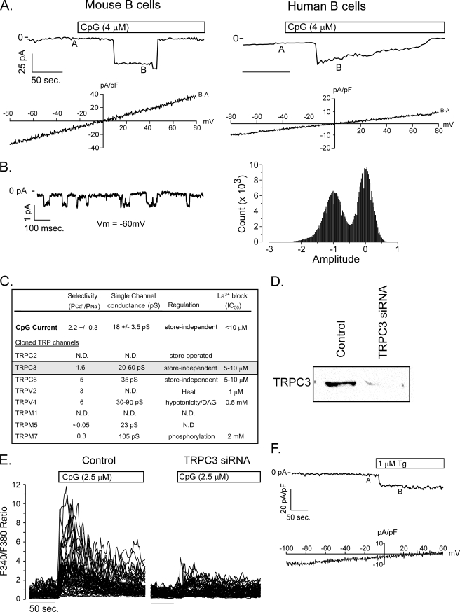
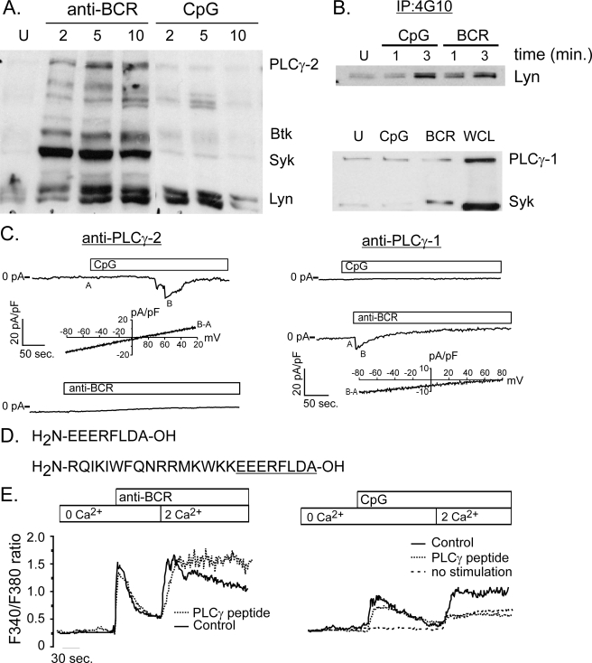
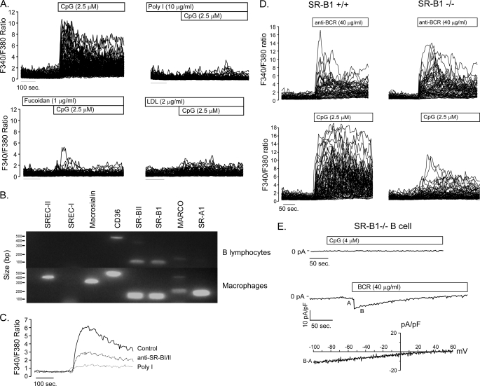
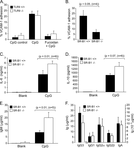
It is well established that CpG promotes pro-inflammatory cytokine and antibody production by B cells via the Toll-like receptor 9 (TLR9)-dependent pathway. However, scavenger receptors (SRs) are also capable of binding such pathogen-derived molecules, yet their contribution to CpG-induced signaling events has not yet been evaluated. Here we identified a novel TLR9-independent mechanism of CpG-induced signaling and immune function that is mediated by the scavenger B1 receptor (SR-B1). Specifically, we show that CpG/SR-B1 triggers calcium entry into primary B lymphocytes via phospholipase C gamma-1-mediated activation of TRPC3 channels and also B cell adhesion to vascular cell adhesion molecule-1. CpG-induced calcium signals and vascular cell adhesion molecule-1 adhesion are TLR9-independent and are mediated exclusively by SR-B1. Although pro-inflammatory cytokine and Ig production induced by CpG require TLR9 expression, we also found that SR-B1 negatively regulates TLR9-dependent production of interleukin-6, interleukin-10, and IgM. Thus, our results provide a novel perspective on the complexity of CpG signaling within B cells by demonstrating that SR-B1 is an alternative pathway for nucleic acid-induced signaling that provides feedback inhibition on specific TLR9-dependent responses of B cells. Consequently, these results have wide implications for understanding the mechanisms regulating immune tolerance to nucleic acids and pathogen-associated molecules.


Figures

FIGURE 1.
FIGURE 1.
CpG DNA elicits TLR9 independent Ca2+ signals in mouse and human primary B lymphocytes. A, BCR engagement (left panel) and CpG (right panel) produce a similar biphasic elevation in cytoplasmic [Ca2+] in purified mouse B cells. B, CpG elicits Ca2+release from intracellular stores in Ca2+-free medium in both mouse and human primary B cells. Subsequent superfusion with Ca2+ containing solution produces a secondary increase in cytoplasmic Ca2+ due to extracellular Ca2+ influx. C, CpG induced Ca2+ release from stores and Ca2+ entry into primary B cells occurs does not require TLR9 expression. CpG produced a similar transient elevation of cytosolic Ca2+ in TLR9+/+ (left panels) and TLR9−/− B cells bathed in Ca2+-free extracellular Ca2+ that decayed to baseline levels within 2 min. Reintroduction of extracellular Ca2+ in the continued presence of CpG, produced a sustained secondary increase in free cytosolic Ca2+ concentration indicative of entry through activated plasma membrane channels. D, the threshold for CpG-induced Ca2+ mobilization is between 250 and 500 nm CpG. GpC mobilizes Ca2+ with a similar dose threshold. In all experiments, intracellular Ca2+ was measured in single cells using fura-2 fluorescence emission ratio imaging as described under “Experimental Procedures.”
FIGURE 2.
FIGURE 2.
CpG induced calcium entry is mediated by TRPC3. A, patch clamp recording of steady-state current elicited by CpG in primary mouse (left) and human (right) B cells at −70 mV holding potential. Voltage ramps (−80 mV to +80 mV) recorded at A were subtracted from ramps applied at B, and the I-V relationship was plotted beneath each macroscopic current. The current reversal potential of ∼0 mV in both cell types indicates that CpG activates a non-selective cation channel in primary B cells. These currents are representative of measurements obtained from more than 25 mouse and 3 of 8 human primary B cells. B, single channel activity of CpG-induced channel at a holding voltage of −60 mV (left) and current amplitude frequency histogram (right). C, the calculated single channel conductance, relative Ca2+ versus Na+ permeability, and store dependence of activation for CpG currents in B lymphocytes and published values for TRP family members (53, 54) expressed by primary murine B cells (5) indicate that CpG-induced currents most closely resemble TRPC3. D, Western analysis demonstrates almost complete suppression of TRPC3 protein expression in primary murine B lymphocytes. Lysates were obtained from 5 million primary murine B lymphocytes 48 h after transfection with TRPC3 siRNA. E, TRPC3 siRNA transfection did not affect BCR-induced Ca2+ signals, but almost completely suppressed CpG-induced responses. E, thapsigargin elicits an inward current in primary B cells (upper panel). The background-subtracted I-V current (BA, bottom panel) exhibits inward rectification consistent with CRAC and not non-selective cation channel activation by thapsigargin.
FIGURE 3.
FIGURE 3.
CpG-induced calcium signals are mediated by PLCγ-1. A, phosphotyrosine (4G10) Western analysis of protein lysates from BCR- and CpG-activated primary murine B cells. BCR engagement induces phosphorylation of key enzymes, including Lyn, Syk, Btk, and PLCγ-2. By contrast, CpG stimulation does not activate Syk, Btk, or PLCγ-2, but does trigger a transient increase in tyrosine phosphorylation of a protein with mobility identical to Lyn kinase. B, Western blot analysis of phosphotyrosine immunoprecipitates confirms Lyn phosphorylation by CpG at levels comparable to that induced by BCR stimulation (upper panel). Although PLCγ-1 is expressed in primary B cells, neither CpG nor BCR engagement triggers a detectable increase in its phosphorylation (bottom panel). An increase in phospho-Syk levels following BCR engagement indicates that cells are activated by this stimulus. C, intracellular dialysis of anti-PLCγ-1 antibody (right) but not PLCγ-2 (left) blocks CpG activation of TRPC3 currents in primary B cells. By contrast, anti-PLCγ-2, but not anti-PLCγ-1 blocks CRAC current activation by anti-BCR antibody. I-V plots below CpG and CRAC macroscopic currents are shown to demonstrate identity of respective currents. D, sequence is shown for peptide that represents the domain within PLCγ-1 that interacts with TRPC3 channels. Also shown (bottom) is the same PLCγ-1 peptide constructed with an N-terminal antennapedia sequence (underlined) to render the peptide membrane permeant (55). E, the membrane-permeant PLCγ-1/TRPC3 peptide was used to examine the role of TRPC3 in BCR- and CpG-induced Ca2+ entry in primary B cells. The peptide did not inhibit BCR-induced Ca2+ release from stores or subsequent Ca2+entry into cells (left). By contrast, this PLCγ-1/TRPC3 interacting domain peptide completely blocked CpG-induced Ca2+ entry (right). The residual Ca2+ entry observed in the presence of peptide was also seen in unstimulated cells following switch from Ca2+ free to Ca2+ containing external solution (dotted line).
FIGURE 4.
FIGURE 4.
Role of scavenger receptor B1 in CpG-mediated calcium signaling. A, CpG-induced Ca2+ signals are blocked by the non-selective scavenger receptor ligands poly-inosine (Poly I), fucoidan, and low density lipoprotein (LDL). B, reverse transcription-PCR analysis demonstrates expression of CD36, MARCO, and splice variants SR-B1 and SR-B2 in primary B lymphocytes. RNA isolated from macrophages served as a positive control for SREC-I, SREC-II, macrosialin, and SR-A1 primers. C, anti-SR-B1 antibody partially blocks CpG-induced Ca2+ signals. D, SR-B1 expression is required for CpG- but not BCR-induced Ca2+ signaling. BCR engagement produces similar Ca2+ signals in SR-B1−/− (upper right) B cells as in SR-B1+/+ controls (upper left); however, SR-B1−/− B lymphocytes are nearly unresponsive to CpG stimulation (lower right). E, CpG fails to elicit a NSCC current in SR-B1−/− lymphocytes, while anti-BCR antibody activates normal CRAC currents in SR-B1−/− B cells, consistent with a requirement for SR-B1 in CpG-mediated TRPC3-dependent Ca2+ entry in primary B cells.
FIGURE 5.
FIGURE 5.
Regulation of IgM, IL-6, and IL-10 production and VCAM-1 adhesion by SR-B1 signaling in B lymphocytes. A, CpG triggers an increase in VCAM-1 adhesion of primary B cells (filled bars). The increase was observed 30 min after CpG stimulation and was completely blocked by the scavenger receptor blocking ligand fucoidan. The ability of CpG to trigger an increase in VCAM-1 adhesion, and inhibition by fucoidan, is independent of TLR9 expression as these responses are similar in TRL9+/+ (filled bars) and TLR9−/− (open bars) B lymphocytes. B, CpG-induced VCAM-1 adhesion is significantly attenuated in B cells lacking SR-B1 expression. C, SR-B1 negatively regulates CpG-induced production of immunoregulatory cytokines IL-6 and IL-10, and IgM by B cells. SR-B1+/+ and SR-B1−/− primary B lymphocytes were stimulated in vitro, culture supernatants were harvested 48 h after stimulation and assayed by enzyme-linked immunosorbent assay. In response to CpG, SR-B1-deficient B cells elaborated roughly 2-fold more IL-6 (C), IL-10 (D), and IgM (E) than did wild-type cells. F, CpG elicited the production of significant amounts of IgG3 and relatively small amounts of IgG1, IgG2α, IgG2β, and IgA. However, unlike the effect on IgM production, SR-B1 deficiency did not regulate production of these other Ig isotypes.

Similar articles

Cited by

References

    1. Berridge M. J. (1993) Nature 361,315–325 - PubMed
    1. Sugawara H., Kurosaki M., Takata M., Kurosaki T. (1997) EMBO J. 16,3078–3088 - PMC - PubMed
    1. Miyakawa T., Maeda A., Yamazawa T., Hirose K., Kurosaki T., Iino M. (1999) EMBO J. 18,1303–1308 - PMC - PubMed
    1. Hagar R. E., Burgstahler A. D., Nathanson M. H., Ehrlich B. E. (1998) Nature 396,81–84 - PMC - PubMed
    1. Liu Q. H., Liu X., Wen Z., Hondowicz B., King L., Monroe J., Freedman B. D. (2005) J. Immunol. 174,68–79 - PubMed

Publication types

MeSH terms