. 2009 Sep;191(17):5446-57.
doi: 10.1128/JB.00602-09. Epub 2009 Jun 19.

CodY activates transcription of a small RNA in Bacillus subtilis

Affiliations

CodY activates transcription of a small RNA in Bacillus subtilis

Heike Preis et al. J Bacteriol. 2009 Sep.

Abstract

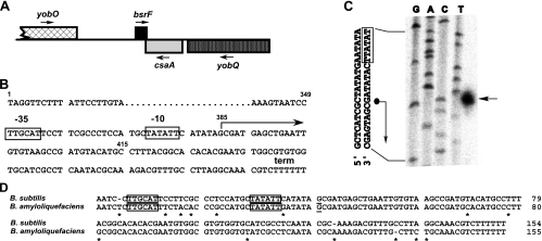
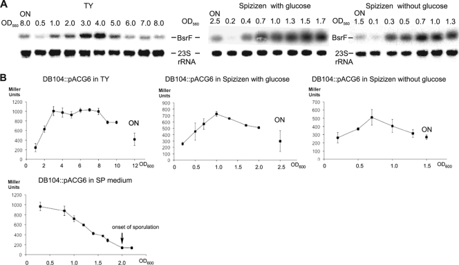
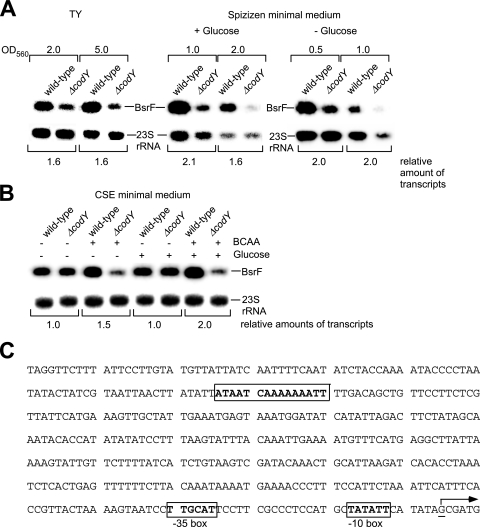
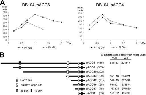
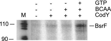
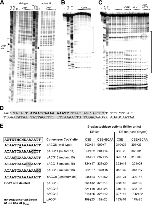
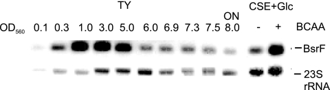
Regulatory small RNAs (sRNAs) in bacterial genomes have become a focus of research over the past 8 years. Whereas more than 100 such sRNAs have been found in Escherichia coli, relatively little is known about sRNAs in gram-positive bacteria. Using a computational approach, we identified two sRNAs in intergenic regions of the Bacillus subtilis genome, SR1 and SR2 (renamed BsrF). Recently, we demonstrated that SR1 inhibits the translation initiation of the transcriptional activator AhrC. Here, we describe detection of BsrF, its expression profile, and its regulation by CodY. Furthermore, we mapped the secondary structure of BsrF. BsrF is expressed in complex and minimal media in all growth phases in B. subtilis and, with a similar expression profile, also in Bacillus amyloliquefaciens. Neither overexpression nor deletion of bsrF affected the growth of B. subtilis. BsrF was found to be long-lived in complex and minimal media. Analysis of 13 putative transcription factor binding sites upstream of bsrF revealed only an effect for CodY. Here, we showed by using Northern blotting, lacZ reporter gene fusions, in vitro transcription, and DNase I footprinting that the transcription of bsrF is activated by CodY in the presence of branched-chain amino acids and GTP. Furthermore, BsrF transcription was increased 1.5- to 2-fold by glucose in the presence of branched-chain amino acids, and this increase was independent of the known glucose-dependent regulators. BsrF is the second target for which transcriptional activation by CodY has been discovered.


Figures

FIG. 1.
FIG. 1.
Location of the bsrF gene. (A) Schematic diagram of the location of the bsrF gene on the B. subtilis chromosome. The direction of transcription is indicated by arrows. Genes transcribed from the plus strand are indicated above the line. (B) Sequence of the bsrF gene. −35 and −10 boxes of the bsrF promoter are indicated. The start site and direction of transcription are indicated by an arrow, and the transcription termination site is indicated by term. The transcription start site is at position 2078305 in the B. subtilis genome. The small numbers indicate the positions of nucleotides referred to in Fig. 8. (C) Mapping of the 5′ end of BsrF. The results of a sequencing reaction with pUCBSR2 (containing the entire bsrF gene under its own promoter) and primer SB847 are shown in the left four lanes. The lane on the right shows the results of a primer extension reaction with total RNA of B. subtilis DB104 and 5′-labeled primer SB847. The arrow indicates the transcriptional start site of BsrF. Part of the sequence is shown on the left. The dot and the arrow indicate the mapped transcriptional start site. (D) Alignment of the bsrF genes of B. subtilis and B. amyloliquefaciens. Promoter boxes are indicated by boxes, and the transcription start site is underlined.
FIG. 2.
FIG. 2.
Expression profile of BsrF. (A) Expression of BsrF in complex and minimal media as monitored by Northern blotting. B. subtilis strain DB104 was grown in different media, and aliquots were removed at the OD560 indicated, immediately frozen in liquid nitrogen, and later used for preparation of total RNA. RNA was treated with glyoxal, separated on 2% agarose gels, blotted onto a nylon membrane, and hybridized with an [α-32P]dATP-labeled BsrF-specific DNA probe. Autoradiograms of the corresponding gels are shown. ON, overnight cultivation (∼15 h). For correction of loading errors, filters were reprobed with [γ-32P]ATP-labeled oligonucleotide SB747 specific for 23S rRNA. (B) Expression of BsrF in complex, minimal, and sporulation media as monitored by measurement of the β-galactosidase activity. B. subtilis strain DB104::pACG6 was grown in different media, and at the OD600 indicated, samples were withdrawn and used for measurement of the β-galactosidase activity. The data are the averages ± standard deviations for three independent measurements. ON, overnight cultivation.
FIG. 3.
FIG. 3.
Determination of the BsrF half-lives in different media. Half-lives were determined as described in Materials and Methods. Samples were taken at the times indicated after rifampin addition or without rifampin. Reprobing was performed with [γ-32P]ATP-labeled oligonucleotide SB767 specific for 5S rRNA. Autoradiograms of the Northern blots are shown. (A) Determination of the BsrF half-life in Spizizen minimal medium without glucose in the presence and absence of Hfq. (B) Determination of the BsrF half-life in TY medium (calculation performed as described above). (C) BsrF binds Hfq. Purified B. subtilis Hfq was added to 5′-end-labeled in vitro-synthesized BsrF and incubated for 15 min at 37°C in TMN buffer, and the complex was separated on a 6% native polyacrylamide gel as described previously (18). t1/2, half-life.
FIG. 4.
FIG. 4.
Secondary structure of BsrF. (A) Secondary structure probing of BsrF with RNases. Purified, 5′-end-labeled BsrF was subjected to limited cleavage with the RNases indicated at the top. The digested RNAs were separated on an 8% denaturing gel. Autoradiograms are shown. The following RNase concentrations used were: RNase T1, 10−2 U/μl (1:50); RNase T2, 10−1 U/μl (1:500); and RNase V1, 10−1 U/μl (1:10). Lane C, control without RNase treatment; lane L, alkaline ladder. undil., undiluted. (B) Proposed secondary structure of BsrF. Two structures consistent with the cleavage data shown in panel A are shown. Major and minor cuts are indicated by symbols. The main stem-loops L1, L2, and L3 and the terminator stem-loop T are indicated.
FIG. 5.
FIG. 5.
CodY affects the amount of BsrF. (A) Effect of CodY on the expression of bsrF in TY and Spizizen minimal media. B. subtilis DB104 was grown to the OD560 indicated, and total RNA was prepared, treated with glyoxal, separated on 2% agarose gels, and subjected to Northern blotting. Autoradiograms of Northern blots are shown. (B) Effect of CodY, glucose, BCAA, and glucose plus BCAA on the expression of bsrF in CSE medium. Cultures were grown until the OD600 was 0.3 (log phase), and samples were prepared and treated as described above for panel A. An autoradiogram of a Northern blot is shown. (C) Sequence of the bsrF upstream region with a putative CodY binding site. The CodY site is enclosed in a box, and the transcription start site is indicated by an underlined G residue and an arrow. −35 and −10 boxes of pBsrF are indicated.
FIG. 6.
FIG. 6.
BsrF transcription is activated in the presence of glucose. (A) Effect of glucose on the bsrF expression depending on a sequence upstream of pBsrF. B. subtilis strains DB104::pACG6 and DB104::pACG4 were grown in Spizizen medium with and without glucose, and at the OD600 indicated, samples were withdrawn and used for β-galactosidase measurement. The data are averages of three independent determinations. (B) Influence of the putative CcpA binding site on the activating effect of glucose on bsrF expression. Diagrams of all constructed pACG derivatives are shown. The numbers of base pairs upstream of the bsrF transcription start site are indicated in parentheses. The β-galactosidase activities in Spizizen medium with and without glucose at an OD600 of 1.3 are indicated on the right. The values are averages for three independent determinations. The −35 and −10 boxes of the bsrF promoter, the CodY binding site, and the hypothetical CcpA binding site are indicated.
FIG. 7.
FIG. 7.
In vitro transcription with B. subtilis RNA polymerase. In vitro transcription in the presence or absence of CodY was performed with B. subtilis RNA polymerase. BCAA and GTP were added at concentrations of 10 mM and 2 mM, respectively, where indicated. The template was prepared by performing PCR with oligonucleotides SB1233 and SB1064 and comprises the same bsrF upstream region as pACG6.
FIG. 8.
FIG. 8.
Interaction of CodY with the bsrF promoter region. (A and B) DNase I footprinting analysis of the interaction of CodY and the bsrF promoter. DNase I footprinting with increasing amounts of purified CodY-His6 (1.0 μM, 1.4 μM, 1.8 μM, 2.2 μM, and 2.6 μM) (indicated by triangles) was performed in the presence of 2 mM GTP and 10 mM BCAA as described in Materials and Methods. Lane C, control without DNase; lane −, control with DNase but without CodY. The binding site is indicated. (A) Coding strand DNA fragment comprising nt 1 to 176 (Fig. 5C). Dots indicate residues replaced in mutant 11. (B) Noncoding strand (DNA fragment comprising nt 57 to 145 [see Fig. 5C]). (C) Analysis in the presence and absence of CodY, BCAA, and GTP (DNA fragment as in panel B). The triangles indicate increasing amounts of CodY (as described above for panel A). The bracket indicates the region protected by CodY. (D) Sequences on the coding and noncoding strands examined. Protected regions are shaded. The predicted CodY consensus binding site is indicated by bold type. (E) Overview of the mutants with mutations in the CodY binding site. Mutated nucleotides are shaded. The corresponding pACG derivatives and the measured β-galactosidase activities are indicated on the right. Mutated pACG derivatives pACG11, pACG15, pACG18, and pACG19 contain the same region upstream of the CodY site as pACG6, whereas pACG9, pACG10, pACG12, pACG13, and pACG4 contain shortened upstream regions (see Fig. 6). For measurement of β-galactosidase activity, B. subtilis strains were grown in CSE medium with 0.1% glucose with or without BCAA (as indicated) until the OD600 was 0.3. All of the values are averages of at least three independent determinations with five different integrants.
FIG. 9.
FIG. 9.
Expression of the bsrF gene in B. amyloliquefaciens. B. amyloliquefaciens strain FZB42 was grown in TY or CSE medium, and aliquots were removed at hourly intervals at the OD560 indicated, immediately frozen in liquid nitrogen, and later used for preparation of total RNA. Northern blotting was performed as described in the legend to Fig. 2 with the same BsrF probe. Autoradiograms of the corresponding gels are shown. ON, overnight cultivation (∼15 h). For correction of loading errors, filters were reprobed with [γ-32P]ATP-labeled oligonucleotide SB1319 specific for B. amyloliquefaciens 23S rRNA.

Similar articles

Cited by

References

    1. Barrick, J. E., N. Sudarsan, Z. Weinberg, W. L. Ruzzo, and R. R. Breaker. 2005. 6S RNA is a widespread regulator of eubacterial RNA polymerase that resembles an open promoter. RNA 11744-784. - PMC - PubMed
    1. Belitsky, B. R., and A. L. Sonenshein. 2008. Genetic and biochemical analysis of CodY binding sites in Bacillus subtilis. J. Bacteriol. 1901224-1236. - PMC - PubMed
    1. Boisset, S., T. Geissmann, E. Huntzinger, P. Fechter, N. Bendridi, M. Possedko, C. Chevalier, A. C. Helfer, Y. Benito, A. Jacquier, C. Gaspin, F. Vandenesch, and P. Romby. 2007. Staphylococcus aureus RNAIII coordinately represses the synthesis of virulence factors and the transcription regulator Rot by an antisense mechanism. Genes Dev. 211353-1366. - PMC - PubMed
    1. Brantl, S. 1994. The copR gene product of plasmid pIP501 acts as a transcriptional repressor at the essential repR promotor. Mol. Microbiol. 14473-483. - PubMed
    1. Brantl, S. 2009. Bacterial chromosome-encoded small regulatory RNAs. Future Microbiol. 485-103. - PubMed

Publication types

MeSH terms

LinkOut - more resources