. 2009 Sep;191(17):5398-408.
doi: 10.1128/JB.00092-09. Epub 2009 Jun 19.

Cell envelope perturbation induces oxidative stress and changes in iron homeostasis in Vibrio cholerae

Affiliations

Cell envelope perturbation induces oxidative stress and changes in iron homeostasis in Vibrio cholerae

Aleksandra E Sikora et al. J Bacteriol. 2009 Sep.

Abstract

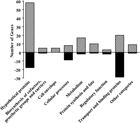
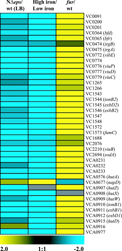
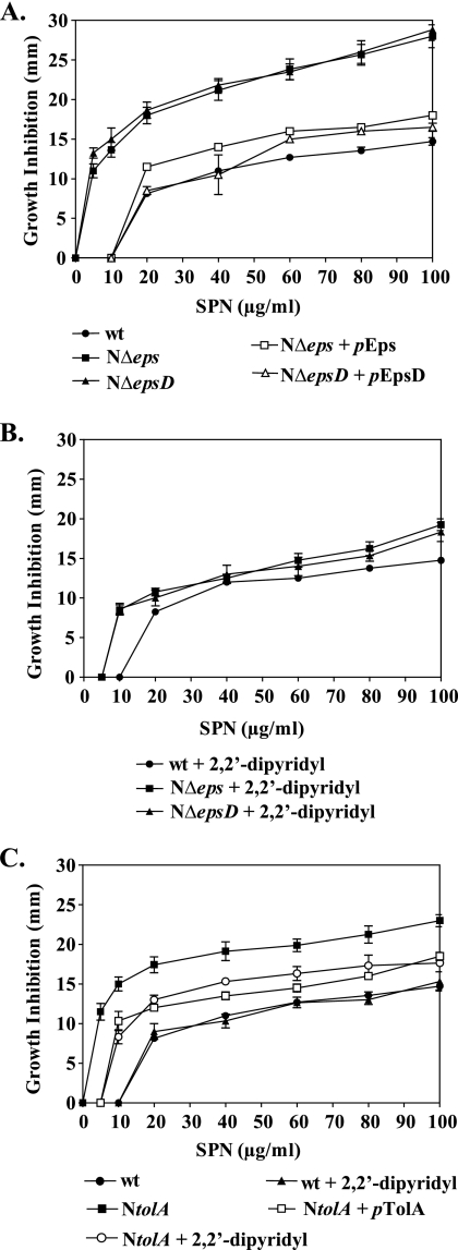
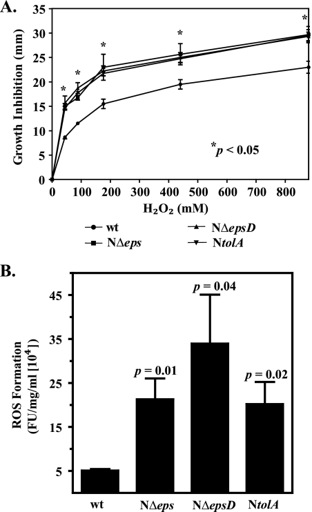
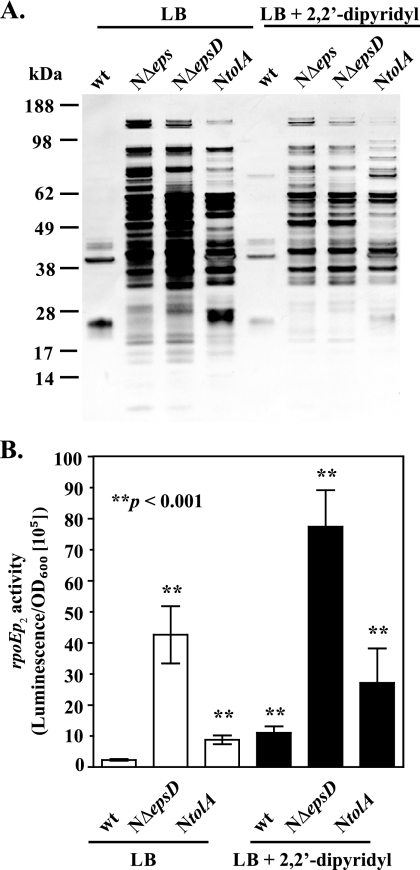
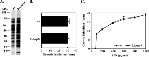
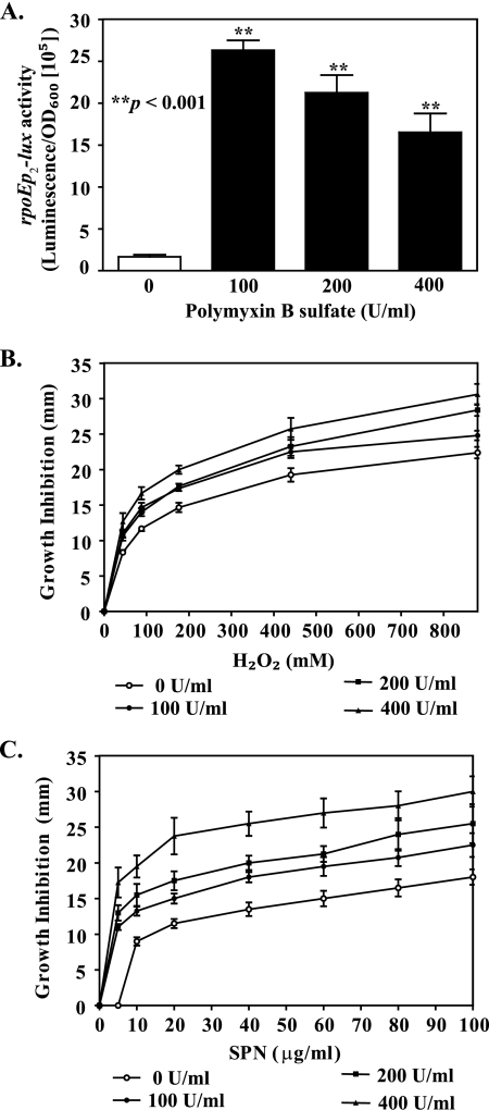
The Vibrio cholerae type II secretion (T2S) machinery is a multiprotein complex that spans the cell envelope. When the T2S system is inactivated, cholera toxin and other exoproteins accumulate in the periplasmic compartment. Additionally, loss of secretion via the T2S system leads to a reduced growth rate, compromised outer membrane integrity, and induction of the extracytoplasmic stress factor RpoE (A. E. Sikora, S. R. Lybarger, and M. Sandkvist, J. Bacteriol. 189:8484-8495, 2007). In this study, gene expression profiling reveals that inactivation of the T2S system alters the expression of genes encoding cell envelope components and proteins involved in central metabolism, chemotaxis, motility, oxidative stress, and iron storage and acquisition. Consistent with the gene expression data, molecular and biochemical analyses indicate that the T2S mutants suffer from internal oxidative stress and increased levels of intracellular ferrous iron. By using a tolA mutant of V. cholerae that shares a similar compromised membrane phenotype but maintains a functional T2S machinery, we show that the formation of radical oxygen species, induction of oxidative stress, and changes in iron physiology are likely general responses to cell envelope damage and are not unique to T2S mutants. Finally, we demonstrate that disruption of the V. cholerae cell envelope by chemical treatment with polymyxin B similarly results in induction of the RpoE-mediated stress response, increased sensitivity to oxidants, and a change in iron metabolism. We propose that many types of extracytoplasmic stresses, caused either by genetic alterations of outer membrane constituents or by chemical or physical damage to the cell envelope, induce common signaling pathways that ultimately lead to internal oxidative stress and misregulation of iron homeostasis.


Figures

FIG. 1.
FIG. 1.
Functional categories of genes differentially expressed in the NΔeps mutant compared to the wild-type strain. The numbers of genes whose expression is differentially expressed in NΔeps mutant compared to wild-type cells are presented according to the functions assigned by The Institute for Genomic Research. Positive and negative numbers stand for the numbers of genes whose expression is induced and repressed, respectively.
FIG. 2.
FIG. 2.
Global transcriptome analysis reveals that iron homeostasis is affected in the T2S mutant of V. cholerae. Genes that were differentially expressed in the NΔeps mutant compared to the wild-type (wt) strain grown in LB medium were identified by SAM analysis using a ≥2.0-fold change in gene expression and a ≤1% FDR as criteria. Expression profiles of genes that are regulated by iron and Fur and that are differentially expressed in the T2S-deficient strain are presented as a heat map using a log2-based color scale (yellow, induced; blue, repressed). A list of genes regulated by iron and Fur was obtained from previously published gene expression data (50).
FIG. 3.
FIG. 3.
The reporter activities of Fur- and iron-responsive genes confirm the results of microarray analysis. Wild-type (wt) V. cholerae N16961 and its NΔepsD and NtolA mutant strains carrying the luciferase transcriptional fusions viuPp-lux (A) and hutAp-lux (B) were grown in LB medium, and luminescence was assessed in technical triplicate when the cultures were 1 h into the stationary phase of growth (OD600, 4). The bars show average data derived from four independent experiments; error bars represent the corresponding SEMs. The calculated P values displayed above the bars indicate that there were statistically significant differences in reporter activities between the wild-type and mutant strains.
FIG. 4.
FIG. 4.
Susceptibility to streptonigrin (SPN), an indirect measurement of the intracellular free iron content. Wild-type (wt) V. cholerae N16961 and T2S mutants (NΔeps, NΔepsD) (A and B) or the NtolA mutant (C) were grown in LB medium to mid-log phase (OD600, about 0.5), and 100 μl of each culture was spread onto LB agar medium alone (A) or supplemented with the iron-specific chelator 2,2′-dipyridyl to a final concentration of 100 μM (B) or 150 μM (C). Cultures of NΔeps, NΔepsD, and NtolA containing plasmid pEps, pEpsD, or pTolA, respectively, were also spread onto LB agar alone and tested for complementation in the presence of 10 μM IPTG (A and C). Sterile filter paper discs immersed with different concentrations of streptonigrin, as indicated, were placed immediately on the surface of each plate. The diameter of the inhibition zone was scored after 24 h of incubation at 37°C. Each datum point represents the mean and SEM from three to nine separate experiments. In comparison to wild-type V. cholerae, both T2S mutants and the NtolA strain showed statistically significant increases in susceptibility to streptonigrin at each of the concentrations tested (P < 0.0001).
FIG. 5.
FIG. 5.
Sensitivity to hydrogen peroxide and endogenous ROS formation are augmented in T2S- and TolA-deficient strains of V. cholerae. (A) Wild-type (wt) V. cholerae N16961 and its isogenic T2S-deficient and NtolA mutants were grown in LB medium until mid-log phase (OD600, about 0.5). Cultures (100 μl) were uniformly distributed on LB agar plates, followed by immediate placing of sterile filter paper discs impregnated with 5 μl of 44, 88, 176, 440, and 880 mM hydrogen peroxide on the surface of the plate. The diameter of the growth inhibition zone was recorded after 24 h of incubation at 37°C. Error bars represent SEMs for four independent experiments, each performed in duplicate. All mutants displayed statistically significantly enhanced susceptibility to hydrogen peroxide relative to that of the parental strain (P < 0.05). (B) Intracellular ROS formation was measured in cells growing logarithmically in LB medium and incubated with the oxidative-stress-sensitive probe H2DCFDA as described in Materials and Methods. Intracellular ROS formation was expressed in arbitrary units of fluorescence intensity (FU), and the emission values were normalized to the protein concentration. Means for four independent experiments with corresponding SEMs are presented. The calculated P values shown above each bar indicate statistically significant increases in ROS levels in T2S- and TolA-deficient strains over those in the wild-type strain.
FIG. 6.
FIG. 6.
Outer membrane integrity is compromised regardless of the iron level. (A) Representative protein profiles of the supernatants isolated from stationary-phase cultures of wild-type (wt) V. cholerae N16961 and its isogenic NΔeps, NΔepsD, and NtolA mutants grown in LB medium without and with the addition of 100 or 150 μM 2,2′-dipyridyl (for the T2S-deficient and NtolA mutants, respectively). Samples were loaded onto SDS-PAGE gels by equivalent OD600 units, and gels were silver stained. The migration of molecular mass markers is indicated on the left. (B) The expression of the rpoEp2-lux fusion gene was determined in wild-type V. cholerae N16961 and its T2S-deficient and NtolA mutants grown at 37°C in LB medium alone or supplemented with 100 or 150 μM 2,2′-dipyridyl (for the T2S-deficient and NtolA mutants, respectively) to mid-log phase (OD600, 0.5). The data are means for three to nine independent experiments (each performed in technical triplicate). Error bars indicate SEMs. The increase in rpoE expression was statistically significant for the NΔeps, NΔepsD, and NtolA strains regardless of the presence or absence of the iron chelator (P < 0.001). Treatment with the iron-specific chelator resulted in the detection of statistically significantly different σE activity in the wild-type strain (P < 0.0001).
FIG. 7.
FIG. 7.
Iron homeostasis and oxidative stress are not affected in a T2S mutant of E. coli O157:H7. (A) Protein profiles of filtered culture supernatants of the E. coli O157:H7 parental wild-type (wt) strain and its EΔetpM mutant grown in LB medium at 37°C to stationary phase (16 h), revealed by SDS-PAGE and silver staining. Samples were matched by equivalent OD600 units. The migration of molecular mass markers is indicated. (B and C) The susceptibilities of the wild-type and EΔetpM mutant strains to hydrogen peroxide (B) and streptonigrin (SPN) (C) were tested by uniformly distributing 100 μl of mid-log-phase cultures (OD600, about 0.5) on LB agar plates and then immediately placing sterile filter paper discs impregnated with either 8.8 M hydrogen peroxide or different concentrations of streptonigrin, as indicated, on the surfaces of the plates. The diameter of the inhibition zone was recorded after 24 h of incubation at 37°C. Means and corresponding SEMs for three independent experiments are reported.
FIG. 8.
FIG. 8.
Extracytoplasmic and oxidative stress responses are triggered in wild-type V. cholerae cells following treatment with polymyxin B sulfate. (A) rpoE activity was monitored for wild-type V. cholerae N16961 grown at 37°C to mid-log phase (OD600, 0.5) in LB medium supplemented with increasing concentrations of polymyxin B sulfate (0, 100, 200, and 400 U/ml). Bars show means and SEMs from four independent experiments. P values indicate that the elevation in the envelope stress response in polymyxin B-treated cultures was statistically significant. (B and C) Wild-type V. cholerae N16961 was grown in LB medium until mid-log phase (OD600, 0.5), and 100 μl of the culture was spread onto LB agar supplemented with different concentrations of polymyxin B sulfate (0, 100, 200, and 400 U/ml). Sterile filter paper discs immersed with various concentrations of hydrogen peroxide (B) or streptonigrin (SPN) (C) were immediately placed on the surfaces of the plates. The diameter of the growth inhibition zone was scored after 24 h of incubation at 37°C. Each datum point represents the mean and SEMs obtained from five independent experiments (P < 0.05).

Similar articles

Cited by

References

    1. Andrews, S. C., A. K. Robinson, and F. Rodriguez-Quinones. 2003. Bacterial iron homeostasis. FEMS Microbiol. Rev. 27215-237. - PubMed
    1. Baba, T., T. Ara, M. Hasegawa, Y. Takai, Y. Okumura, M. Baba, K. A. Datsenko, M. Tomita, B. L. Wanner, and H. Mori. 2006. Construction of Escherichia coli K-12 in-frame, single-gene knockout mutants: the Keio collection. Mol. Syst. Biol. 22006.0008. - PMC - PubMed
    1. Bader, M. W., W. W. Navarre, W. Shiau, H. Nikaido, J. G. Frye, M. McClelland, F. C. Fang, and S. I. Miller. 2003. Regulation of Salmonella typhimurium virulence gene expression by cationic antimicrobial peptides. Mol. Microbiol. 50219-230. - PubMed
    1. Benov, L., and I. Fridovich. 1995. Superoxide dismutase protects against aerobic heat shock in Escherichia coli. J. Bacteriol. 1773344-3346. - PMC - PubMed
    1. Berry, M. C., G. C. McGhee, Y. Zhao, and G. W. Sundin. 2009. Effect of a waaL mutation on lipopolysaccharide composition, oxidative stress survival, and virulence in Erwinia amylovora. FEMS Microbiol. Lett. 29180-87. - PubMed

Publication types