. 2009 Aug;8(8):1298-306.
doi: 10.1128/EC.00078-09. Epub 2009 Jun 19.

Functional genomics of adhesion, invasion, and mycelial formation in Schizosaccharomyces pombe

Affiliations

Functional genomics of adhesion, invasion, and mycelial formation in Schizosaccharomyces pombe

James Dodgson et al. Eukaryot Cell. 2009 Aug.

Abstract

Investigation into the switch between single-celled and filamentous forms of fungi may provide insights into cell polarity, differentiation, and fungal pathogenicity. At the molecular level, much of this investigation has fallen on two closely related budding yeasts, Candida albicans and Saccharomyces cerevisiae. Recently, the much more distant fission yeast Schizosaccharomyces pombe was shown to form invasive filaments after nitrogen limitation (E. Amoah-Buahin, N. Bone, and J. Armstrong, Eukaryot. Cell 4:1287-1297, 2005) and this genetically tractable organism provides an alternative system for the study of dimorphic growth. Here we describe a second mode of mycelial formation of S. pombe, on rich media. Screening of an S. pombe haploid deletion library identified 12 genes required for mycelial development which encode potential transcription factors, orthologues of S. cerevisiae Sec14p and Tlg2p, and the formin For3, among others. These were further grouped into two phenotypic classes representing different stages of the process. We show that galactose-dependent cell adhesion and actin assembly are both required for mycelial formation and mutants lacking a range of genes controlling cell polarity all produce mycelia but with radically altered morphology.


Figures

FIG. 1.
FIG. 1.
Deficient mycelial growth in a laboratory strain of S. pombe 972. (A) A standard strain of 972 (left) formed invasive structures after incubation at 30°C for 14 days on rich YES medium, but the laboratory strain (right) did not. (B) Standard 972 (left) formed invasive structures on nitrogen-limited LNB medium more rapidly and vigorously than did the laboratory strain (right) after 7 days at 30°C. (C) Standard 972 (left panel) formed invasive structures on LNB after incubation for 14 days at a range of temperatures, while the laboratory strain (right panel) did so only at 30°C. All colonies are shown before (above) and after (below) vigorous washing to reveal cells that invaded the medium.
FIG. 2.
FIG. 2.
Morphology of invasive structures formed on rich YES medium. (A) After 11 weeks of incubation, threadlike filaments form large and complex structures (10× objective; scale bar, 100 μm). (B) High magnification shows that the filaments are composed of chains of cells (63× objective; scale bar, 10 μm). (C) After 3 days of incubation, individual foci produce short filaments (arrow) that are elongated and bent (20× objective; scale bar, 10 μm). (D) After 7 days of incubation on YES medium, numerous lenticular colonies embedded in the agar are observed that are composed of nonfilamentous cells (2.5× objective; scale bar, 100 μm).
FIG. 3.
FIG. 3.
Effect of deleting pka1 on mycelial growth on YES medium. Strains were plated at high cell density, incubated on YES medium at 30°C, and imaged as before. (A) After 7 days, only wild-type 972 formed detectable invasive structures. (B) After 40 days, both strains formed visible invasive structures. (C) Mycelia formed by the pka1 deletion strain (upper images) are morphologically distinct from the wild type (lower images), forming invasive structures composed of elongated cells without branching. Scale bars, 50 μm (20× objective) and 10 μm (63× objective).
FIG. 4.
FIG. 4.
Screening of the deletion library for strains defective in invasive growth. (I) Eight strains were inoculated onto thick YES plates. (II) After 2 days at 30°C, a large cell mass from each strain was transferred. (III) After a further 7 days at 30°C, the surface cells were removed by vigorous washing and the plates were inspected for remaining cells that had invaded the medium.
FIG. 5.
FIG. 5.
Distinction between adhesive and nonadhesive phenotypes of noninvasive strains. Strains were plated at high cell density on YES medium, incubated at 30°C for 14 days, gently rinsed, and then vigorously washed. Gentle rinsing (center) removed all of the cells of the strain with a deletion of snf5, but some surface cells of the strain with a deletion of for3 and wild-type 972 remained attached to the agar. Vigorous washing (right) removed all of the remaining surface cells, revealing invasive structures formed by 972 but none formed by the mutant strains.
FIG. 6.
FIG. 6.
Galactose-specific inhibition of cell-to-surface adhesion. Strain 972 was plated at high cell density on LNB medium with supplements as shown. After 7 days at 30°C, the colonies were first gently rinsed with water (middle panels) to show adherent cells and then washed vigorously (lower panels) to show invasive structures. Only the addition of 1 M galactose prevented adhesion to the surface.
FIG. 7.
FIG. 7.
Effect of latrunculin (Lat) A on mycelial growth. Strain 972 was plated at high density on YES medium supplemented as shown and incubated at 30°C for 7 days. At all concentrations of latrunculin A, normal adhesion to the surface (middle panels) was observed. Vigorous washing (lower panels) showed no invasive structures with 1 μM latrunculin A and reduced invasion at lower concentrations.
FIG. 8.
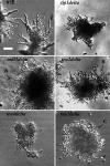
FIG. 8.
Strains with deletions of genes involved in microtubule organization and as microtubule-based cortical markers and effectors (Δtea1, Δtea2, Δtea4, Δtip1, and Δmal3) were invasive but showed aberrant filament formation. Only a few filaments were formed, and these frequently appeared twisted (arrow A), swollen (arrow B), and truncated (arrow C). Scale bar, 40 μm.

References

    1. Abeliovich, H., E. Grote, P. Novick, and S. Ferro-Novick. 1998. Tlg2p, a yeast syntaxin homolog that resides on the Golgi and endocytic structures. J. Biol. Chem. 27311719-11727. - PubMed
    1. Amoah-Buahin, E., N. Bone, and J. Armstrong. 2005. Hyphal growth in the fission yeast Schizosaccharomyces pombe. Eukaryot. Cell 41287-1297. - PMC - PubMed
    1. Bach, I., C. Carriere, H. P. Ostendorff, B. Andersen, and M. G. Rosenfeld. 1997. A family of LIM domain-associated cofactors confer transcriptional synergism between LIM and Otx homeodomain proteins. Genes Dev. 111370-1380. - PubMed
    1. Cao, F., S. Lane, P. P. Raniga, Y. Lu, Z. Zhou, K. Ramon, J. Y. Chen, and H. P. Liu. 2006. The Flo8 transcription factor is essential for hyphal development and virulence in Candida albicans. Mol. Biol. Cell 17295-307. - PMC - PubMed
    1. Chang, F., and M. Peter. 2003. Yeasts make their mark. Nature Cell Biol. 5294-299. - PubMed

Publication types

MeSH terms

Substances