. 2009 Aug;8(8):1228-34.
doi: 10.1128/EC.00090-09. Epub 2009 Jun 19.

Special Sm core complex functions in assembly of the U2 small nuclear ribonucleoprotein of Trypanosoma brucei

Affiliations

Special Sm core complex functions in assembly of the U2 small nuclear ribonucleoprotein of Trypanosoma brucei

Christian Preusser et al. Eukaryot Cell. 2009 Aug.

Abstract

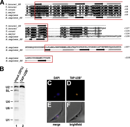
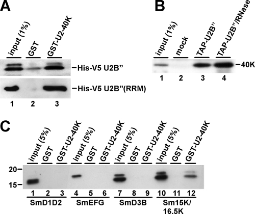
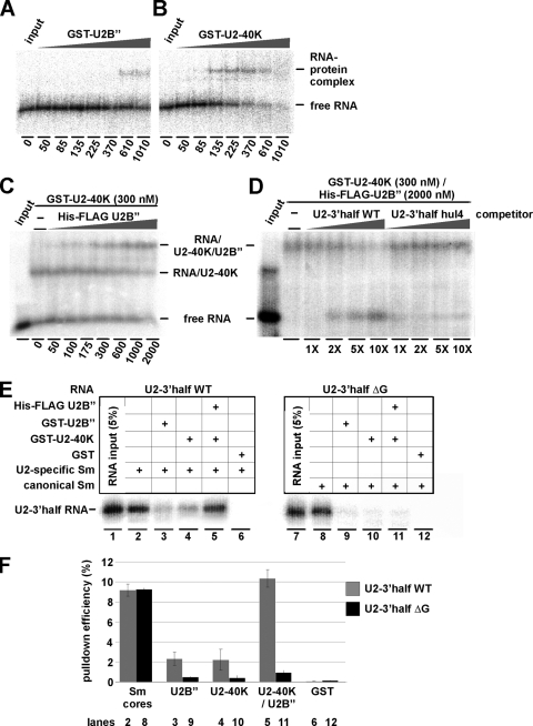
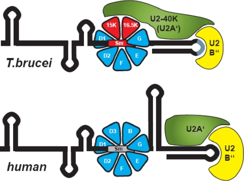
The processing of polycistronic pre-mRNAs in trypanosomes requires the spliceosomal small ribonucleoprotein complexes (snRNPs) U1, U2, U4/U6, U5, and SL, each of which contains a core of seven Sm proteins. Recently we reported the first evidence for a core variation in spliceosomal snRNPs; specifically, in the trypanosome U2 snRNP, two of the canonical Sm proteins, SmB and SmD3, are replaced by two U2-specific Sm proteins, Sm15K and Sm16.5K. Here we identify the U2-specific, nuclear-localized U2B'' protein from Trypanosoma brucei. U2B'' interacts with a second U2 snRNP protein, U2-40K (U2A'), which in turn contacts the U2-specific Sm16.5K/15K subcomplex. Together they form a high-affinity, U2-specific binding complex. This trypanosome-specific assembly differs from the mammalian system and provides a functional role for the Sm core variation found in the trypanosomal U2 snRNP.


Figures

FIG. 1.
FIG. 1.
Sequence alignment, U2 snRNA binding specificity, and nuclear localization of T. brucei U2B″. (A) Multiple sequence alignment of trypanosomatid and human U2B″ sequences. ClustalW alignments of the U2B″ protein sequences from T. brucei, Trypanosoma cruzi, and Leishmania major and human U2B″ are shown, with the RRMs boxed. Residues that are conserved or whose physicochemical character is conserved are highlighted in black and gray, respectively. The secondary structures of U2B″ from T. brucei and Homo sapiens are shown above and below the alignment, respectively. α-Helices are represented as tubes and ß strands as arrows, while disordered regions are shown as tildes (∼). The GeneDB or NCBI accession numbers of U2B″ sequences are as follows: T. brucei, Tb927.3.3480; T. cruzi, Tc00.1047053507951.140; L. major, LmjF29.0865; and H. sapiens, NP_003083. The total numbers of amino acids are given on the right. (B) U2 snRNA specificity of T. brucei U2B″. Extract was prepared from a T. brucei cell line stably expressing TAP-tagged U2B″, followed by IgG pulldown of TAP-tagged complexes and Northern blot analysis of copurifying RNAs, detecting U2, SL, U4, U6, and U1 snRNAs (as indicated on the left; lane 2). For comparison, 20% of the total input was analyzed (lane 1). (C through F) Nuclear localization of T. brucei U2B″. TAP-U2B″-expressing cells were fixed (F) and stained with DAPI (C). In parallel, TAP-tagged U2B″ was detected by anti-protA primary antibody and Alexa 594 secondary antibody (D). (E) Merged view of DAPI and TAP-U2B″ staining.
FIG. 2.
FIG. 2.
Protein-protein interactions of U2-40K, U2B″, and the U2-specific Sm proteins. (A) U2-40K and U2B″ interact in vitro. Immobilized GST-40K protein (lane 3) or GST as a control (lane 2) were incubated with lysate from E. coli cells expressing His-V5-tagged U2B″ protein (top panel) or the RRM domain only of His-V5 U2B″ (bottom panel). After washing, bound proteins were eluted and analyzed by SDS-PAGE and Western blotting, using anti-V5 antibodies. (B) U2-40K and U2B″ interact in vivo. Lysates were prepared from a T. brucei cell line stably expressing TAP-tagged U2B″ protein without (lane 3) or with (lane 4) an additional RNase treatment or from the wild-type strain (mock; lane 2), followed by precipitation by IgG Sepharose. TAP-U2B″-associated proteins were analyzed by SDS-PAGE and Western blotting with anti-40K antibodies. (C) U2-40K interacts specifically with the U2-specific Sm proteins. Immobilized GST-40K protein (lanes 3, 6, 9, and 12) or GST as a control (lanes 2, 5, 8, and 11) were incubated with purified His-tagged SmD1/D2, SmE/F/G, SmD3/B, or Sm16.5K/15K proteins (His-tagged proteins are underlined). After washing, bound proteins were eluted and analyzed by SDS-PAGE and Western blotting, using anti-His6 antibodies.
FIG. 3.
FIG. 3.
U2 snRNA interactions of U2-40K, U2B″, and the U2-specific Sm proteins. (A through D) Analysis of U2B″ and U2-40K protein binding to U2-3′half RNA. Increasing amounts of GST-U2B″ (50 to 1,010 nM, as indicated below) (A), or GST-U2-40K protein (50 to 1,010 nM) (B), or His-FLAG U2B″ (50 to 2,000 nM) (C) were combined with 32P-labeled U2-3′half snRNA (5 nM). The reactions shown in panel C also contained a constant amount of GST-U2-40K (300 nM). (D) In addition, to test for the role of the 3′-terminal loop of U2 snRNA, the formation of the RNP complex of GST-U2-40K (300 nM), His-FLAG-U2B″ (2,000 nM), and 32P-labeled TbU2-3′half wild-type RNA (2 nM) was analyzed in the presence of unlabeled competitor RNA [TbU2-3′half wild type or TbU2-3′half hul4 RNA at a 1- to 10-fold molar excess, as indicated below]. RNP complexes were separated from free RNA by non-denaturing gel electrophoresis. In each panel, free RNA was analyzed as well (input lanes). (E and F) snRNA binding specificity of the U2-40K/B″ complex. 32P-labeled wild-type and ΔG94 mutant (ΔG) T. brucei U2-3′half RNAs (nucleotides 67 through 148 of U2 snRNA) were reconstituted in vitro with recombinant canonical (lanes 8 through 12) or U2-specific (lanes 2 through 6) Sm core proteins, followed by the addition of GST-U2B″, GST-U2-40K, His-FLAG-U2B″/GST-U2-40K, or GST alone (as indicated) and further incubation for 30 min at 30°C. The respective RNA inputs are analyzed in lanes 1 and 7 (5% of each) (E). To control for efficient Sm core assembly, His-tag pulldown assays were performed (lanes 2 and 8). For the other reconstitutions, coprecipitated RNAs were recovered by GST pulldown and analyzed by denaturing gel electrophoresis (E). (F) The efficiencies of the reconstitutions were quantitated and diagrammed, using as a measure the percentages of precipitated RNAs to their respective inputs with standard deviations (n = 3). Gray bars, wild-type U2-3′half RNA; black bars, ΔG mutant RNA. The lane numbers refer to the gel shown in panel E.
FIG. 4.
FIG. 4.
Model of the interactions in the core of the trypanosomal U2 snRNP (top), in comparison with the human counterpart (bottom). RNA secondary structures are indicated by black lines; boxed regions represent the Sm sites (the U2-specific Sm site is in red). Protein homologies are represented by the same colors. Direct binding of the trypanosome U2B″ to U2 snRNA hairpin loop IV is drawn in analogy to the human RNP structure, consistent with the competition experiment shown in Fig. 3D. The interactions between trypanosome U2-40K (U2A′) and U2B″ (Fig. 2A and B), as well as between U2-40K (U2A′) and the U2-specific Sm16.5K/15K subcomplex (Fig. 2C) are schematically represented. It is not known which of the two U2-specific Sm proteins is contacted by U2-40K (U2A′). Note the trypanosome-specific characteristics, the U2-40K interaction with the U2-Sm proteins, the lack of stem-loop III, several deviations of the loop IV nucleotides conserved in other species (gray line) (discussed in reference 7), and the unusually small, single-RRM U2B″. This model addresses only the 3′-terminal domain of the U2 snRNP, including the U2-specific Sm site region.

Similar articles

Cited by

References

    1. Bell, M., and A. Bindereif. 1999. Cloning and mutational analysis of the Leptomonas seymouri U5 snRNA gene: function of the Sm site in core RNP formation and nuclear localization. Nucleic Acids Res. 273986-3994. - PMC - PubMed
    1. Bentley, R. C., and J. D. Keene. 1991. Recognition of U1 and U2 small nuclear RNAs can be altered by a 5-amino-acid segment in the U2 small nuclear ribonucleoprotein (snRNP) B″ protein and through interactions with U2 snRNP-A′ protein. Mol. Cell. Biol. 111829-1839. - PMC - PubMed
    1. Berriman, M., et al. 2005. The genome of the African trypanosome Trypanosoma brucei. Science 309416-422. - PubMed
    1. Boelens, W., D. Scherly, R. P. Beijer, E. J. Jansen, N. A. Dathan, I. W. Mattaj, and W. J. van Venrooij. 1991. A weak interaction between the U2A′ protein and U2 snRNA helps to stabilize their complex with the U2B″ protein. Nucleic Acids Res. 19455-460. - PMC - PubMed
    1. Cross, M., B. Wieland, Z. Palfi, A. Günzl, U. Röthlisberger, H. W. Lahm, and A. Bindereif. 1993. The trans-spliceosomal U2 snRNP protein 40K of Trypanosoma brucei: cloning and analysis of function domains reveals homology to a mammalian snRNP protein. EMBO J. 121239-1248. - PMC - PubMed

Publication types

MeSH terms

LinkOut - more resources