. 2009 Jul 15;183(2):1005-12.
doi: 10.4049/jimmunol.0803706. Epub 2009 Jun 19.

B cell intrinsic MyD88 signals drive IFN-gamma production from T cells and control switching to IgG2c

Affiliations

B cell intrinsic MyD88 signals drive IFN-gamma production from T cells and control switching to IgG2c

Tom A Barr et al. J Immunol. .

Abstract

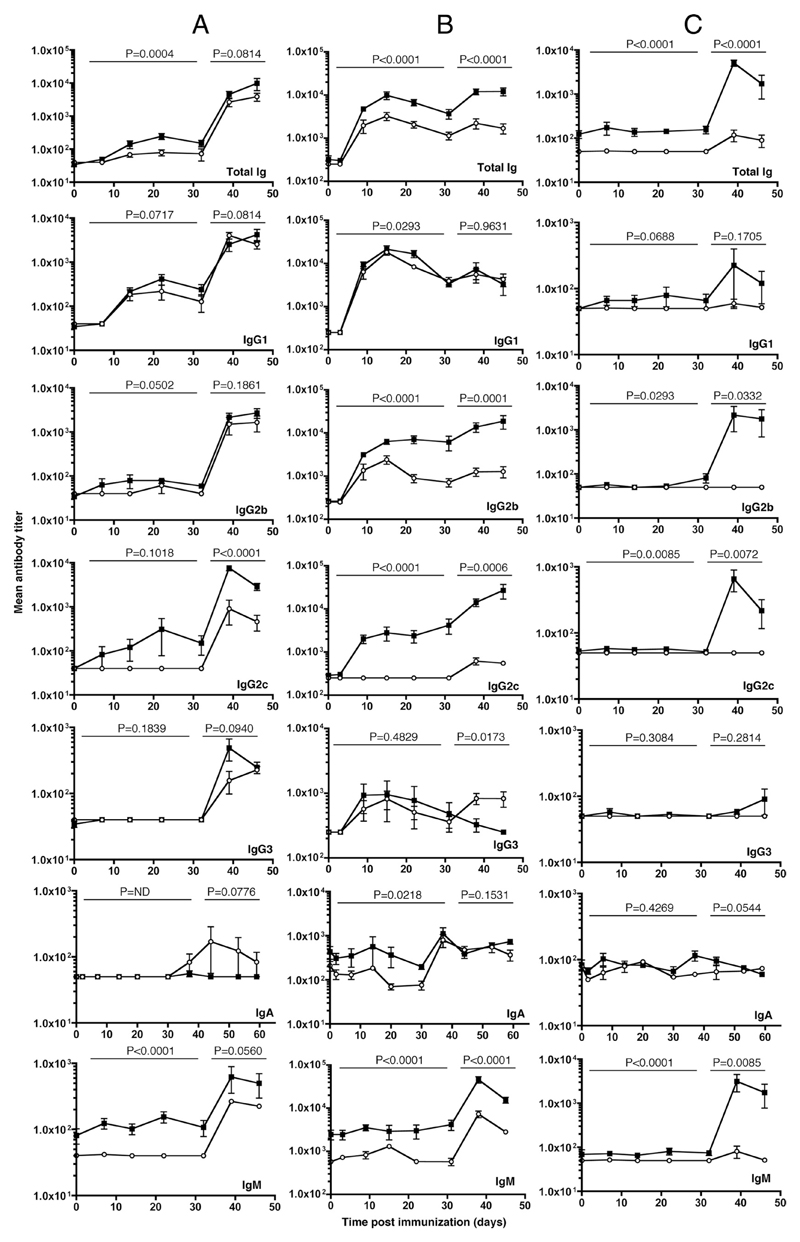
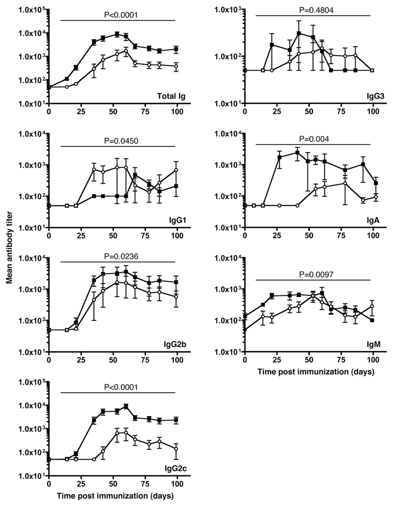
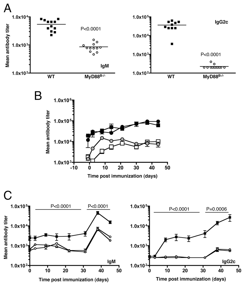
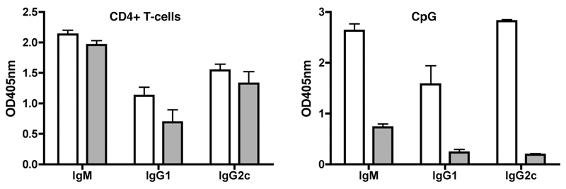
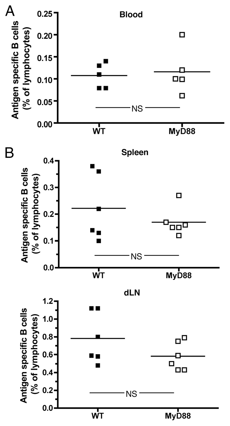
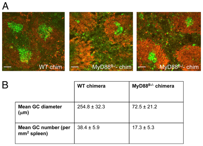
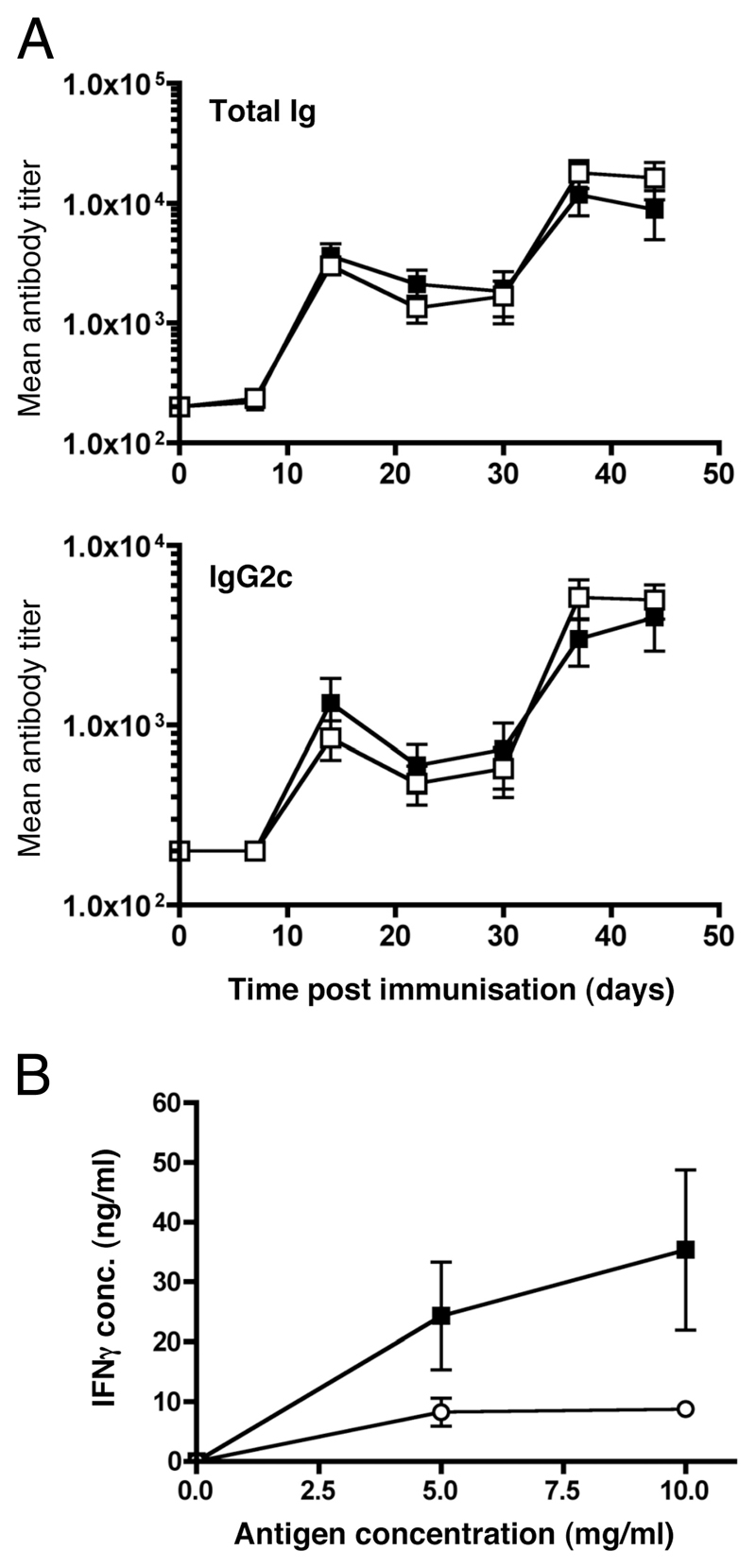
The question of whether Ab responses to T-dependent Ags require B cell intrinsic signaling via the main TLR adaptor (MyD88) has become embroiled in confusion. In part this may be related to the methods used to analyze B cell intrinsic signaling. We have used a mixed bone marrow chimera model to generate mice in which the B cell compartment is completely deficient in MyD88 expression, while the other hematopoietic lineages are largely normal. These mice were immunized with T-dependent Ags or infected with Salmonella. We found that the Ag-specific IgG2c primary response was absolutely dependent on MyD88 signaling to B cells, while other Ig classes were not (IgG1 and IgG3) or much less so (IgG2b, IgA). The MyD88(B-/-) chimeric mice exhibited an impairment of development of IFN-gamma effector T cells, a likely contributory factor in the lack of IgG2c. We also found that B cell intrinsic MyD88 signals are required for the production of natural Abs. The data emphasize the nonredundant role of B cells as programmers of T cell differentiation in vivo.


Conflict of interest statement

Disclosures

The authors have no financial conflict of interest.

Figures

Figure 1
Figure 1
B cell-intrinsic MyD88 signals are required for IgM and IgG2c responses to TD Ags. Mice were immunized s.c. with 100 μg of OVA (A) or DNP-OVA (B) emulsified in IFA with 10 μg of LPS. To measure the response to TI-1 Ag, mice were immunized with 10 μg of LPS in IFA (C). Animals were boosted at day 30 with a second dose of 100 μg of OVA or DNP-OVA (A and B) or 10 μg of LPS. Ag-specific Ab titers were measured by ELISA for each Ab isotype at the time points indicated. In each case, filled symbols represent WT control chimeras and open symbols MyD88B−/− cell chimeras. Values indicate mean titers for groups of six animals and error bars represent SEM. Two-way ANOVA analysis was performed to determined significance of variation for the primary and boosted response. Values of p are given above each graph. Data presented are representative of three independent experiments.
Figure 2
Figure 2
Ab responses to S. typhimurium are largely dependent B cell-intrinsic MyD88 signals. Groups of six mice were infected i.v. with 1 X 10⁶ CFU of the aroA attenuated SL3261 strain of S. enterica. Mice were then bled weekly and Ag-specific Ab titers measured by ELISA (on sonicate Ag) for each Ab isotype as indicated. Values indicate mean titers for groups of six animals and error bars the SEM. Two-way ANOVA tests were performed to determined significance of variation for responses as indicated by p values given at the top of each graph. Data presented are representative of three independent experiments.
Figure 3
Figure 3
Reconstitution of natural Ab does not restore deficient Ab responses in MyD88B−/− mice. A, Preimmune levels of serum IgM and IgG2c in groups of 12 WT chimeras (■) and 12 MyD88B−/− chimeras (○) were quantitated by ELISA. B, A cohort of these mice received three injections of naive serum days −1, 0, and +2 relative to immunization with DNP-OVA (in IFA plus LPS) on day 0. The total Ig levels in the serum were determined by ELISA in each group as indicated. C, DNP-specific Ab responses were quantified at various time points following immunization. In each case, black symbols represent WT control chimeras, white symbols MyD88B−/− chimeras, and gray symbols MyD88B−/− chimeras transfused with naive serum. A, Each point represents the titer from an individual mouse and the mean titer for the group is indicated by the bar. B and C, Values plotted are means based on titers from five individual mice. Statistical analysis is by Student’s t test with p values as indicated on the graph.
Figure 4
Figure 4
MyD88-deficient B cells can produce IgG2c and IgM when cocultured with T cells. CD19-positive B cells sorted from MyD88−/− mice were cultured at 2 X 10⁶ cells/ml for 5 days with purified CD4-positive OT-II cells and pOVA at 0.5 μg/ml (left) or CpG at 5 μg/ml (right). In each case, □ represent WT B cells and ▩ represent MyD88−/− B cells. Secreted Ab was quantified by capture ELISA on harvested supernatants. Values indicate mean for triplicate cultures on cells pooled from three animals. Data are representative of two independent experiments.
Figure 5
Figure 5
The frequency of Ag-specific B cells is normal in. MyD88B−/− chimeras Mice were immunized s.c. with 100 μg of PE emulsified in IFA with 10 μg of LPS. Mice were bled at day 20 and the number of B220+PE+ (i.e., Ag-specific) B cells in the blood was quantified by FACS (A). Twenty days after boosting at day 30 with a second dose of 100 μg of PE, spleen and lymph nodes were collected and B220+PE+ B cells were enumerated (B). ■ symbols represent WT mice and □ symbols represent MyD88B−/− chimeras. Six mice per group were used and the mean cell frequency for each group is indicated by the bar. No significant differences between WT and MyD88B−/− were observed. Statistical analysis was by Student’s t test, where a p > 0.05 was taken as NS. Data shown are representative of three independent experiments. dLN, draining lymph node.
Figure 6
Figure 6
Lack of MyD88 in B cells impairs the GC response. Spleens collected at day 14 postimmunization with DNP-OVA (in IFA plus LPS). GC were quantified in 5-μm cryostat sections stained with peanut agglutinin (green) and with IgM (red). Representative images from WT and MyD88B−/− chimeras (chim) are shown at the top. Images were captured using Openlab imaging software and the number of GC per unit area of spleen tissue was enumerated and the GC diameter was measured using Volocity software. A summary of data from spleens from seven mice in each group from two separate experiments is shown. Numbers are means ± SE.
Figure 7
Figure 7
Impaired switching to IgG2c in vivo is not due to a lack of IFN-γ production by B cell, but is associated with a suboptimal IFN-γ response by T cells. A, Groups of 7 IFN-γB−/− or WT chimeric mice were immunized with DNP-OVA (in IFA plus LPS). Mice were bled at day 14 and DNP-specific Abs were determined by ELISA (total Ig and IgG2c). Each symbol represents the titer from an individual mouse. ■ symbols represent WT chimeric mice and □ symbols represent IFN-γB−/− chimeras. The mean titer is indicated by the bar. B, CD4-positive T cells were magnetically sorted from the spleens of WT (■) or MyD88B−/− (□) chimeric mice 7 days after immunization with SL3261. These cells were then cocultured with Salmonella Ag (C5SENaOH) at a range of concentrations in the presence of WT-irradiated APC. Following 3 days of incubation, supernatants were harvested and levels of secreted IFN-γ were quantified by ELISA. Presented data are means based on groups of four mice and are representative of three independent experiments. Error bars show SEM.

References

    1. Sporri R, Reis e Sousa C. Inflammatory mediators are insufficient for full dendritic cell activation and promote expansion of CD4+ T cell populations lacking helper function. Nat Immunol. 2005;6:163–170. - PubMed
    1. Schnare M, Barton GM, Holt AC, Takeda K, Akira S, Medzhitov R. Toll-like receptors control activation of adaptive immune responses. Nat Immunol. 2001;2:947–950. - PubMed
    1. Pasare C, Medzhitov R. Toll-dependent control mechanisms of CD4 T cell activation. Immunity. 2004;21:733–741. - PubMed
    1. Pasare C, Medzhitov R. Control of B-cell responses by Toll-like receptors. Nature. 2005;438:364–368. - PubMed
    1. Ruprecht CR, Lanzavecchia A. Toll-like receptor stimulation as a third signal required for activation of human naive B cells. Eur J Immunol. 2006;36:810–816. - PubMed

Publication types