. 2009 Jul 15;183(2):792-6.
doi: 10.4049/jimmunol.0900173. Epub 2009 Jun 19.

Cutting edge: TNF-alpha mediates sensitization to ATP and silica via the NLRP3 inflammasome in the absence of microbial stimulation

Affiliations

Cutting edge: TNF-alpha mediates sensitization to ATP and silica via the NLRP3 inflammasome in the absence of microbial stimulation

Luigi Franchi et al. J Immunol. .

Abstract

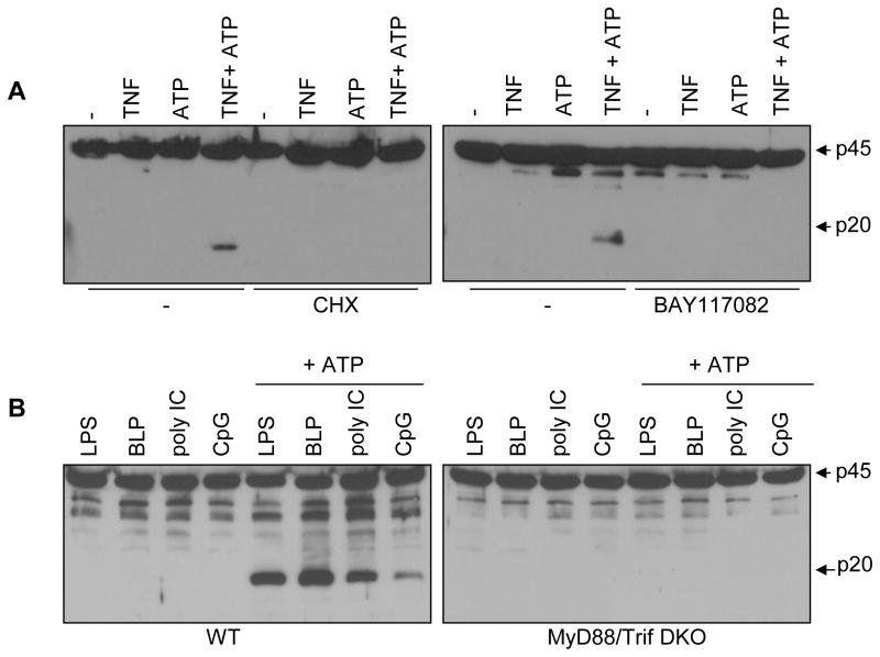
The Nlrp3 inflammasome is critical for the activation of caspase-1 in response to danger signals and particulate matter. However, its role in sterile inflammation remains unclear because prestimulation of phagocytic cells with microbial molecules is required for caspase-1 activation. We show here that exposure of macrophages and dendritic cells to TNF-alpha promotes ATP- or silica-mediated caspase-1 activation and IL-1beta secretion in the absence of microbial stimulation. The effect of TNF-alpha was abolished in macrophages deficient in TNF receptor I and II, Nlrp3, or ASC, whereas that induced by TLR ligands required MyD88/Trif. In addition to TNF-alpha, IL-1alpha and IL-1beta promoted caspase-1 activation via Nlrp3 in response to ATP. Remarkably, macrophages tolerized to TNF-alpha, but not to LPS, retained full sensitivity to ATP stimulation via Nlrp3. These results provide a mechanism by which danger signals and particulate matter mediate inflammation via the Nlrp3 inflammasome in the absence of microbial infection.


Conflict of interest statement

Disclosures

The authors declare that no competing interest exist

Figures

Figure 1
Figure 1
TNF-α induces caspase-1 activation in response to ATP and silica in the absence of microbial stimulation. A, BMDM were stimulated for 6 hrs with TNF (100 ng/ml), CD40L (10μg/ml), agonistic anti-Fas antibody (10μg/ml), IFN-γ (100 U/ml) or LPS (10 ng/ml). When indicated ATP (5 mM) was added for the last 30 minutes B, BMDM were stimulated for 6 hrs with PMA (100 ng/ml), RANKL (1 μg/ml), IL-1α (10 ng/ml), IL-1β (10 ng/ml). When indicated ATP (5 mM) was added for the last 30 minutes. C, BMDM were stimulated with TNF-α and then stimulated with ATP, silica or infected with S. Typhimurium (Salm). (AC) Extracts were prepared from cell and culture supernatants and immunoblotted with caspase-1 antibody. Arrows denote procaspase-1 (p45) and its processed p20 subunit. Results are representative of three separate experiments.
Figure 2
Figure 2
TNF-α promotes IL-1β secretion and caspase-1 activation via TNFR-I/II and the Nlrp3 inflammasome. A–B, BMDC from wild-type (WT), TnfrI/II deficient (TNFRI/II-DKO) and Nlrp3 deficient (Nlrp3-KO) mice were stimulated with TNF-α for 6 hrs and then stimulated, or not, with ATP for 30 minutes. IL-1β was measured 4 hrs after stimulation in cell-free supernatants by ELISA. Values represent mean ± SD of triplicate cultures. * p < 0.01 between WT and mutant macrophages. C, BMDM were stimulated as in panels A and B. Extracts were immunoblotted with caspase-1 antibody. Arrows denote procaspase-1 (p45) and its processed p20 subunit. (A–B) Results are representative of three separate experiments.
Figure 3
Figure 3
TNF-mediated activation of caspase-1 requires gene transcription. A, BMDM were pretreated with 50 μM of cyclohexemide (CHX) or the NF-κB inhibitor BAY 11-7082 (20μM) for 1 hour, and then stimulated with TNF-α for 6 hrs. When indicated ATP was added for the last 30 minutes. B, BMDM from WT or MyD88//Trif/ mice (DKO) were stimulated for 6 hrs with LPS (1ug/ml), BLP (1ug/ml), poly IC (1ug/ml) or CpG (1ug/ml). When indicated ATP (5 mM) was added for the last 30 minutes. A and B, extracts were immunoblotted with caspase-1 antibody. Arrows denote procaspase-1 (p45) and its processed p20 subunit Results are representative of three separate experiments.
Figure 4
Figure 4
TNF-α, but not LPS, induces ATP-mediated IL-1β secretion and caspase-1 activation via Nlrp3 in tolerized macrophages. A–D, BMDM were left unstimulated or stimulated with TNF-α (100 ng/ml) (A–B) or LPS (C and D) for 6 or 24 hrs and then stimulated with ATP (5 mM) for 30 min. A–C, IL-1β was measured after stimulation in cell-free supernatants by ELISA. Values represent mean ± SD of triplicate cultures. n. d. refers to undetectable. * p < 0.01 between WT and mutant macrophages. B–D, Extracts were immunoblotted with caspase-1 antibody. Arrows denote procaspase-1 (p45) and its processed p20 subunit. (A–D) Results are representative of three separate experiments.

Similar articles

Cited by

References

    1. Kono H, Rock KL. How dying cells alert the immune system to danger. Nat Rev Immunol. 2008;8:279–289. - PMC - PubMed
    1. Ishii KJ, Koyama S, Nakagawa A, Coban C, Akira S. Host innate immune receptors and beyond: making sense of microbial infections. Cell Host Microbe. 2008;3:352–363. - PubMed
    1. Franchi L, Warner N, Viani K, Nunez G. Function of Nod-like receptors in microbial recognition and host defense. Immunol Rev. 2009;227:106–128. - PMC - PubMed
    1. Chen CJ, Kono H, Golenbock D, Reed G, Akira S, Rock KL. Identification of a key pathway required for the sterile inflammatory response triggered by dying cells. Nat Med. 2007;13:851–856. - PubMed
    1. Petrilli V, Dostert C, Muruve DA, Tschopp J. The inflammasome: a danger sensing complex triggering innate immunity. Curr Opin Immunol. 2007;19:615–622. - PubMed

Publication types