. 2009 Jul 1;183(1):438-44.
doi: 10.4049/jimmunol.0900204.

The transmembrane E3 ligase GRAIL ubiquitinates and degrades CD83 on CD4 T cells

Affiliations

The transmembrane E3 ligase GRAIL ubiquitinates and degrades CD83 on CD4 T cells

Leon L Su et al. J Immunol. .

Abstract

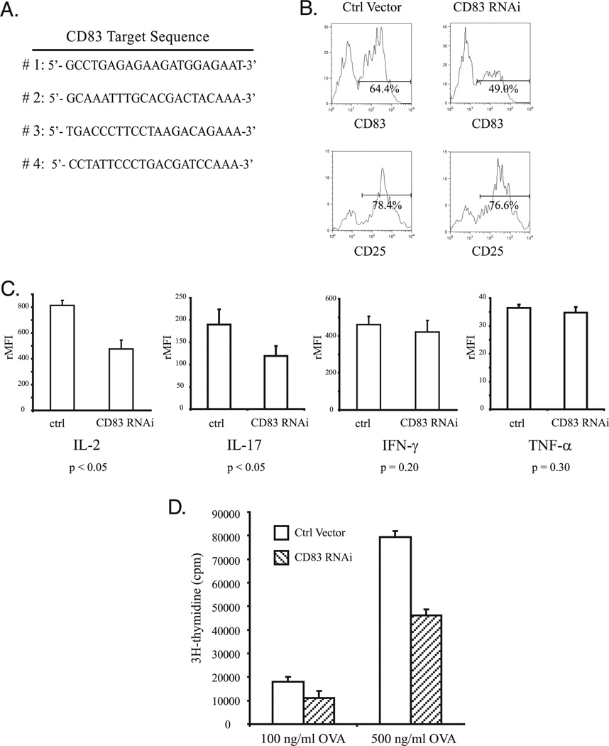
Ubiquitination of eukaryotic proteins regulates a broad range of cellular processes, including T cell activation and tolerance. We have previously demonstrated that GRAIL (gene related to anergy in lymphocytes), a transmembrane RING finger ubiquitin E3 ligase, initially described as induced during the induction of CD4 T cell anergy, is also expressed in resting CD4 T cells. In this study, we show that GRAIL can down-modulate the expression of CD83 (previously described as a cell surface marker for mature dendritic cells) on CD4 T cells. GRAIL-mediated down-modulation of CD83 is dependent on an intact GRAIL extracellular protease-associated domain and an enzymatically active cytosolic RING domain, and proceeds via the ubiquitin-dependent 26S proteosome pathway. Ubiquitin modification of lysine residues K168 and K183, but not K192, in the cytoplasmic domain of CD83 was shown to be necessary for GRAIL-mediated degradation of CD83. Reduced CD83 surface expression levels were seen both on anergized CD4 T cells and following GRAIL expression by retroviral transduction, whereas GRAIL knock-down by RNA interference in CD4 T cells resulted in elevated CD83 levels. Furthermore, CD83 expression on CD4 T cells contributes to T cell activation as a costimulatory molecule. This study supports the novel mechanism of ubiquitination by GRAIL, identifies CD83 as a substrate of GRAIL, and ascribes a role for CD83 in CD4 T cell activation.


Conflict of interest statement

Disclosures

The authors have no financial conflict of interest.

Figures

FIGURE 1
FIGURE 1
GRAIL down-regulation of CD83 requires an intact RING domain and N-terminal PA domain. A, Sequence alignment of HSV-1 ICP0 and murine GRAIL RING domains. Conserved residues required for zinc binding are boxed. B, HEK293 cells were transfected with murine CD83 plasmid (0.5 µg) along with either control vector, wild-type GRAIL, H2N2, or APA GRAIL IRES eGFP plasmid (1.0 µg each). CD83 surface expression levels were evaluated on eGFP+ cells by FACS analysis 36 h post-transfection. Rat IgG1 isotype control included for each sample. (mean fluorescence intensities: vector, 359; GRAIL, 43.8; H2N2, 351; ΔPA, 253.) C, HEK293 cells were transfected as described in B. Cell lysates were prepared and subjected to SDS-PAGE. The levels of CD83-myc and GRAIL-V5 were detected by Western blot (wb) using anti-myc-HRP (top) and anti-V5-HRP (middle) Ab, respectively. Blots were reprobed for cyclophilin-B (CypB) as a protein loading control (bottom). V, Vector; GR, GRAIL; H2, H2N2 GRAIL; ΔPA, N-terminal PA deletion.
FIGURE 2
FIGURE 2
N-terminal PA domain of GRAIL facilitates CD83 binding. A, HEK293 cells were transfected with CD83 myc/His-tagged vector (1.0 µg) along with wild-type and mutant GRAIL-V5-tagged vectors (1.0 µg each). Thirty-six hours post-transfection, interaction between GRAIL and CD83 was determined by immunoprecipitation (ip) with anti-V5 Ab, followed by Western blot (wb) probed with anti-His Ab (top left), or immunoprecipitation with anti-myc Ab, followed by Western blot probed with anti-V5 Ab (top right). Blots were reprobed with appropriate Ab to show presence of immunoprecipitated protein (bottom). B, HEK293 cells were transfected with 0.5 µg of ICOS-myc-tagged plasmid, along with vector or GRAIL-V5 tagged plasmid (1.0 µg each). Protein levels were analyzed as described in Fig. 1C. V, Vector; GR, GRAIL; H2, H2N2 GRAIL; ΔPA, N-terminal PA deletion.
FIGURE 3
FIGURE 3
GRAIL-mediated degradation of CD83 proceeds via an ubiquitin-mediated process. HEK293 cells were transfected with 0.5 µg of CD83-myc, 0.5 µg of Flag-Ubiquitin, and either 1.0 µg of vector, GRAIL, or H2N2 plasmid, as indicated. Before lysate preparation, cells were either left untreated or treated with MG132 (25 µMf) for 2 h. To eliminate the possibility of protein coprecipitation in the samples, lysates were boiled in lysis buffer containing 1% SDS for 5 min before a 10-fold dilution and subsequent immunoprecipitation (ip) using anti-myc Abs. The blot was probed with anti-Flag-HRP to detect the polyubiquitinated CD83 species (top). The blot was reprobed with anti-myc to show CD83 immunoprecipitation levels (bottom). wb, Western blot; ip, immunoprecipitation.
FIGURE 4
FIGURE 4
GRAIL-specific degradation of CD83 requires membrane proximal lysine residues. A, Protein sequence of carboxyl-terminal domain of CD83-myc, with lysine residues underlined. Position of lysine to arginine (K→R) substitutions noted. B, HEK293 cells were transfected with either wild-type CD83 or CD83 bearing single K→R mutations (0.5 µg each) in the absence or presence of 1.0 µg vector encoding wild-type (wt) GRAIL. Lysates were prepared and levels of CD83 were detected by Western blot (wb) with anti-myc Ab 36 h post-transfection (top). All blots were reprobed with anti-V5 Ab to assess GRAIL expression. The expression of cyclophilin-B (CypB) was used as an internal loading control. C, HEK293 cells were transfected with either CD83 bearing double (left), or triple (right) K→R mutations (0.5 µg each) in the absence or presence of wild-type GRAIL plasmid (1.0 µg). Protein levels of CD83 were determined as described in B. TM, Transmembrane; V, vector; GR, GRAIL.
FIGURE 5
FIGURE 5
GRAIL expression results in decreased CD83 expression in CD4 T cells. A, GRAIL levels in CD4 T cells decrease upon TCR engagement. CD4+ T effector cells were prepared by CD4 T cell MACS depletion and 3-day anti-CD3/CD28 stimulation, followed by 4-day rest in medium. Cells were then subjected to plate-bound anti-CD3 (0.5 µg) and soluble anti-CD28 (0.5 µg) stimulation for times indicated. Cell lysates were prepared and subjected to SDS-PAGE. GRAIL protein level was detected using a monoclonal GRAIL Ab (top). Blot was reprobed with anti-cyclophilin-B as protein loading control (bottom). B TCR stimulation induces CD83 expression on CD4 T cells. CD4 T cells were prepared as described in A. Cells were stained for CD83 surface expression at 0-, 24-, and 48-h TCR stimulation. Isotype control for 24-h stimulation sample included. (MFIs: 0 h 8.1; 24 h 31.4; 48 h 48.5) C Ectopic GRAIL expression reduces CD83 expression levels. CD4 T cells were isolated by AutoMACS separation stimulated for 24 h then transduced with retrovirus containing vector control (V) GRAIL (GR) or H2N2 (H2). Levels of CD83 surface expression on transduced GFP+ cells were evaluated by FACS analysis at 24 h post-transduction. Percentage of CD83 surface expression on GFP+ cells denoted by gated region: V 25.0%; GR 20.2%; H2 26.0%. D Percentage of CD83 surface expression on GRAIL and H2N2-transduced cells normalized to vector control. Average of three independent experiments. E Ionomycin-anergized CD4 T cells display increased GRAIL expression. CD4 T cells were prepared as described in A then either left untreated or treated with ionomycin (1 µM) for 18 h. Lysates were prepared and subjected to SDS-PAGE. Blots were probed with polyclonal GRAIL anti-serum (top) and reprobed with anti-actin (bottom) to ensure equal loading. F Anergized CD4 T cells display reduced surface expression of CD83. Ionomycin-anergized CD4 T cells were prepared as described in E then subjected to plate-bound anti-CD3 (0.5 µg) and soluble anti-CD28 (0.5 µg) stimulation for 24 h and CD83 levels were evaluated by FACS. (Stimulated non-ionomycin treated = activated MFI 31.5; stimulated ionomycin treated = anergized MFI 22.3; isotype control MFI 8.8). G GRAIL RNAi results in increased CD83 expression levels. CD4 T cells were electroporated with GRAIL shRNA vectors (GR RNAi no.1 GR RNAi no.2) or pSiren Luciferase control. Twenty-four hours post-electroporation cells were either left unstimulated (left) or stimulated with plate-bound anti-CD3 (0.5 µg/ml) and soluble anti-CD28 (0.5 µg/ml) for 24 h (right) then CD83 expression on ZsGreen+ cells were determined by FACS. Percentage of CD83 surface expression on ZsGreen+ cells denoted by gated region: unstimulated (left): Ctrl 15.4%;RNAi no.1 36.0%; RNAi no. 2 31.1%; stimulated (right): Ctrl 41.1%; RNAi no. 1 65.5%; RNAi no. 2 55.6%.
FIGURE 6
FIGURE 6
CD83 expression on murine CD4+ T cells is important in TCR-mediated T cell proliferation. A, Target sequence of murine CD83 for RNAi oligo design. Four different hairpin loop-containing sense and antisense oligonucleotides were synthesized, annealed, and cloned into the pSiren-Ret-roQ-ZsGreen RNAi vector. B, Specific CD83 knockdown in CD4+ T cells. A pSiren Luciferase RNAi control, or a mixture of the CD83 pSiren RNAi vectors (4 µg total) were electroporated into DO11 OVA-specific CD4+ T cells. Cells were stimulated with pOVA (100 ng/ml) presented by irradiated APC 24 h post-electroporation. Surface expression of CD83 (top) and CD25 (bottom) on ZsGreen+ CD4 cells were determined by FACS analysis (Gated regions). C, Cytokine expression profile of CD83 RNAi-electroporated CD4+ T cells. DO11 OVA-specific CD4 T cells were electroporated with either control or CD38 shRNA vectors (6 µg total). Zs-Green+ cells were FACS sorted 24 h post-electroporation and subsequently stimulated with irradiated APCs in the presence of 500 ng/ml OVA peptide for 48 h. Supernatants were collected and subjected to Luminex cytokine bead assay. The p values were determined using paired t test. D, CD83 knockdown on CD4+ T cells diminished TCR-mediated T cell proliferation. DO11 OVA-specific CD4 T cells were treated with CD83 RNAi and stimulated with irradiated APCs as described in C. Forty-eight hours post-pOVA peptide stimulation, cells were pulsed with [3H]thymidine for 6 h, harvested, and filter was counted. Results are an average of four individual wells.

Similar articles

Cited by

References

    1. Pickart CM. Mechanisms underlying ubiquitination. Annu. Rev. Biochem. 2001;70:503–533. - PubMed
    1. Anandasabapathy N, Ford GS, Bloom D, Holness C, Paragas V, Seroogy C, Skrenta H, Hollenhorst M, Fathman CG, Soares L. GRAIL: an E3 ubiquitin ligase that inhibits cytokine gene transcription is expressed in anergic CD4+ T cells. Immunity. 2003;18:535–547. - PubMed
    1. Seroogy CM, Soares L, Ranheim EA, Su L, Holness C, Bloom D, Fathman CG. The gene related to anergy in lymphocytes, an E3 ubiquitin ligase, is necessary for anergy induction in CD4 T cells. J. Immunol. 2004;173:79–85. - PubMed
    1. Lin J, Kattah M, Lineberry N, Su L, Utz PJ, Fathman CG, Wu L. Rapamycin, anti-IL-2, and CTLA4-Ig inhibit naïve CD4+ T cell proliferation through a common mechanism of action regulating GRAIL expression. J. Immunol. In press. 2009 - PMC - PubMed
    1. Lineberry NB, Su LL, Lin JT, Coffey GP, Seroogy CM, Fathman CG. Cutting edge: the transmembrane E3 ligase GRAIL ubiq-uitinates the costimulatory molecule CD40 ligand during the induction of T cell anergy. J. Immunol. 2008;181:1622–1626. - PMC - PubMed

Publication types

MeSH terms