

Generation of protective T cell-independent antiviral antibody responses in SCID mice reconstituted with follicular or marginal zone B cells
- PMID: 19542462
- PMCID: PMC2774789
- DOI: 10.4049/jimmunol.0900068
Generation of protective T cell-independent antiviral antibody responses in SCID mice reconstituted with follicular or marginal zone B cells
Abstract
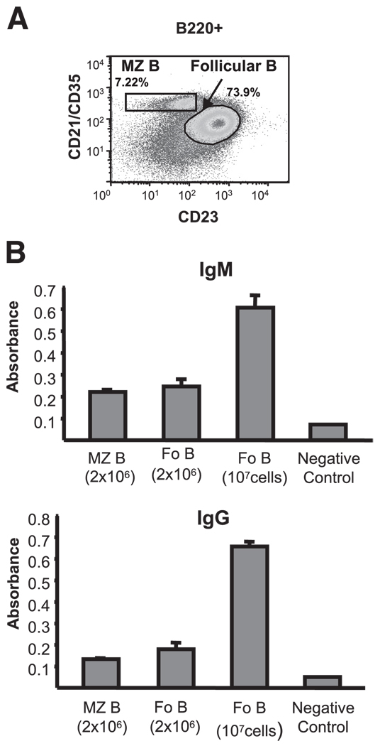
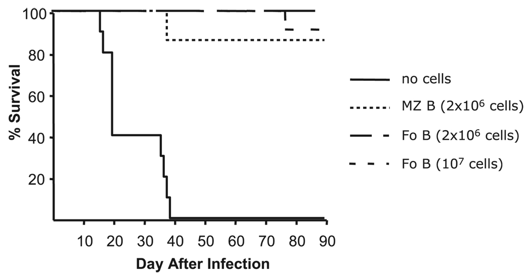
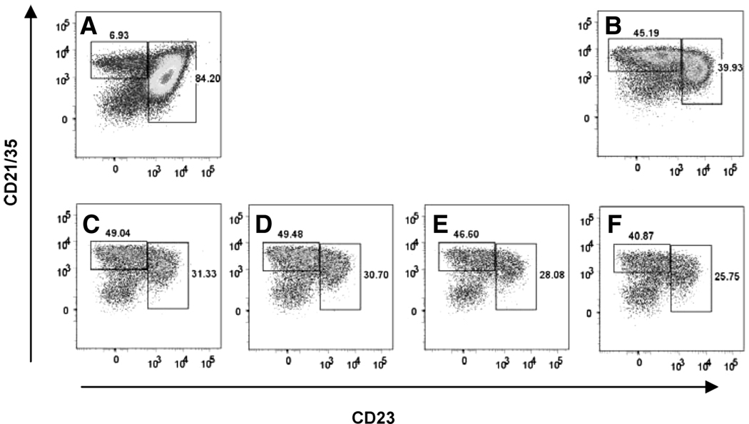
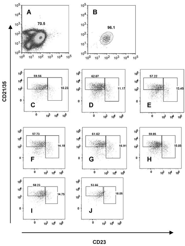
B cells generated in the bone marrow of adult mice enter the periphery as transitional B cells and subsequently differentiate into one of two phenotypically and functionally distinct subsets, marginal zone (MZ) or follicular (Fo) B cells. Recent reports indicate, however, that in response to environmental cues, such as lymphopenia, mature Fo B cells can change to display phenotypic markers characteristic of MZ B cells. Previously, we found that splenic B cells transferred to SCID mice responded to polyoma virus (PyV) infection with T cell-independent (TI) IgM and IgG secretion, reducing the viral load and protecting mice from the lethal effect of the infection. The contribution of MZ and Fo B cell subsets to this antiviral TI-2 response, however, has not been addressed. In this study, we show that both sort-purified MZ and Fo B cells generate protective TI Ab responses to PyV infection when transferred into SCID mice. Moreover, the transferred Fo B cells in the spleens of the PyV-infected SCID mice change phenotype, with many of them displaying MZ B cell characteristics. These findings demonstrate the plasticity of the B cell subsets in virus-infected hosts and show for the first time that B cells derived exclusively from Fo B cells can effectively function in antiviral TI-2 responses.
Conflict of interest statement
The authors have no financial conflict of interest.
Figures
Similar articles
-
The role of CD40-CD154 interaction in antiviral T cell-independent IgG responses.J Immunol. 2000 Jun 1;164(11):5877-82. doi: 10.4049/jimmunol.164.11.5877. J Immunol. 2000. PMID: 10820268
-
T cell-independent antibody-mediated clearance of polyoma virus in T cell-deficient mice.J Exp Med. 1996 Feb 1;183(2):403-11. doi: 10.1084/jem.183.2.403. J Exp Med. 1996. PMID: 8627153 Free PMC article.
-
Rapid response of marginal zone B cells to viral particles.J Immunol. 2004 Oct 1;173(7):4308-16. doi: 10.4049/jimmunol.173.7.4308. J Immunol. 2004. PMID: 15383559
-
The dual function of the splenic marginal zone: essential for initiation of anti-TI-2 responses but also vital in the general first-line defense against blood-borne antigens.Clin Exp Immunol. 2002 Oct;130(1):4-11. doi: 10.1046/j.1365-2249.2002.01953.x. Clin Exp Immunol. 2002. PMID: 12296846 Free PMC article. Review.
-
The splenic marginal zone in humans and rodents: an enigmatic compartment and its inhabitants.Histochem Cell Biol. 2006 Dec;126(6):641-8. doi: 10.1007/s00418-006-0210-5. Epub 2006 Jul 1. Histochem Cell Biol. 2006. PMID: 16816939 Review.
Cited by
-
Division of labor during primary humoral immunity.Immunol Res. 2013 Mar;55(1-3):277-86. doi: 10.1007/s12026-012-8372-9. Immunol Res. 2013. PMID: 22945808 Free PMC article. Review.
-
Specific humoral immunity versus polyclonal B cell activation in Trypanosoma cruzi infection of susceptible and resistant mice.PLoS Negl Trop Dis. 2010 Jul 6;4(7):e733. doi: 10.1371/journal.pntd.0000733. PLoS Negl Trop Dis. 2010. PMID: 20625554 Free PMC article.
-
Single-cell transcriptome profiling and the use of AID deficient mice reveal that B cell activation combined with antibody class switch recombination and somatic hypermutation do not benefit the control of experimental trypanosomosis.PLoS Pathog. 2021 Nov 11;17(11):e1010026. doi: 10.1371/journal.ppat.1010026. eCollection 2021 Nov. PLoS Pathog. 2021. PMID: 34762705 Free PMC article.
-
T Cell-Independent Gamma Interferon and B Cells Cooperate To Prevent Mortality Associated with Disseminated Chlamydia muridarum Genital Tract Infection.Infect Immun. 2018 Jun 21;86(7):e00143-18. doi: 10.1128/IAI.00143-18. Print 2018 Jul. Infect Immun. 2018. PMID: 29661927 Free PMC article.
-
A cluster of broadly neutralizing IgG against BK polyomavirus in a repertoire dominated by IgM.Life Sci Alliance. 2023 Jan 30;6(4):e202201567. doi: 10.26508/lsa.202201567. Print 2023 Apr. Life Sci Alliance. 2023. PMID: 36717250 Free PMC article.
References
-
- Monroe JG, Dorshkind K. Fate decisions regulating bone marrow and peripheral B lymphocyte development. Adv. Immunol. 2007;95:1–50. - PubMed
-
- Cariappa A, Tang M, Parng C, Nebelitskiy E, Carroll M, Georgopoulos K, Pillai S. The follicular versus marginal zone B lymphocyte cell fate decision is regulated by Aiolos, Btk, and CD21. Immunity. 2001;14:603–615. - PubMed
-
- Lopes-Carvalho T, Kearney JF. Development and selection of marginal zone B cells. Immunol. Rev. 2004;197:192–205. - PubMed
-
- Samardzic T, Marinkovic D, Danzer CP, Gerlach J, Nitschke L, Wirth T. Reduction of marginal zone B cells in CD22-deficient mice. Eur. J. Immunol. 2002;32:561–567. - PubMed
-
- Kuroda K, Han H, Tani S, Tanigaki K, Tun T, Furukawa T, Taniguchi Y, Kurooka H, Hamada Y, Toyokuni S, Honjo T. Regulation of marginal zone B cell development by MINT, a suppressor of Notch/RBP-J signaling pathway. Immunity. 2003;18:301–312. - PubMed
Publication types
MeSH terms
Substances
Grants and funding
LinkOut - more resources
Full Text Sources
