. 2009 Jul 1;183(1):560-7.
doi: 10.4049/jimmunol.0900241.

Major role of gamma delta T cells in the generation of IL-17+ uveitogenic T cells

Affiliations

Major role of gamma delta T cells in the generation of IL-17+ uveitogenic T cells

Yan Cui et al. J Immunol. .

Abstract

We show that in vitro activation of interphotoreceptor retinoid-binding protein (IRBP)-specific T cells from C57BL/6 mice immunized with an uveitogenic IRBP peptide (IRBP(1-20)) under TH17-polarizing conditions is associated with increased expansion of T cells expressing the gammadelta TCR. We also show that highly purified alphabeta or gammadelta T cells from C57BL/6 mice immunized with IRBP(1-20) produced only small amounts of IL-17 after exposure to the immunizing Ag in vitro, whereas a mixture of the same T cells produced greatly increased amounts of IL-17. IRBP-induced T cells from IRBP-immunized TCR-delta(-/-) mice on the C57BL/6 genetic background produced significantly lower amounts of IL-17 than did wild-type C57BL/6 mice and had significantly decreased experimental autoimmune uveitis-inducing ability. However, reconstitution of the TCR-delta(-/-) mice before immunization with a small number of gammadelta T cells from IRBP-immunized C57BL/6 mice restored the disease-inducing capability of their IRBP-specific T cells and greatly enhanced the generation of IL-17(+) T cells in the recipient mice. Our study suggests that gammadelta T cells are important in the generation and activation of IL-17-producing autoreactive T cells and play a major role in the pathogenesis of experimental autoimmune uveitis.


Figures

FIGURE 1
FIGURE 1
Activation of IL-17+ IRBP-specific T cells is associated with expansion of γδ T cells. T cells prepared from the spleens and draining lymph nodes of IRBP1–20-immunized B6 mice at day 13 postimmunization. were stimulated in vitro with immunizing Ag (10 μg/ml) and APCs (irradiated spleen cells) for 2 days. The activated T cell blasts were then separated by Ficoll gradient centrifugation and cultured in IL-2 (Th1-polarized)- or IL-23 (Th17-polarized)-containing medium for 5 days. Finally, the cells were stained for intracellular IL-17 and IFN-γ using FITC-anti-IL-17 and PE-anti-IFN-γ Abs (A), for the αβ TCR and CD3 (B), and for the γδ TCR and IL-17 (C). Approximately 15–20% of the Th17-polarized T cells did not express the αβ TCR but expressed the γδ TCR. Results are representative of multiple experiments. FL-1H and FL-2H, fluorescence.
FIGURE 2
FIGURE 2
γδ T cells isolated from IRBP-immunized B6 mice predominantly express Vγ4Vδ4. Splenic T cells from naive or IRBP1–20, MOG35–55, or CFA-immunized B6 mice were stained using a panel of mAbs specific for γδ TCR V segments (Vγ4, Vγ1, Vδ4, Vδ5, and Vδ6.3) and an Ab specific for the mouse pan-TCR δ chain (GL3), followed by FACS analysis. The results shown are representative of those in five experiments. FL-1H, fluorescence.
FIGURE 3
FIGURE 3
Role of γδ and αβ T cells in the Ag-specific IL-17 response. A–C, Purification of αβ TCR+ and γδ TCR+ T cells: Purified αβ TCR+ and γδ TCR+ T cells were prepared from IRBP1–20-immunized B6 mice using magnetic beads. A, One-step positive selection using GL3 (mAb specific for pan-mouse γδ TCR) resulted in 95% pure γδ T cells. B, Further depletion of αβ T cells resulted in 99% pure γδ T cells. C, 99% pure αβ T cells were prepared similarly. D–F, The partially purified (D) or highly purified (E) γδ T cells or the indicated mixtures (F), with the total number of cells in all 4 × 10⁵ T cells/ well, were incubated with the indicated Ag and APCs in 96-well plates, and the culture supernatants were tested for IL-17. The results shown are representative of those in three experiments.
FIGURE 4
FIGURE 4
Test of interaction between αβ and γδ T cells using culture inserts. A–C, In a 24-well plate, a total of 2 × 10⁶/well in vivo primed αβ and γδ T cells from IRBP1–20-immunized B6 mice (99% pure) were tested alone (B and C) or as a 9:1 mixture (A) of αβ and γδ TCR+ T cells for production of IL-17 or IFN-γ after in vitro stimulation with IRBP1–20 and APCs. Only the cells in the lower chambers are cocultured with irradiated splenocytes. D, 90% αβ and 10% γδ TCR+ T cells were cultured in a same well but separated by inserts. αβ T cells were in the upper chamber, and γδ T cells were in the lower chamber; E, αβ T cells were seeded in the lower chamber, and γδ T cells were seeded in the upper chamber. Results are representative of those in three experiments.
FIGURE 5
FIGURE 5
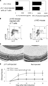
Transfer of γδ T cells into TCR-δ−/− mice restores their ability to generate IRBP-specific T cells. The IFN-γ- and IL-17-producing abilities of the IRBP-specific T cells of TCR-δ−/− mice, either untreated or injected with 2 × 10⁵ γδ T cells were determined 2 day after in vitro stimulation with immunizing Ag (A). Intracellular staining of IL-17+ T cells were assessed after 2 days of in vitro stimulation with immunizing Ag and another 3-day culture in medium containing IL-23 (10 ng/ml; B). The disease-inducing ability of the IRBP-specific T cells (2 × 10⁶) from the TCR-δ−/− mice, either untreated or injected with γδ T cells, was also compared after 48 h of in vitro stimulation with the immunizing Ag (C and D). Results are representative of those in two separate experiments. KO, Knockout.

Similar articles

Cited by

References

    1. Faure JP. Autoimmunity and the retina. Curr. Top. Eye Res. 1980;2:215–302. - PubMed
    1. Wacker WB, Donoso LA, Kalsow CM, Yankeelov JA, Jr., Organisciak DT. Experimental allergic uveitis: isolation, characterization, and localization of a soluble uveitopathogenic antigen from bovine retina. J. Immunol. 1977;119:1949–1958. - PubMed
    1. Caspi RR, Roberge FG, McAllister CG, El-Saied M, Kuwabara T, Gery I, Hanna E, Nussenblatt RB. T cell lines mediating experimental autoimmune uveoretinitis (EAU) in the rat. J. Immunol. 1986;136:928–933. - PubMed
    1. Fox GM, Redmond TM, Wiggert B, Kuwabara T, Chader GJ, Gery I. Dissociation between lymphocyte activation for proliferation and for the capacity to adoptively transfer uveoretinitis. J. Immunol. 1987;138:3242–3246. - PubMed
    1. Shao H, Liao T, Ke Y, Shi H, Kaplan HJ, Sun D. Severe chronic experimental autoimmune uveitis (EAU) of the C57BL/6 mouse induced by adoptive transfer of IRBP1–20-specific T cells. Exp. Eye Res. 2006;82:323–331. - PubMed

Publication types

MeSH terms