. 2009 Jul 1;183(1):568-77.
doi: 10.4049/jimmunol.0802361.

IRAK4 kinase activity is required for Th17 differentiation and Th17-mediated disease

Affiliations

IRAK4 kinase activity is required for Th17 differentiation and Th17-mediated disease

Kirk A Staschke et al. J Immunol. .

Abstract

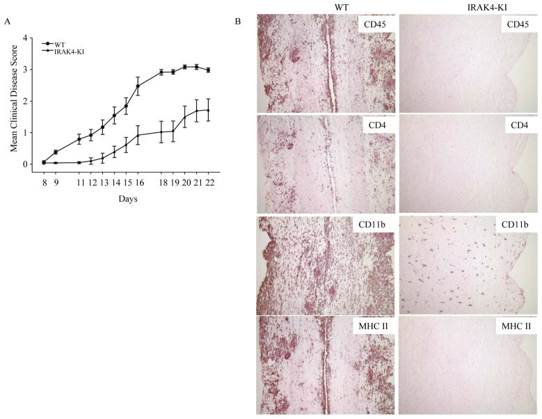
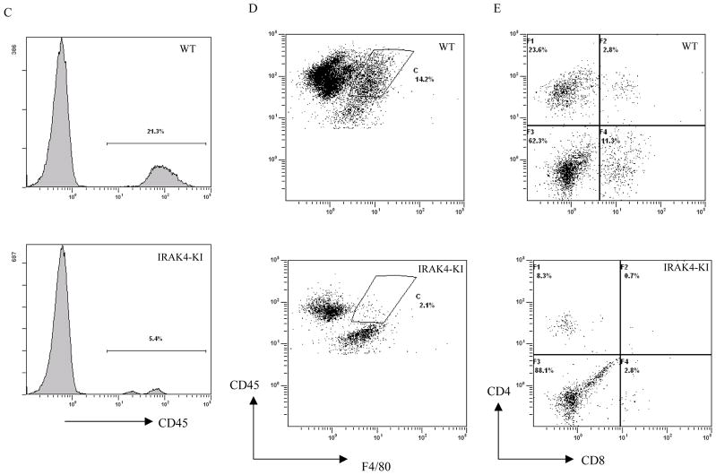
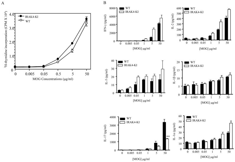
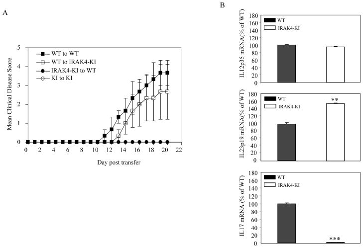
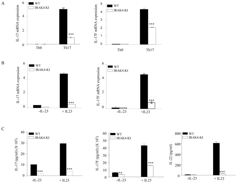
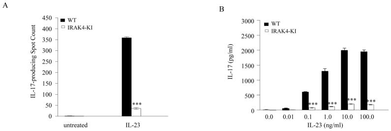
Both IL-23- and IL-1-mediated signaling pathways play important roles in Th17 cell differentiation, cytokine production, and autoimmune diseases. The IL-1R-associated kinase 4 (IRAK4) is critical for IL-1/TLR signaling. We show here that inactivation of IRAK4 kinase in mice (IRAK4 KI) results in significant resistance to experimental autoimmune encephalomyelitis due to a reduction in infiltrating inflammatory cells into the CNS and reduced Ag-specific CD4(+) T cell-mediated IL-17 production. Adoptive transfer of myelin oligodendrocyte glycoprotein 35-55-specific IRAK4 KI Th17 cells failed to induce experimental autoimmune encephalomyelitis in either wild-type or IRAK4 KI recipient mice, indicating the lack of autoantigen-specific Th17 cell activities in the absence of IRAK4 kinase activity. Furthermore, the absence of IRAK4 kinase activity blocked induction of IL-23R expression, STAT3 activation by IL-23, and Th17 cytokine expression in differentiated Th17 cells. Importantly, blockade of IL-1 signaling by IL-1RA inhibited Th17 differentiation and IL-23-induced cytokine expression in differentiated Th17 cells. The results of these studies demonstrate that IL-1-mediated IRAK4 kinase activity in T cells is essential for induction of IL-23R expression, Th17 differentiation, and autoimmune disease.


Figures

Fig. 1
Fig. 1. Reduced induction and progression of EAE in IRAK4 KI mice
(A) EAE was induced following immunization of MOG35-55 (300 μg/mouse, n = 15) in CFA on days 0 and 7, as described in Methods and Materials. The severity of EAE is presented as mean disease scores in each group. The data represent one of two independent experiments. There was also higher percentage of disease incidence (EAE score >1.0) after combined from two independent experiments in WT mice (23/25, 92%) than IRAK4 KI mice (11/26, 42%). (B) Mononuclear cells isolated from the pooled spinal cords of four individuals from IRAK4 KI (disease score: 0, 0, 0.5, 2.8) or WT mice (disease score 3.5, 3.0, 3.0, 3.0) on day 21 were stained for CD45. The CD45+ cells were gated and the percentage of positive cells is indicated. (C–D) Mononuclear cells isolated from the same pooled spinal cords as in panel B were stained for anti-CD45 plus F4/80 for infiltrated macrophages (C) or with anti-CD4 antibody and anti-CD8 antibody for infiltrated T cells (D). Percentages of CD4+-, CD8+-T cells or F4/80+-macrophages were counted on gated CD45+ cells. The data represent one of two independent experiments with similar results.
Figure 2
Figure 2. Impaired IL-17 production in MOG35-55-primed lymphocytes from IRAK4 KI mice
Splenocytes were isolated from five each of IRAK4 KI and WT mice on days 21 following immunization with MOG35-55 peptide in CFA. The MOG35-55-primed cells were then cultured in the presence of the indicated concentration of MOG35-55. (A). T cell proliferation was determined using tritium-labeled thymidine incorporation assay. The results were plotted as mean c.p.m. ± SEM from triplicates. The data represent one of two independent experiments. (B) The MOG35-55-stimulated splenocyte culture supernatants were collected at 72 hr. ELISA was used to measure the antigen-stimulated production of IFN-γ, IL-2, IL-5, IL-1α, IL-1β and IL-17 in the cultures. Values shown represent the mean and error bars represent SD. The data represent one of two independent experiments. Asterisk** denotes 0.001 < p < 0.01, * denotes 0.01< p < 0.05, ns denotes not significant
Figure 3
Figure 3. Impaired IL-17 expression in CNS of IRAK4 KI mice
(A). Differentiated MOG35-55-specific Th17 cells from both MOG35-55-immunized IRAK4 KI and WT mice were adoptively transferred into recipient mice of either WT or IRAK4 KI mice. The progression of disease was scored according to our EAE disease score as described in Materials and Methods. (B). Reduced IL-17 mRNA expression in spinal cord of IRAK4 KI mice with EAE. Spinal cord was used for preparing RNA on day 21 following MOG35-55 immunization of both WT and IRAK4 KI mice. The mRNA expression of indicated genes was assessed by real-time PCR. The mRNA expression of WT mice was set to 100%. The data represent one of two independent experiments with similar results. Asterisk ** denotes 0.001<p < 0.01, *** denotes p<0.001.
Figure 4
Figure 4. Reduced expression of Th17 cytokines in differentiated Th17 cells from IRAK4 KI mice
Naïve CD4+ T cells were purified from both WT and IRAK4 KI mice and differentiated into Th17 cells with plate-bound anti-CD3, anti-CD28 mAbs in the presence IL-1β, IL-6, and TGF-β for 4 days, or T cells were incubated with only plate-bound mAbs anti-CD3 and anti-CD28 in the presence of neutralizing mAbs to IL-4 and IFNγ (Th0 cells). (A). Total mRNAs were isolated from 4-day differentiated Th17 cells or Th0 cells. Both IL-17 and IL-17F mRNA expression were assessed by real-time PCR. The expression level was normalized to β-actin mRNA. The data represent one of three independent experiments. (B). Differentiated Th17 cells were washed, replated in medium, and incubated in the presence or absence of recombinant IL-23 for 16 hrs. Total mRNA isolation and IL-17/IL-17F mRNA expression was performed as in (A). The data represent one of three independent experiments. (C) Cell supernatants from the same treatment groups from (B) were then filtered into fresh 96-well tissue culture plates, and different cytokine levels were measured by ELISA assay. Values shown represent the mean and error bars represent SD. The data represent one of three independent experiments. Asterisk ** denotes 0.001<p<0.01, *** denotes p < 0.001.
Figure 5
Figure 5. Reduced IL-23 receptor expression and IL-23-stimulated Stat3 activation in Th17 cells of IRAK4 KI mice
Purified native CD4+ T cells from both WT and IRAK4 KI mice were differentiated into Th17 cells in vitro with plate-bound anti-CD3 and anti-CD28 mAbs in the presence of IL-1β, IL-6, and TGF-β for 4 days, or T cells were incubated with only plate-bound mAbs anti-CD3 and anti-CD28 in the presence of neutralizing mAbs to IL-4 and IFNγ (Th0 cells). (A) Total mRNA was purified from both Th0 and Th17 cells and the expression of IL-23 receptor mRNA was assessed by real-time PCR and normalized to β-actin mRNA. The data represent one of three independent experiments with similar results. (B) Differentiated Th17 cells were washed and then treated with recombinant human IL-23 for 16 hrs. Total RNA isolation, IL-23R mRNA expression was assessed as in (A) and the data represent one of three independent experiments. (C). Differentiated Th17 cells were washed, starved in serum free medium for 4 hrs and then stimulated with recombinant IL-23 for 15 min. Cell lysates were run on Western blot and probed with mAbs to either total Stat3 and phospho-Stat3 (Y705). Loading control was probed with β-actin mAb. (D). Fold change of phospho-STAT3 in Th17 cell lysate stimulated with IL-23 compared with untreated cell lysate. The phospho-Stat3 was measured by ELISA and the phospho-Stat3 in untreated samples was set to be 1.0. The data represent one of two independent experiments.
Figure 6
Figure 6. IL-1RA blocks Th17 cell differentiation
(A). Purified naïve WT CD4+ T cells were differentiated into Th17 cells with plate-bound anti-CD3 and anti-CD28 mAbs, IL-6, and TGF-β in the presence or absence of IL-1RA (1 μg/ml) for 4 days. Total mRNA was then purified and the expression of IL-17, IL-17F, and IL-23R was assessed by real-time PCR and normalized to β-actin mRNA. The data represent one of three independent experiments. (B). A portion of differentiated Th17 cells in the absence of IL-1RA from (A) were washed, re-plated in medium, and incubated in the presence or absence of recombinant IL-23 or IL-23 and IL-1RA for 16 hrs. Total mRNAs were isolated from cells and IL-17, IL-17F, and IL-23R mRNA expression was quantitated by real-time PCR. The expression level was normalized to β-actin mRNA. The data represent one of three independent experiments. Asterisk *** denotes p < 0.001.

References

    1. Weaver CT, Hatton RD, Mangan PR, Harrington LE. IL-17 family cytokines and the expanding diversity of effector T cell lineages. Annu Rev Immunol. 2007;25:821–852. Review. - PubMed
    1. Weaver CT, Harrington LE, Mangan PR, Gavrieli M, Murphy KM. Th17: an effector CD4 T cell lineage with regulatory T cell ties. Immunity. 2006;24:677–688. Review. - PubMed
    1. Tzartos JS, Friese MA, Craner MJ, Palace J, Newcombe J, Esiri MM, Fugger L. Interleukin-17 production in central nervous system-infiltrating T cells and glial cells is associated with active disease in multiple sclerosis. Am J Pathol. 2008;172:146–155. - PMC - PubMed
    1. Matusevicius D, Kivisäkk P, He B, Kostulas N, Ozenci V, Fredrikson S, Link H. Interleukin-17 mRNA expression in blood and CSF mononuclear cells is augmented in multiple sclerosis. Mult Scler. 1999;5:101–104. - PubMed
    1. Kim HR, Kim HS, Park MK, Cho ML, Lee SH, Kim HY. The clinical role of IL-23p19 in patients with rheumatoid arthritis. Scand J Rheumatol. 2007;36:259–264. - PubMed

Publication types

MeSH terms

Substances