

Genetic dissection of the mouse CNS using magnetic resonance microscopy
- PMID: 19542887
- PMCID: PMC2734144
- DOI: 10.1097/WCO.0b013e32832d9b86
Genetic dissection of the mouse CNS using magnetic resonance microscopy
Abstract
Purpose of review: Advances in magnetic resonance microscopy (MRM) make it practical to map gene variants responsible for structural variation in brains of many species, including mice and humans. We review results of a systematic genetic analysis of MRM data using as a case study a family of well characterized lines of mice.
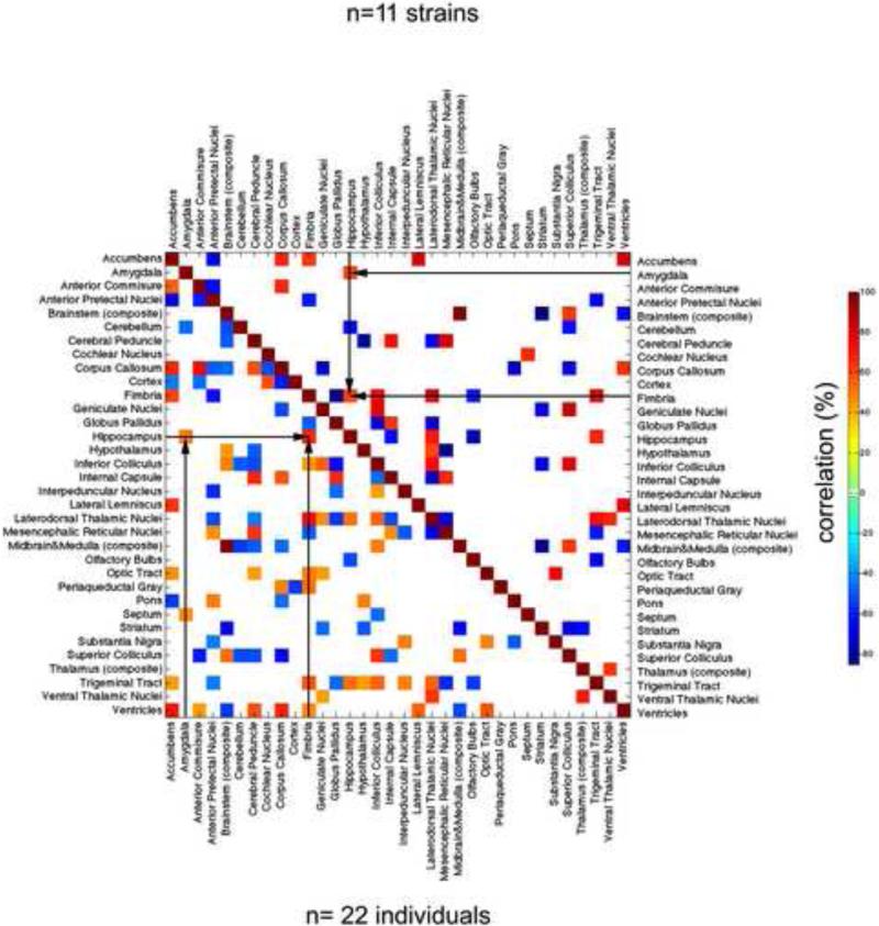
Recent advances: MRM has matured to the point that we can generate high contrast, high-resolution images even for species as small as a mouse, with a brain merely 1/3000th the size of humans. We generated 21.5-micron data sets for a diverse panel of BXD mouse strains to gauge the extent of genetic variation, and as a prelude to comprehensive genetic and genomic analyses. Here we review MRM capabilities and image segmentation methods; heritability of brain variation; covariation of the sizes of brain regions; and correlations between MRM and classical histological data sets.
Summary: The combination of high throughput MRM and genomics will improve our understanding of the genetic basis of structure-function correlations. Sophisticated mouse models will be critical in converting correlations into mechanisms and in determining genetic and epigenetic causes of differences in disease susceptibility.
Figures
Similar articles
-
Genetic dissection of the mouse brain using high-field magnetic resonance microscopy.Neuroimage. 2009 May 1;45(4):1067-79. doi: 10.1016/j.neuroimage.2009.01.021. Epub 2009 Jan 24. Neuroimage. 2009. PMID: 19349225 Free PMC article.
-
Evaluation of five diffeomorphic image registration algorithms for mouse brain magnetic resonance microscopy.J Microsc. 2017 Nov;268(2):141-154. doi: 10.1111/jmi.12594. Epub 2017 Jun 14. J Microsc. 2017. PMID: 28613391
-
Mouse brain magnetic resonance microscopy: Applications in Alzheimer disease.Microsc Res Tech. 2015 May;78(5):416-24. doi: 10.1002/jemt.22489. Epub 2015 Mar 21. Microsc Res Tech. 2015. PMID: 25810274 Review.
-
Intensity inhomogeneity correction using N3 on mouse brain magnetic resonance microscopy.J Neuroimaging. 2013 Oct;23(4):502-7. doi: 10.1111/jon.12041. Epub 2013 Jul 29. J Neuroimaging. 2013. PMID: 23895576
-
Small animal imaging with magnetic resonance microscopy.ILAR J. 2008;49(1):35-53. doi: 10.1093/ilar.49.1.35. ILAR J. 2008. PMID: 18172332 Free PMC article. Review.
Cited by
-
Merged magnetic resonance and light sheet microscopy of the whole mouse brain.Proc Natl Acad Sci U S A. 2023 Apr 25;120(17):e2218617120. doi: 10.1073/pnas.2218617120. Epub 2023 Apr 17. Proc Natl Acad Sci U S A. 2023. PMID: 37068254 Free PMC article.
-
Post-genomic behavioral genetics: From revolution to routine.Genes Brain Behav. 2018 Mar;17(3):e12441. doi: 10.1111/gbb.12441. Epub 2017 Dec 21. Genes Brain Behav. 2018. PMID: 29193773 Free PMC article. Review.
-
Visualization of synaptic domains in the Drosophila brain by magnetic resonance microscopy at 10 micron isotropic resolution.Sci Rep. 2015 Mar 10;5:8920. doi: 10.1038/srep08920. Sci Rep. 2015. PMID: 25753480 Free PMC article.
-
Development and advancements in rodent MRI-based brain atlases.Heliyon. 2024 Mar 8;10(6):e27421. doi: 10.1016/j.heliyon.2024.e27421. eCollection 2024 Mar 30. Heliyon. 2024. PMID: 38510053 Free PMC article. Review.
-
In vivo microscopic voxel-based morphometry with a brain template to characterize strain-specific structures in the mouse brain.Sci Rep. 2017 Mar 7;7(1):85. doi: 10.1038/s41598-017-00148-1. Sci Rep. 2017. PMID: 28273899 Free PMC article.
References
Publication types
MeSH terms
Grants and funding
LinkOut - more resources
Full Text Sources
Medical
Research Materials
