

Review
doi: 10.1038/nrn2608.
Circuits controlling vertebrate locomotion: moving in a new direction
Affiliations
- PMID: 19543221
- PMCID: PMC2847453
- DOI: 10.1038/nrn2608
Item in Clipboard
Review
Circuits controlling vertebrate locomotion: moving in a new direction
Nat Rev Neurosci.
2009 Jul.
Abstract
Neurobiologists have long sought to understand how circuits in the nervous system are organized to generate the precise neural outputs that underlie particular behaviours. The motor circuits in the spinal cord that control locomotion, commonly referred to as central pattern generator networks, provide an experimentally tractable model system for investigating how moderately complex ensembles of neurons generate select motor behaviours. The advent of novel molecular and genetic techniques coupled with recent advances in our knowledge of spinal cord development means that a comprehensive understanding of how the motor circuitry is organized and operates may be within our grasp.
Figures
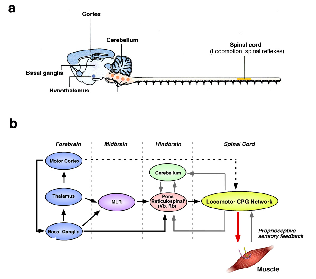
(a) Schematic of the rodent central nervous system showing the neural structures that are part of in the motor pathways controlling simple behaviors such as mastication, respiration and locomotion. Adapted from Ref. . (b) Motor pathways in aquatic and terrestrial vertebrates share a similar neuroanatomical structure. Local control of muscle movements is effected by pools of motor neurons in the spinal cord that are part of a dispersed locomotor CPG network. The motor commands are modulated by proprioceptive sensory feedback via sensory afferents. Descending reticulospinal, rubrospinal and vestibulospinal pathways control the locomotor network in the spinal cord, although the reticulospinal pathway is the primary pathway for intiating locomotion. The reticulospinal pathway can be activated by the mesencephalic locomotor region (MLR), which has inputs from the basal ganglia and thalamus. The cerebellum coordinates motor behaviors by mediating sensory and internal feedback and optimizing the motor pattern to the task at hand. It also coordinates spinal motor actions with the supraspinal motor pathways. Connections from the motor cortex refine and initiate motor actions. The black lines indicate direct command pathways, the grey lines indicate feed-back pathways. VS, vestibulospinal; RbS, rubrospinal.
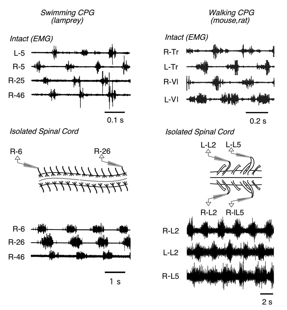
(a) Examples of spinal motor activity during swimming in the lamprey. (Top) Electromyograph (EMG) recordings of different myotomes at located at different axial levels. (Bottom) Ventral root recordings from the isolated spinal cord exhibit a slow pattern of rhythmic motor activity. N.B. The motor outputs of the intact animal and isolated spinal cord show the same patterns of motor coordination and segmental lag. (b) Walking motor behavior. (Top) EMG recordings showing muscle activity in the cat hindlimb. (Bottom) Isolates spinal cord preparation from P0 mouse. Electroneurogram (ENG) recordings from L2 and L5 ventral roots following the induction of walking by NMDA and serotonin (5-HT). The ENG traces give a measure of flexor-related (L2) and extensor-related (L5) motor activity. Adapted from Ref. .
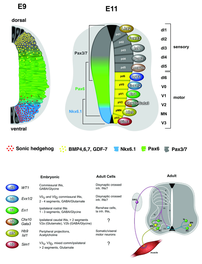
(a) Schematic cross sections through the developing mouse spinal cord showing the patterning and specification of early spinal cord progenitors and their neuronal progeny. At E9, a gradient of Sonic hedgehog (red) ventrally and BMP/GDF7 (yellow) dorsally provide instructive positional signals to dividing progenitors in the ventricular zone. This leads to the restricted activation of patterning factors in discrete dorsventral domains, which are represented by Nkx6.1 (ventral), Pax6 (intermediate) and Pax3 and Pax7 (dorsal). At E11, eleven early classes of postmitotic neuron are present in the embryonic spinal cord. dI1-dI5 neurons that are derived from dorsal progenitors (grey) primarily contribute to sensory spinal pathways, while dI6, MN and V0-V3 neurons from ventral progenitors (yellow) are elements of the locomotor circuitry. Some of the postmitotic transcription factors that mark each of the eleven early generic populations are indicated. (b) Six classes of embryonic neurons are proposed to give rise to the core elements of the spinal locomotor circuitry. The neurotransmitter phenotypes and axonal projections of these embryonic neurons are indicated along with some of the known adult cells types that are derived from each population. (Right) Schematic of the adult spinal cord showing the position and projections of somatic motor neurons (MN, yellow), V1-derived Renshaw cells (RC, green) and Ia inhibitory interneurons (Ia, green) and V0 commissural neurons (blue). The laminae of the spinal cord are indicated by Roman numerals.
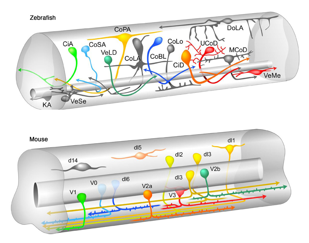
Similar neuronal cell types are present in the embryonic spinal cords of aquatic and terrestrial vertebrates. The putative zebrafish homologues of V0, V1, V2 and V3 locomotor interneurons are indicated by the same colour. V0 and CoSA neurons (light blue), V1 and CiA neurons (light green), V2a and CiD neurons (orange), V2b and VeLD neurons (dark green), V3 and UCoD/VeMe neurons (red). Schematic of embryonic zebrafish spinal cord is courtesy of David McLean and Joe Fetcho (Cornell University).
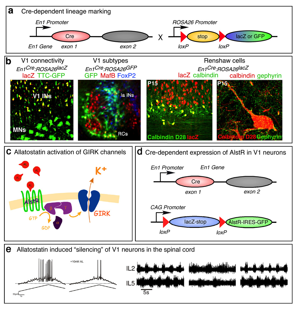
(a) Example of fate mapping and lineage tracing using the Cre-loxP system. Mice carrying an En1Cre allele, in which the first exon of the En1 gene was replaced with sequences encoding Cre, were crossed with ROSA26 reporter mice that express either GFP or lacZ after removal of a loxP-flanked stop sequence. (b) Left panel: Intramuscular injections of the retrograde tracer TTC-EGFP in En1Cre:ROSA26lacZ mice shows that V1 neurons are synaptically connected to motor neurons . Middle left panel: V1 neuron subtypes differentially express MafB (Renshaw cells, red) and FoxP2 (Non-Ia/RC interneurons, blue). Right panels: Identification of Renshaw cells as V1-derived neurons. Renshaw cells are marked by the expression of calbindin (left panel). Renshaw cells uniquely possess large gephyrin clusters on their soma and proximal dendrites (right panel). (c) Schematic showing allatostatin-dependent activation of inwardly rectifying GIRK channels in mammalian neurons. (d) Cre-dependent expression of the allatostatin receptor in V1 neurons in mice. (e) Selective expression of AlstR-GFP in postmitotic V1 neurons at E11 results in a reduction in V1 neuron excitability (left) Allatostatin receptor-mediated silencing of V1 neurons in the locomoting spinal cord results in a slowing of the locomotor rhythm, which phenocopies the defect seen when V1 neurons are ablated (right; see Ref. for details).
References
-
- Dickinson MH, Farley CT, Full RJ, Koehl MAR, Kram R, Lehman S. How animals move: an integrative view. Science. 2000;288:100–106. - PubMed
-
- Grillner S. Locomotion in vertebrates: central mechanisms and reflex interactions. Physiol. Rev. 1975;55:247–304. - PubMed
-
- Orlovsky GN, Deliagina TG, Grillner S. From mollusc to man. New York: Oxford University Press; 1999. Neural Control of Locomotion. This text provides a good introduction and overview of the neural control of locomotion.
-
- Marder E, Bucher D.Central Pattern generators and the control of rhythmic movements Curr. Biol 200111R986–R996.This is an outstanding review that outlines many of the basic principles that operate in rhythmic motor systems. It nicely reviews the invertebrate and vertebrate literature to give an integrative overview of how CPGs are organized and operate. - PubMed
-
- Grillner S.The motor infrastructure: from ion channels to neuronal networks Nat. Rev. Neurosci 20034573–586.This excellent review focuses primarily on the lamprey and outlines the critical findings and principles that underlie the rhythmic motor patterns that control swimming movements. It provides a detailed overview of the swimming CPG and should be read in conjunction with this review. - PubMed
Publication types
MeSH terms
Grants and funding
LinkOut - more resources
Full Text Sources
