. 2009 Jul;11(7):896-902.
doi: 10.1038/ncb1899. Epub 2009 Jun 21.

Centromere assembly requires the direct recognition of CENP-A nucleosomes by CENP-N

Affiliations

Centromere assembly requires the direct recognition of CENP-A nucleosomes by CENP-N

Christopher W Carroll et al. Nat Cell Biol. 2009 Jul.

Abstract

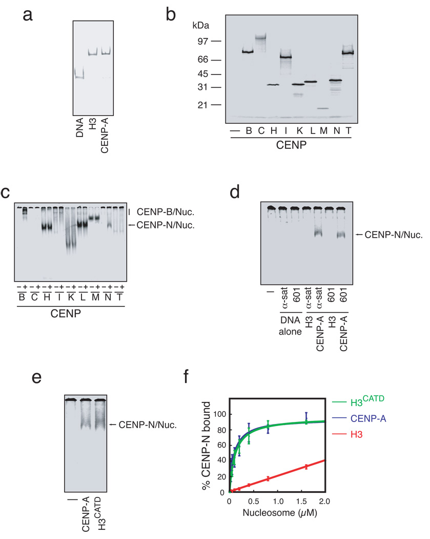
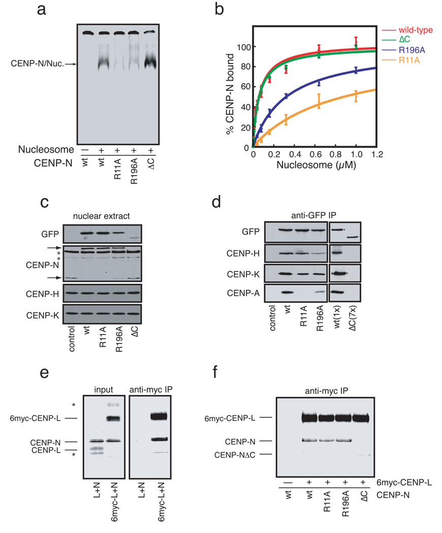
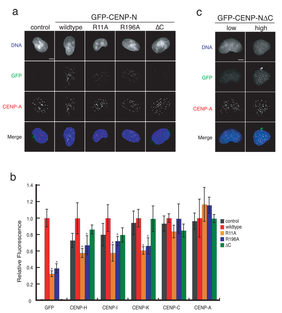
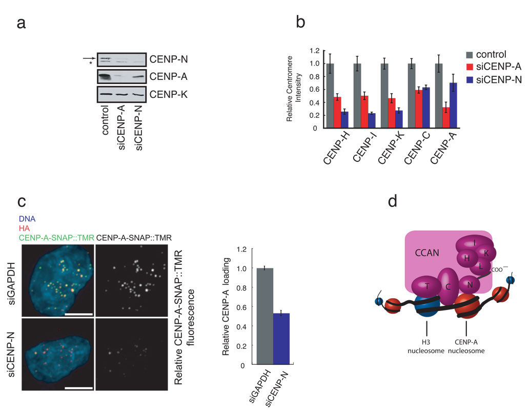
Centromeres are specialized chromosomal domains that direct kinetochore assembly during mitosis. CENP-A (centromere protein A), a histone H3-variant present exclusively in centromeric nucleosomes, is thought to function as an epigenetic mark that specifies centromere identity. Here we identify the essential centromere protein CENP-N as the first protein to selectively bind CENP-A nucleosomes but not H3 nucleosomes. CENP-N bound CENP-A nucleosomes in a DNA sequence-independent manner, but did not bind soluble CENP-A-H4 tetramers. Mutations in CENP-N that reduced its affinity for CENP-A nucleosomes caused defects in CENP-N localization and had dominant effects on the recruitment of CENP-H, CENP-I and CENP-K to centromeres. Depletion of CENP-N using siRNA (short interfering RNA) led to similar centromere assembly defects and resulted in reduced assembly of nascent CENP-A into centromeric chromatin. These data suggest that CENP-N interprets the information encoded within CENP-A nucleosomes and recruits other proteins to centromeric chromatin that are required for centromere function and propagation.


Figures

Figure 1
Figure 1. CENP-N binds CENP-A nucleosomes
a) Reconstitution of conventional and centromeric nucleosomes. Free 186bp human alpha satellite DNA (DNA) or mononucleosomes containing either histone H3 or CENP-A were assembled by salt dialysis and resolved by native gel electrophoresis. b) In vitro expression of centromere proteins. Centromere proteins to be used as substrates in nucleosome binding assays were expressed in coupled transcription and translation reactions containing with 35S-methionine. c) CENP-A nucleosome binding assay. 35S-labeled centromere proteins (~1 nM) were incubated in the presence (+) or absence (−) of reconstituted CENP-A nucleosomes (50 nM) and separated by native gel electrophoresis. Both CENP-B and CENP-N exhibit CENP-A nucleosome dependent changes in migration. d) CENP-N binding to nucleosomes depends upon CENP-A and not DNA sequence. α-satellite DNA (α-sat) or 601 DNA (601), or H3-or CENP-A-containing nucleosomes reconstituted with α-satellite or 601 DNA were incubated with 35S-labeled CENP-N and resolved by native gel electrophoresis. The faster migrating band (arrow) indicates nucleosome bound CENP-N. No nucleosome or DNA control (−). e) Binding of CENP-N to CENP-A nucleosomes occurs through the CATD region of CENP-A. No nucleosome control (−) and CENP-A or H3CATD nucleosomes reconstituted with α-satellite DNA were bound to 35S-CENP-N and assayed as in a. f) CENP-N binds with equal affinity to CENP-A and H3CATD nucleosomes. CENP-N binding was assayed as in (a) with increasing nucleosome concentration and quantified based upon the 35S-CENP-N signal in the gel (N=3, error bars represent SEM).
Figure 2
Figure 2. Identification and characterization CENP-N mutants defective in CENPA-nucleosome binding
a) CENP-N mutants exhibit a range of affinities for CENP-A nucleosomes. CENP-N (wt) or the indicated CENP-N mutants were expressed and an equal concentration (~1nM) of each mutant was assayed for its ability to bind CENP-A nucleosomes (300nM). The control reaction (−) lacked CENP-A nucleosomes. b) Doseresponse experiments for each CENP-N mutant were performed as in (a) with increasing concentrations of CENP-A nucleosome added to each reaction (N=3, error bars represent SEM). c) Expression of CENP-N mutants in HEK293 cells. Nuclear extracts from stable HEK293 cell lines expressing GFP-CENP-N (wt) or the indicated GFP-CENP-N mutant were separated by SDS-PAGE and western blotted with the indicated antibodies. Two nonspecific bands (*) are present in the anti-CENP-N western blot. Upper and lower arrows indicate positions of GFP-CENP-N and endogenous CENP-N, respectively. Quantitation of each bands is presented in Figure S4a. d) Coimmunoprecipitation of CENP-H, K, and A from micrococcal-nuclease solubilized chromatin with CENP-N mutants. Anti-GFP immunoprecipitates from each cell line were probed with the indicated antibodies. 7X more nuclear extract from the CENP-NΔC cell line was required to achieve equal levels of CENP-N in the immunoprecipitations. Quantitation of each bands is presented in Figure S4b. e) CENP-N binds to CENP-L. 6xmyc-CENP-L, CENPL and CENP-N were expressed and labeled with 35S-methionine and the indicated proteins were mixed at equal stoichiometry. 20% of each mixture was resolved as input (left panel). The remaining 80% was immunoprecipitated with anti-myc antibodies (right panel). Two background bands (*) are present in the input reactions. f) CENP-L binding requires the C-terminus of CENP-N. Immunoprecipitations are identical to those in (e), except that equal amounts of wildtype CENP-N (wt) or the indicated CENP-N mutant was used in each reaction.
Figure 3
Figure 3. CENP-N mutants exhibit centromere assembly defects
a) Representative images of control HEK293 cells and stable cell lines expressing wildtype or the indicated CENP-N mutant. Scale bar represents 5 µm. b) CENP-N mutants cause centromere assembly defects. The centromere fluorescence intensity of the indicated centromere protein at individual centromeres in stable cell lines was measured (see Methods). Error bars represent SEM for three independent experiments including >300 centromeres from >20 cells for each cell line. Asterisks indicate p<0.05 significance compared to wildtype. c) The C-terminus of CENP-N is not required for centromere localization. Representative images from transiently transfected HeLa cells show the localization of GFP-CENP-NΔC in low and high expressing cells. Scale bar represents 5 µm.
Figure 4
Figure 4. Depletion of CENP-N affects centromere assembly
a) CENP-N depletion leads to a reduction in CENP-A levels. HeLa cells were treated with the indicated siRNA for 56 hours and the abundance of the indicated centromere protein was determined by western blotting. Arrow (top panel) indicates the position of CENP-N. A background band (*) was present in the anti-CENP-N western blot. CENP-K is included as a loading control. b) CENP-N depletion causes centromere assembly defects. HeLa cells were treated as in (a), except that the centromere fluorescence intensity of the indicated protein was measured. Error bars represent SEM for three independent experiments including >150 centromeres from >10 cells for each experiment. c) Reduced CENP-A assembly after CENP-N RNAi. CENP-A-SNAP cells were synchronized, transfected with siRNA’s against CENP-N or GAPDH and assayed for CENP-A loading by specifically labeling nascent CENP-A-SNAP using TMR-Star as outlined in Figure S5c. Representative images are shown (left panel). HA labels steady state pool of CENP-A-SNAP and is used as centromere marker. Scale bar is 5 µm. Fluorescence intensity of 200 centromeres from 20 cells was quantified in 3 experiments and normalized to the GAPDH siRNA control. Error bars represent SEM. d) A model depicting the multiple roles of CENP-N in CENP-A nucleosome recognition, centromere assembly through the recruitment of CCAN proteins via the CENP-N carboxy-terminal region, and propagation of centromeric chromatin through the CCAN dependent regulation of CENP-A nucleosome assembly.

Comment in

References

    1. Cheeseman IM, Desai A. Molecular architecture of the kinetochore-microtubule interface. Nat Rev Mol Cell Biol. 2008;9:33–46. - PubMed
    1. Choo KH. Domain Organization at the Centromere and Neocentromere. Dev Cell. 2001;1:165–177. - PubMed
    1. Carroll CW, Straight AF. Centromere formation: from epigenetics to self-assembly. Trends Cell Biol. 2006;16:70–78. - PubMed
    1. Okada M, et al. The CENP-H-I complex is required for the efficient incorporation of newly synthesized CENP-A into centromeres. Nat Cell Biol. 2006;8:446–457. - PubMed
    1. Foltz DR, et al. The human CENP-A centromeric nucleosome-associated complex. Nat Cell Biol. 2006;8:458–469. - PubMed

Publication types

LinkOut - more resources