. 2009 Jun 19;4(6):e5978.
doi: 10.1371/journal.pone.0005978.

Divergent cardiopulmonary actions of heme oxygenase enzymatic products in chronic hypoxia

Affiliations

Divergent cardiopulmonary actions of heme oxygenase enzymatic products in chronic hypoxia

Sally H Vitali et al. PLoS One. .

Abstract

Background: Hypoxia and pressure-overload induce heme oxygenase-1 (HO-1) in cardiomyocytes and vascular smooth muscle cells (VSMCs). HO-1(-/-) mice exposed to chronic hypoxia develop pulmonary arterial hypertension (PAH) with exaggerated right ventricular (RV) injury consisting of dilation, fibrosis, and mural thrombi. Our objective was to identify the HO-1 product(s) mediating RV protection from hypoxic injury in HO-1(-/-) mice.

Methodology/principal findings: HO-1(-/-) mice were exposed to seven weeks of hypoxia and treated with inhaled CO or biliverdin injections. CO reduced right ventricular systolic pressure (RVSP) and prevented hypoxic pulmonary arteriolar remodeling in both HO-1(-/-) and control mice. Biliverdin had no significant effect on arteriolar remodeling or RVSP in either genotype. Despite this, biliverdin prevented RV failure in the hypoxic HO-1(-/-) mice (0/14 manifested RV wall fibrosis or thrombus), while CO-treated HO-1(-/-) mice developed RV insults similar to untreated controls. In vitro, CO inhibited hypoxic VSMC proliferation and migration but did not prevent cardiomyocyte death from anoxia-reoxygenation (A-R). In contrast, bilirubin limited A-R-induced cardiomyocyte death but did not inhibit VSMC proliferation and migration.

Conclusions/significance: CO and bilirubin have distinct protective actions in the heart and pulmonary vasculature during chronic hypoxia. Moreover, reducing pulmonary vascular resistance may not prevent RV injury in hypoxia-induced PAH; supporting RV adaptation to hypoxia and preventing RV failure must be a therapeutic goal.


Conflict of interest statement

Competing Interests: The authors have declared that no competing interests exist.

Figures

Figure 1
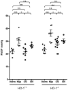
Figure 1. Right ventricular systolic pressure (RVSP) after 7 weeks of hypoxia in HO-1−/− and HO-1+/− control mice.
Mean values +/− SEM are indicated with each dot representing RVSP measurement for one animal. n.s. = not significant, * = p<0.05, *** = p<0.005 as assessed by Mann-Whitney U test.
Figure 2
Figure 2. Pulmonary arteriolar remodeling in HO-1−/− animals exposed to chronic hypoxia for seven weeks and treated with CO or Biliverdin, as indicated.
(A) Quantification of percent wall thickness. Bars represent means +/− SEM. Each dot represents one animal and ten vessels were averaged for each animal. * = p<0.05 as assessed by Mann-Whitney U test. (B) Representative pulmonary arterioles of HO-1−/− mice stained with H&E for each of the different conditions and treatments above.
Figure 3
Figure 3. Right ventricular weight in HO-1−/− and HO-1+/− control mice after 7 weeks of hypoxia.
RV weight is indexed to animal weight. Means +/− SEM are shown for the number of animals listed below each column. n.s. = not significant, *** = p<0.005 as assessed by Mann-Whitney U test. N = normoxia, H = hypoxia.
Figure 4
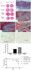
Figure 4. Right ventricular wall injury in HO-1−/− mice exposed to chronic hypoxia for seven weeks.
(A) Biventricular (short-axis) sections of the heart at the papillary muscle level stained with H&E reveal RV dilation and thrombus in the hypoxic HO-1−/− controls and the CO-treated hypoxic HO-1−/− mice but not the hypoxic HO-1−/− mice treated with BV. (B,C) Masson's trichrome stained right ventricular sections from an untreated HO-1−/− mouse at 40× (B) and 100× (C) power. A fibrotic area of RV wall is evident with overlying mural thrombus. (D,E) Carbon monoxide-treated HO-1−/− mouse RVs stained with Masson's trichrome at 100× (D) and 200× (E) power showing an area of RV wall fibrosis and overlying mural thrombus. (F,G) TUNEL-stained RV (F) and LV (G) wall from an area of full-thickness wall fibrosis in an untreated HO-1−/− mouse at 200× magnification. The LV has TUNEL stained luminal debris but is otherwise negative for TUNEL-staining. (H) Quantification of the percentage of HO-1−/− animals in each group developing an area of Masson's trichrome-positive fibrosis spanning the full thickness of the RV wall. (I) Survival curve of HO-1 hemizygous and null mice in 8.5–9% oxygen for seven weeks in response to treatment with CO or Biliverdin.
Figure 5
Figure 5. PASMC proliferation and migration in response to hypoxia.
(A) Effect of bilirubin and CO on PASMC proliferation in response to hypoxia by BrdU incorporation. Average fold change of relative light units over unstimulated (no PDGF) controls from four experiments are shown. (B) Effect of bilirubin and CO on PASMC migration in hypoxia. Average fold change of relative light units over unstimulated (no PDGF) controls from three experiments is shown. * = p,0.05, *** = p<0.005 as assessed by Mann-Whitney U test.
Figure 6
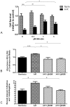
Figure 6. Bilirubin protects cardiomyocytes from anoxia-reoxygenation (A-R) injury and has an anti-apoptotic effect.
(A) Cell survival ratio relative to untreated normoxic controls as assessed by calcein/ethidium staining. Mean +/− SEM for 6 wells per condition is shown and graph is representative of three independent experiments. (B) Lactate dehydrogenase (LDH) measured by absorbance at 490 in the media of cardiomyocytes exposed to A-R. Means +/− SEM are shown. (C) Flow cytometric analysis of Annexin V-positive, 7AAD-negative cardiomyocytes after A-R. Bars represent the percentage of 10,000 counted cells that fell into this category. Data are an average of three different experiments. * = p<0.05, ** = p<0.01, *** = p<0.005 as assessed by Mann-Whitney U test.

Similar articles

Cited by

References

    1. Duckers HJ, Boehm M, True AL, Yet SF, San H, et al. Heme oxygenase-1 protects against vascular constriction and proliferation. Nat Med. 2001;7:693–698. - PubMed
    1. Zhang M, Zhang BH, Chen L, An W. Overexpression of heme oxygenase-1 protects smooth muscle cells against oxidative injury and inhibits cell proliferation. Cell Res. 2002;12:123–132. - PubMed
    1. Foo RS, Siow RC, Brown MJ, Bennett MR. Heme oxygenase-1 gene transfer inhibits angiotensin II-mediated rat cardiac myocyte apoptosis but not hypertrophy. J Cell Physiol. 2006;209:1–7. - PubMed
    1. Hu CM, Chen YH, Chiang MT, Chau LY. Heme oxygenase-1 inhibits angiotensin II-induced cardiac hypertrophy in vitro and in vivo. Circulation. 2004;110:309–316. - PubMed
    1. Tongers J, Fiedler B, Konig D, Kempf T, Klein G, et al. Heme oxygenase-1 inhibition of MAP kinases, calcineurin/NFAT signaling, and hypertrophy in cardiac myocytes. Cardiovasc Res. 2004;63:545–552. - PubMed

Publication types