

Genotyping and phylogenetic analysis of Yersinia pestis by MLVA: insights into the worldwide expansion of Central Asia plague foci
- PMID: 19543392
- PMCID: PMC2694983
- DOI: 10.1371/journal.pone.0006000
Genotyping and phylogenetic analysis of Yersinia pestis by MLVA: insights into the worldwide expansion of Central Asia plague foci
Abstract
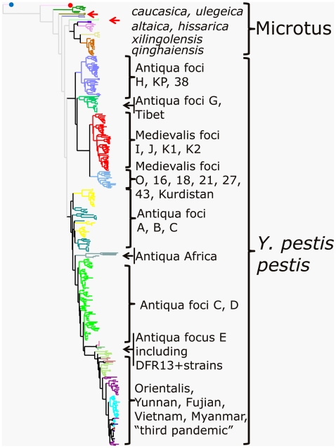
Background: The species Yersinia pestis is commonly divided into three classical biovars, Antiqua, Medievalis, and Orientalis, belonging to subspecies pestis pathogenic for human and the (atypical) non-human pathogenic biovar Microtus (alias Pestoides) including several non-pestis subspecies. Recent progress in molecular typing methods enables large-scale investigations in the population structure of this species. It is now possible to test hypotheses about its evolution which were proposed decades ago. For instance the three classical biovars of different geographical distributions were suggested to originate from Central Asia. Most investigations so far have focused on the typical pestis subspecies representatives found outside of China, whereas the understanding of the emergence of this human pathogen requires the investigation of strains belonging to subspecies pestis from China and to the Microtus biovar.
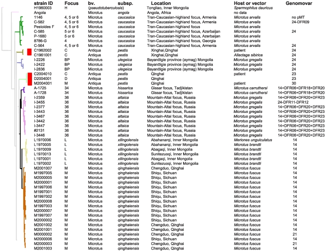
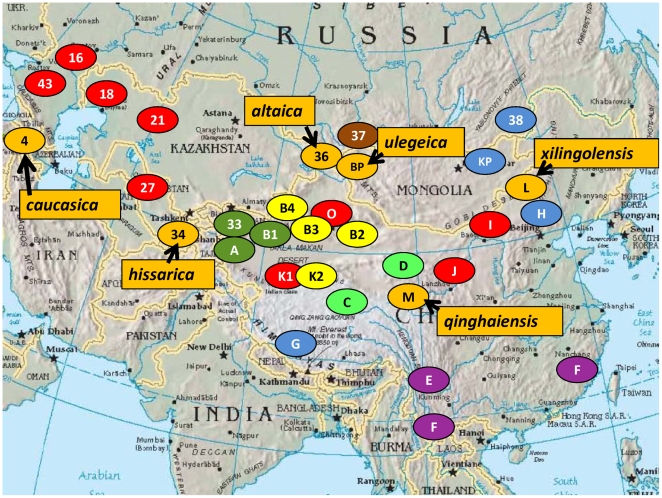
Methodology/principal findings: Multi-locus VNTR analysis (MLVA) with 25 loci was performed on a collection of Y. pestis isolates originating from the majority of the known foci worldwide and including typical rhamnose-negative subspecies pestis as well as rhamnose-positive subspecies pestis and biovar Microtus. More than 500 isolates from China, the Former Soviet Union (FSU), Mongolia and a number of other foci around the world were characterized and resolved into 350 different genotypes. The data revealed very close relationships existing between some isolates from widely separated foci as well as very high diversity which can conversely be observed between nearby foci.
Conclusions/significance: The results obtained are in full agreement with the view that the Y. pestis subsp. pestis pathogenic for humans emerged in the Central Asia region between China, Kazakhstan, Russia and Mongolia, only three clones of which spread out of Central Asia. The relationships among the strains in China, Central Asia and the rest of the world based on the MLVA25 assay provide an unprecedented view on the expansion and microevolution of Y. pestis.
Conflict of interest statement
Figures
Similar articles
-
Yersinia pestis lineages in Mongolia.PLoS One. 2012;7(2):e30624. doi: 10.1371/journal.pone.0030624. Epub 2012 Feb 17. PLoS One. 2012. PMID: 22363455 Free PMC article.
-
[Standard algorithm of molecular typing of Yersinia pestis strains].Zh Mikrobiol Epidemiol Immunobiol. 2012 May-Jun;(3):25-35. Zh Mikrobiol Epidemiol Immunobiol. 2012. PMID: 22830271 Russian.
-
Genotyping of Indian Yersinia pestis strains by MLVA and repetitive DNA sequence based PCRs.Antonie Van Leeuwenhoek. 2009 Oct;96(3):303-12. doi: 10.1007/s10482-009-9347-2. Epub 2009 May 16. Antonie Van Leeuwenhoek. 2009. PMID: 19449123
-
Review of genotyping methods for Yersinia pestis in Madagascar.PLoS Negl Trop Dis. 2024 Jun 27;18(6):e0012252. doi: 10.1371/journal.pntd.0012252. eCollection 2024 Jun. PLoS Negl Trop Dis. 2024. PMID: 38935608 Free PMC article. Review.
-
Plague in the genomic area.Clin Microbiol Infect. 2012 Mar;18(3):224-30. doi: 10.1111/j.1469-0691.2012.03774.x. Clin Microbiol Infect. 2012. PMID: 22369155 Review.
Cited by
-
Yersinia pestis lineages in Mongolia.PLoS One. 2012;7(2):e30624. doi: 10.1371/journal.pone.0030624. Epub 2012 Feb 17. PLoS One. 2012. PMID: 22363455 Free PMC article.
-
Rapid identification and typing of Yersinia pestis and other Yersinia species by matrix-assisted laser desorption/ionization time-of-flight (MALDI-TOF) mass spectrometry.BMC Microbiol. 2010 Nov 12;10:285. doi: 10.1186/1471-2180-10-285. BMC Microbiol. 2010. PMID: 21073689 Free PMC article.
-
Genetic diversity and spatial-temporal distribution of Yersinia pestis in Qinghai Plateau, China.PLoS Negl Trop Dis. 2018 Jun 25;12(6):e0006579. doi: 10.1371/journal.pntd.0006579. eCollection 2018 Jun. PLoS Negl Trop Dis. 2018. PMID: 29939993 Free PMC article.
-
Plague in Iran: its history and current status.Epidemiol Health. 2016 Jul 24;38:e2016033. doi: 10.4178/epih.e2016033. eCollection 2016. Epidemiol Health. 2016. PMID: 27457063 Free PMC article. Review.
-
Yersinia pseudotuberculosis ST42 (O:1) Strain Misidentified as Yersinia pestis by Mass Spectrometry Analysis.Genome Announc. 2014 Jun 12;2(3):e00435-14. doi: 10.1128/genomeA.00435-14. Genome Announc. 2014. PMID: 24926044 Free PMC article.
References
-
- Baltazard M. Epidemiology of plague. WHO Chronicle. 1960;14:419–426.
-
- Mollaret HH, Karimi Y, Eftekhari M, Baltazard M. [Burrowing Plague.]. Bull Soc Pathol Exot Filiales. 1963;56:1186–1193. - PubMed
-
- Mollaret HH. [Experimental Preservation Of Plague In Soil.]. Bull Soc Pathol Exot Filiales. 1963;56:1168–1182. - PubMed
Publication types
MeSH terms
Substances
LinkOut - more resources
Full Text Sources
Other Literature Sources
Medical
