

A case of primary adrenal diffuse large B-cell lymphoma achieving complete remission with rituximab-CHOP chemotherapy
- PMID: 19543522
- PMCID: PMC2698205
- DOI: 10.3346/jkms.2009.24.3.525
A case of primary adrenal diffuse large B-cell lymphoma achieving complete remission with rituximab-CHOP chemotherapy
Abstract
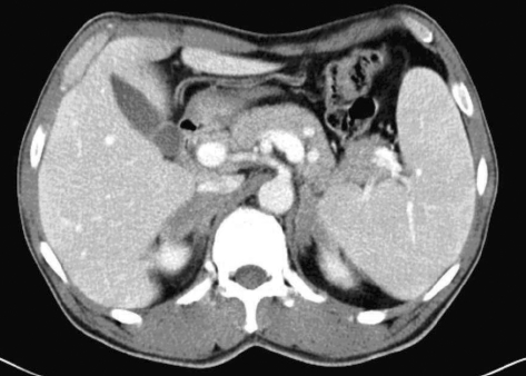
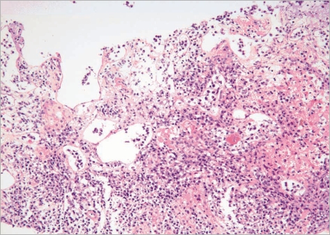
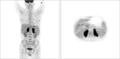
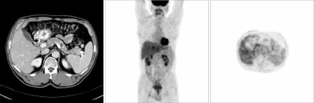
Primary adrenal lymphoma is a very rare extranodal lymphoma; its clinical features consist of a high incidence of bilateral adrenal involvement and diffuse large B-cell lymphoma. We report a patient with primary bilateral adrenal diffuse large B-cell lymphoma who achieved complete remission with R-CHOP (rituximab-cyclophosphamide, doxorubicin, vincristine, and prednisone) chemotherapy. A 52-yr-old man presented with fever and progressive fatigue for 3 months. Computed tomography (CT) scans of the abdomen and pelvis demonstrated large bilateral adrenal masses, and a needle biopsy of the left adrenal mass revealed diffuse large B-cell lymphoma. After 6 cycles of R-CHOP chemotherapy, CT scans showed no residual disease. To our knowledge, this is the second report to date of a patient with primary bilateral adrenal diffuse large B-cell lymphoma who achieved complete remission using R-CHOP chemotherapy.
Keywords: Complete Remission; Lymphoma, Large B-Cell, Diffuse; Primary Adrenal Lymphoma; R-CHOP.
Figures
Similar articles
-
Primary bilateral adrenal lymphoma revealed by hemophagocytic syndrome.Ann Endocrinol (Paris). 2011 Jun;72(3):247-50. doi: 10.1016/j.ando.2011.04.006. Ann Endocrinol (Paris). 2011. PMID: 21640976
-
Non-Hodgkin's lymphoma involving a femur bone and bilateral adrenal glands alone with adrenal insufficiency.BMJ Case Rep. 2017 Jan 31;2017:bcr2016218222. doi: 10.1136/bcr-2016-218222. BMJ Case Rep. 2017. PMID: 28143809 Free PMC article.
-
CHOP chemotherapy plus rituximab compared with CHOP alone in elderly patients with diffuse large-B-cell lymphoma.N Engl J Med. 2002 Jan 24;346(4):235-42. doi: 10.1056/NEJMoa011795. N Engl J Med. 2002. PMID: 11807147 Clinical Trial.
-
Diffuse large B-cell lymphoma solely involving bilateral adrenal glands and stomach: report of an extremely rare case with review of the literature.Int J Clin Exp Pathol. 2014 Oct 15;7(11):8190-7. eCollection 2014. Int J Clin Exp Pathol. 2014. PMID: 25550871 Free PMC article. Review.
-
Primary adrenal diffuse large B cell lymphoma arising within an adrenal myelolipoma: A case report and review of the literature.Curr Probl Cancer. 2021 Dec;45(6):100724. doi: 10.1016/j.currproblcancer.2021.100724. Epub 2021 Mar 6. Curr Probl Cancer. 2021. PMID: 33744015 Review.
Cited by
-
Primary Non Hodgkin's Lymphoma of Left Adrenal Gland - A Rare Presentation.J Clin Diagn Res. 2015 Apr;9(4):XD01-XD03. doi: 10.7860/JCDR/2015/8079.5745. Epub 2015 Apr 1. J Clin Diagn Res. 2015. PMID: 26023630 Free PMC article.
-
Bilateral primary adrenal diffuse large B cell lymphoma: A rare presentation.Radiol Case Rep. 2023 Oct 28;19(1):158-163. doi: 10.1016/j.radcr.2023.09.064. eCollection 2024 Jan. Radiol Case Rep. 2023. PMID: 37954675 Free PMC article.
-
Extranodal diffuse large B-cell lymphomas: A retrospective case series and review of the literature.Hematol Rep. 2018 Apr 3;10(1):7070. doi: 10.4081/hr.2018.7070. eCollection 2018 Mar 2. Hematol Rep. 2018. PMID: 29721249 Free PMC article.
-
Small round tumour cells (CD38, CD 79a positive) in the adrenal gland.Urol Case Rep. 2017 Oct 7;16:22-24. doi: 10.1016/j.eucr.2017.09.009. eCollection 2018 Jan. Urol Case Rep. 2017. PMID: 29021967 Free PMC article. No abstract available.
-
Bilateral primary adrenal lymphoma presenting with adrenal insufficiency.Case Rep Endocrinol. 2012;2012:638298. doi: 10.1155/2012/638298. Epub 2012 Sep 4. Case Rep Endocrinol. 2012. PMID: 22988530 Free PMC article.
References
-
- Rosenberg SA, Diamond HD, Jaslowitz B, Craver LF. Lymphosarcoma: a review of 1269 cases. Medicine (Baltimore) 1961;40:31–84. - PubMed
-
- Kumar R, Xiu Y, Mavi A, El-Haddad G, Zhuang H, Alavi A. FDG-PET imaging in primary bilateral adrenal lymphoma: a case report and review of the literature. Clin Nucl Med. 2005;30:222–230. - PubMed
-
- Grigg AP, Connors JM. Primary adrenal lymphoma. Clin Lymphoma. 2003;4:154–160. - PubMed
-
- Utsunomiya M, Takatera H, Itoh H, Tsujimura T, Itatani H. Bilateral primary non-Hodgkin's lymphoma of the adrenal glands with adrenal insufficiency: a case report. Hinyokika Kiyo. 1992;38:311–314. - PubMed
-
- Kato H, Itami J, Shiina T, Uno T, Arimizu N, Fujimoto H, Mikata A, Tamaru J, Araki A. MR imaging of primary adrenal lymphoma. Clin Imaging. 1996;20:126–128. - PubMed
Publication types
MeSH terms
Substances
Supplementary concepts
LinkOut - more resources
Full Text Sources
Medical
Research Materials
