. 2009 Jun 19;4(6):e5959.
doi: 10.1371/journal.pone.0005959.

Immune regulatory neural stem/precursor cells protect from central nervous system autoimmunity by restraining dendritic cell function

Affiliations

Immune regulatory neural stem/precursor cells protect from central nervous system autoimmunity by restraining dendritic cell function

Stefano Pluchino et al. PLoS One. .

Abstract

Background: The systemic injection of neural stem/precursor cells (NPCs) provides remarkable amelioration of the clinico-pathological features of experimental autoimmune encephalomyelitis (EAE). This is dependent on the capacity of transplanted NPCs to engage concurrent mechanisms of action within specific microenvironments in vivo. Among a wide range of therapeutic actions alternative to cell replacement, neuroprotective and immune modulatory capacities of transplanted NPCs have been described. However, lacking is a detailed understanding of the mechanisms by which NPCs exert their therapeutic plasticity. This study was designed to identify the first candidate that exemplifies and sustains the immune modulatory capacity of transplanted NPCs.

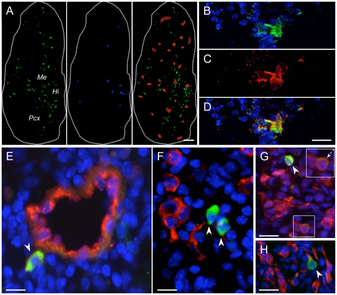
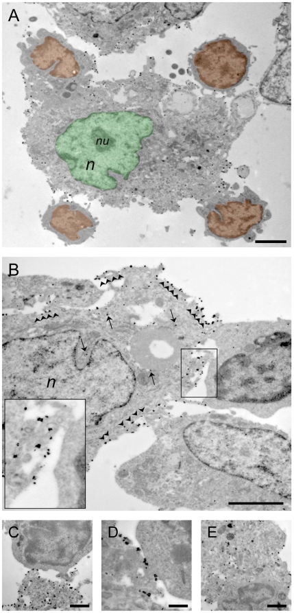
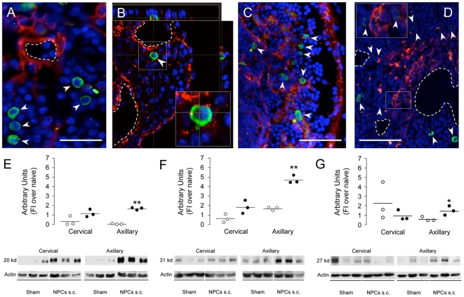
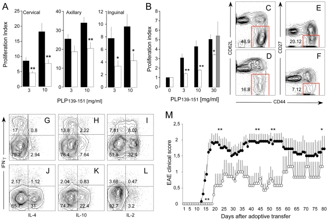
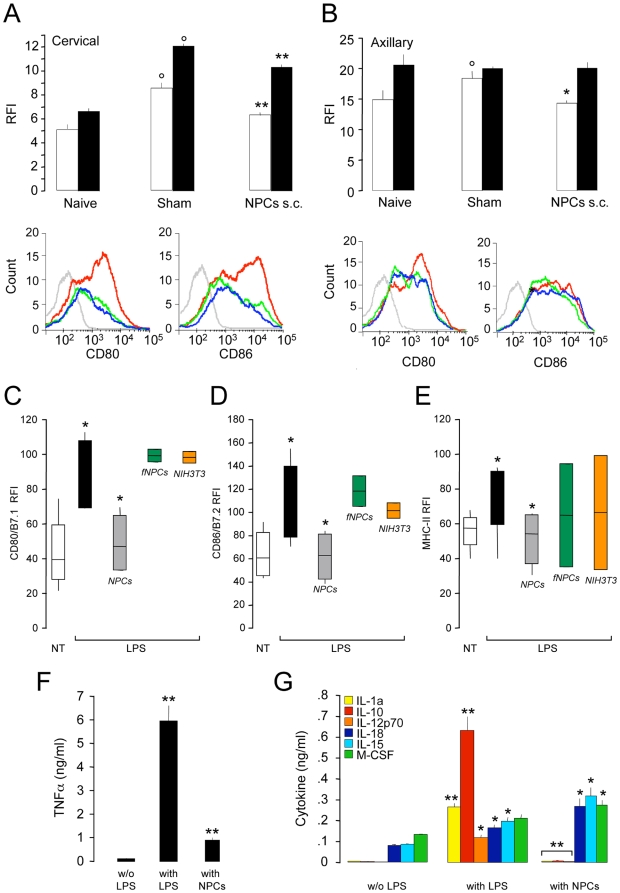
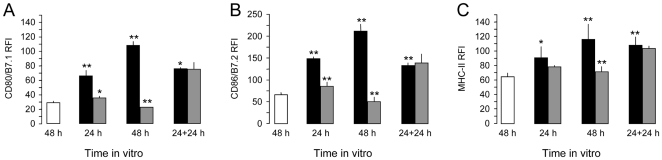
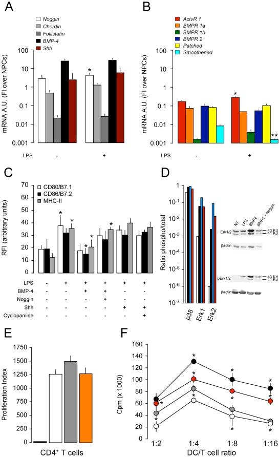
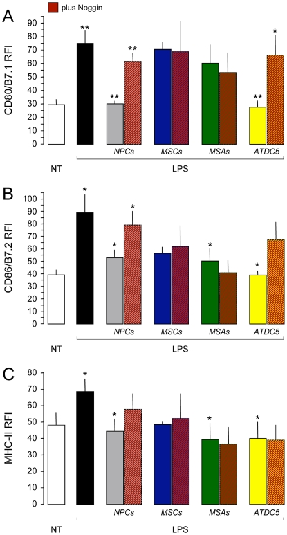
Methodology/principal findings: To achieve the exclusive targeting of the peripheral immune system, SJL mice with PLP-induced EAE were injected subcutaneously with NPCs and the treatment commenced prior to disease onset. NPC-injected EAE mice showed significant clinical improvement, as compared to controls. Exogenous NPCs lacking the expression of major neural antigens were reliably (and for long-term) found at the level of draining lymph nodes, while establishing sophisticated anatomical interactions with lymph node cells. Importantly, injected NPCs were never found in organs other than lymph nodes, including the brain and the spinal cord. Draining lymph nodes from transplanted mice showed focal up-regulation of major developmental stem cell regulators, such as BMP-4, Noggin and Sonic hedgehog. In lymph nodes, injected NPCs hampered the activation of myeloid dendritic cells (DCs) and steadily restrained the expansion of antigen-specific encephalitogenic T cells. Both ex vivo and in vitro experiments identified a novel highly NPC-specific-BMP-4-dependent-mechanism hindering the DC maturation.

Conclusion/significance: The study described herein, identifies the first member of the TGF beta/BMP family of stem cell regulators as a novel tolerogenic factor released by NPCs. Full exploitation of this pathway as an efficient tool for vaccination therapy in autoimmune inflammatory conditions is underway.


Conflict of interest statement

Competing Interests: The authors have declared that no competing interests exist.

Figures

Figure 1
Figure 1. S.c.-injected NPCs accumulate and persist in secondary lymphoid organs from R-EAE mice while lacking the expression of major antigens of the neural lineage.
A, Sagittal reconstruction of a representative axillary lymph node from a R-EAE mouse injected s.c. with syngeneic NPCs. GFP+ NPCs (green) are detected throughout the entire lymph node tissue although they predominantly accumulate as focal clusters in the hilum (Hi), medulla (Me), and paracortex (Pcx). Some of the NPCs are in close contact with CD11b+ macrophages (red). Twenty % of NPCs express the early neuronal differentiation marker doublecortin (DCX) (blue). Scale bar: 300 µm. B–D, Occasionally, GFP+ NPCs (B) expressing the neural cell marker nestin (C) are detected in perivascular lymph node areas. The panel in D is a merged image of the pictures in B and C; E, Representative image of a cervical lymph node where NPCs (green, arrowhead) are found to establish close anatomical interaction(s) with von Willebrand factor antigen-expressing endothelial cells (red); F, Confocal microscopy image of a para-aortic lymph node where NPCs (green, arrowheads) are found in close cell-to-cell contact with CD11c+ DCs (red). G and H, Axillary lymph node sections showing persistence of NPCs (green, arrowheads) in areas colonized by F4/80+ phagocytes (G, red) and MHC class-II+ immune cells (H, red). Occasionally, F4/80+ cells being immunoreactive also for GFP (G, dashed arrow in the boxed area) are identified. NPCs are in green in A–H. Nuclei in B, D and E–H are counterstained with DAPI. Scale bars in B–H: 40 µm. Data refer to R-EAE mice injected s.c. with NPCs at 3 and 10 dpi and sacrificed 72 days after cell injection.
Figure 2
Figure 2. Electron microscopic in vivo appearance of s.c.-injected NPCs in draining lymph nodes.
A, Transmission electron microscopy (TEM) images a large-size immunogold-labelled GFP+ NPC within a cervical lymph node of a R-EAE mouse at 72 days after cell injection. These GFP+ cells display morphological and ultrastructural features similar to individual NPCs from neurospheres in vitro (see also Figure S3). The NPC (pseudocolor green) takes contact with four individual lymph node cells (pseudocolor orange). The NPC shows an irregular and invaginated nucleus (n) with a single nucleolus (nu), and its cytoplasm contains abundant organelles; B, Individual GFP+ NPCs establishing anatomical contacts with two GFP lymph node cells–one of which (upper right) possesses the morphological characteristics of lymph blasts–through polarized microvilli (boxed area). Note the electrodense grain precipitates within the cytoplasm (arrows) and on the membrane (arrowheads and boxed area); C–E, NPCs in lymph nodes establish cell-to-cell contacts with lymph blasts through cytoplasmic expansions (C) or elongated intercellular junctions (D). Transplanted NPCs are occasionally found in deeper contact and enclosing resident lymph cells (E). Images refer to representative draining lymph nodes from R-EAE mice injected s.c. with NPCs at 3 and 10 dpi. Scale bars in A and B: 2 µm, in C and E: 1 µm, and in D: 500 nm.
Figure 3
Figure 3. Establishment of atypical perivascular lymph node niches.
A–D, Representative confocal microscope images of the persistence of s.c.-injected NPCs (arrowheads, green) within lymph node perivascular areas in R-EAE mice at 75 dpi. Focal expression of tenascin C (A, red), BMP-4 (B, red), BMP-7 (C, red) and Shh (D, red) are shown. Dashed lines represent blood vessels. Nuclei in A–D are counterstained with DAPI. Scale bars: A, 50 µm; C and D, 100 µm. E–G, Western blot analysis of BMP-4 (E), Noggin (F) and Shh (G) expression in cervical and axillary lymph nodes from sham-treated (white dots) and NPC-injected (black dots) R-EAE mice at 2 weeks after treatment. Results from individual mice (n = 3/group) are represented as dots and expressed as protein Arbitrary Units (AU, fold induction over average naive) (±SEM). Data refer to R-EAE mice injected s.c. with NPCs at 3 and 10 dpi and sacrificed at two weeks after cell injection. *p≤0.05; **p≤0.005, vs. sham-treated controls.
Figure 4
Figure 4. NPCs inhibit generation of effector T cells.
A, Ex vivo proliferation of lymph node cells (LNCs) from sham- (black bars) and NPC-treated (white bars) R-EAE mice. Mice (n = 15 mice/group) were treated with either the carrier solution or NPC s.c. at 3 dpi and sacrificed at 10 dpi; B, Response to PLP139–151 of LNCs co-cultured with NPCs (LNCs/NPCs) either in the same well (white bars) or in a trans-well system (grey bars). Note the significant suppression of proliferation, compared to control LNCs (black bars). No significant interference with T cell response is observed in trans-well experiments. Data in A and B are mean Proliferation Index (over basal proliferation) (±SEM) from a total of n≥3 independent experiments. *p≤0.05; **p≤0.005, vs. control LNCs; C–F, LNCs/NPCs (D and F) show reduced percentages of CD44high/CD62L and CD44high/CD27 CD4+ effector memory T cells, compared to control LNCs (C and E); G–L, LNCs/NPCs show reduced percentages of IFN-γ- (y axis) and IL-2-producing (x axis in L) CD4+ T cells, while higher percentages IL-4- (x axis in J), IL-10-producing (x axis in K) CD4+ T cells, compared to control LNCs (G, H and I, respectively); M, Adoptive transfer to naive SJL recipients of PLP139–151-specific LNCs having being either co-cultured (LNCs/NPCs, white circles, n = 8 mice) or not (LNCs, black circles, n = 13 mice) with NPCs. Note the significant impairment of EAE development (e.g., delay of the disease onset and reduction of the clinical score), when disease is adoptively transferred with LNCs/NPCs. Data are mean clinical score (±SEM). *p≤0.05; **p≤0.005.
Figure 5
Figure 5. NPCs restrain DC maturation and cytokine production.
A and B, Ex vivo FACS analysis of DCs from the lymph nodes of R-EAE mice treated with either the carrier solution or NPC s.c. at 3 dpi and sacrificed at 7 dpi. The injection of NPCs hinders the up-regulation of CD80 (white bars) and CD86 (black bars) on maturating DCs from cervical and axillary lymph nodes. Histograms in A and B are representative of the FACS analysis for CD80 and CD86 on CD11c+ lymph node DCs from cervical and axillary lymph nodes of naive (blue lines) and R-EAE mice either sham- (red lines) or NPC-treated (green lines). The grey lines are the fluorescence minus one (FMO) control staining. Data are mean relative fluorescence intensity (RFI) over FMO (±SEM) from a total of n = 2 independent experiments (n = 3 mice/group/experiment). °p≤0.05, vs. control DCs. *p≤0.05 and **p≤0.0005, vs. sham-treated controls; C–E, Live NPCs (grey bars), but not PFA-fixed NPCs (fNPCs, green bars) and live NIH3T3 mouse embryonic fibroblasts (orange bars), inhibit the up-regulation of CD80/B7.1 (C), CD86/B7.2 (D), and MHC-II (E) on DCs maturating in vitro with LPS. Cells were co-cultured with DCs (1∶1 NPC/DC ratio) in a trans-well system. White bars are not treated (NT) DCs, while black bars are control (non co-cultured) DCs. Data were obtained from a total of n≥3 independent experiments. Data in A–E are mean RFI over unstained (±SEM); F and G, Co-cultured NPCs significantly inhibit the release of TNFα (F), while completely suppressing the release of IL-1α, IL-12p70 and IL-10 (G), from DCs maturating in vitro with LPS. NPCs also favour the release of IL-15, IL-18 and M-CSF (G). Data are mean cytokine ng/ml (±SEM) from a total of n≥3 independent experiments. C–G, *p≤0.05; **p≤0.005, vs. control DCs.
Figure 6
Figure 6. The NPC-mediated restrainance of DC maturation is reversible upon the establishment of appropriate maturation condition in vitro.
The impairment of the expression of CD80/B7.1 (A), CD86/B7.2 (B) and MHC-II (C) by DCs maturating in vitro with LPS and being co-cultured with NPCs (DCs/NPCs, grey bars) is restored if NPCs are removed from the upper chamber after 24 hours and new medium with LPS is added for further 24 hours. White bars are not treated (NT) DCs, while black bars are control (non co-cultured) DCs. Data are mean RFI (±SEM) over unstained DCs from a total of n = 2 independent experiments. *p≤0.05; **p≤0.005, when either control DCs are compared to NT or DC/NPCs are compared to control DCs.
Figure 7
Figure 7. BMP-4-dependent hindrance of DC maturation.
A and B, qRT-PCR analysis of BMPs/BMP antagonists, BMP receptors and Shh/Shh receptors in DCs. Note the significant up-regulation of mRNA levels for Noggin (A) and ActvR 1 (B) and down-regulation of the Shh receptor Smoothened (B) upon LPS activation of DCs. The mRNA levels of the other two BMP antagonists Chordin and Follistatin (A) remain unchanged after LPS. Data are mean mRNA arbitrary units (FI over NPCs) (±SEM) from a total of n≥3 independent experiments. *p≤0.05; **p≤0.005, vs. control DCs; C, Soluble recombinant BMP-4, but not recombinant Shh, inhibits the up-regulation of CD80/B7.1, CD86/B7.2, and MHC-II on DCs maturating with LPS. Almost complete reversion of the hindrance of DC maturation is obtained when soluble recombinant Noggin is added to the culture system. The addition of the Shh antagonist Cyclopamine did not exert any effect. Data are mean RFI (±SEM) from a total of n≥3 independent experiments. *p≤0.05, vs. control DCs; D, Immunoblot analysis of the TLR ligand-dependent activation of the MAPK p38 and Erk1/2 in DCs maturating with LPS in vitro. Increased phosphorilation of Erk1/2–and to a lower extent of p38MAPK–is obtained when BMP-4 (blue bars) is added to LPS-treated DCs (black bars). The effect is reverted by the addition of the BMP-4 antagonist Noggin (red bars). White bars are not treated (NT) DCs. Values are ratios of phosphorilated over total protein. Relative levels of protein expression were normalized to β actin; E, Proliferation to CD3/CD28 of lymph node CD4+ T cells from naive SJL mice in presence of BMP-4 (grey bar) or Shh (orange bar). Neither of the two morphogens interferes with CD4+ T cell response. The white bar is the proliferation to CD3/CD28 only, while the black bar is the basal proliferation (no CD3/CD28). Data are mean proliferation index (over basal proliferation) (±SEM) from a total of n = 3 independent experiments; F, Proliferation of CD4+ T cells from PLP139–151-immunized mice at first re-call with the antigen in vitro that are co-cultured with naïve DCs which have been pulsed with PLP139–151 during the last 18 hours of LPS-induced maturation (positive control, black circles). Significant reduction of antigen-specific proliferation of CD4+ T cells is observed when DCs are co-cultured with NPCs (white circles), while recovery of the antigen presenting efficiency is observed when the BMP-4 antagonist Noggin is added to the DC/NPC co-culture (red circles). Grey circles are T cells co-cultured with non-activated DCs. Data are mean counts per minute (cpm) (±SEM) from a total of n≥3 independent experiments. *p≤0.05, vs. positive control.
Figure 8
Figure 8. The BMP-4-dependent hindrance of DC maturation is specific for NPCs.
A–C, NPCs (grey bars), but not bone marrow-derived MSCs (blue bars), or vessel associated MSAs (green bars), inhibit the up-regulation of CD80/B7.1 (A), CD86/B7.2 (B), and MHC-II (C) on DCs maturating in vitro with LPS. Almost complete recovery of the co-stimulatory molecule expression is obtained on DC/NPC only, when soluble recombinant Noggin is added to the co-culture system. Cells were co-cultured with DCs (1∶1 cell/DC ratio) in a trans-well system. White bars are not treated (NT) DCs, while black bars are control (non co-cultured) DCs. BMP-secreting ATDC5 condrogenic cells (orange bars) are used as positive control. Data were obtained from a total of n = 2 independent experiments. Data in are mean RFI over unstained (±SEM). *p≤0.05; **p≤0.005, when either control DCs are compared to NT, co-cultures are compared to control DCs or co-cultures plus Noggin are compared to co-cultures.

References

    1. Franklin RJ, Ffrench-Constant C. Remyelination in the CNS: from biology to therapy. Nat Rev Neurosci. 2008;9:839–855. - PubMed
    1. Ben-Hur T, Goldman SA. Prospects of cell therapy for disorders of myelin. Ann N Y Acad Sci. 2008;1142:218–249. - PubMed
    1. Pluchino S, Quattrini A, Brambilla E, Gritti A, Salani G, et al. Injection of adult neurospheres induces recovery in a chronic model of multiple sclerosis. Nature. 2003;422:688–694. - PubMed
    1. Einstein O, Karussis D, Grigoriadis N, Mizrachi-Kol R, Reinhartz E, et al. Intraventricular transplantation of neural precursor cell spheres attenuates acute experimental allergic encephalomyelitis. Mol Cell Neurosci. 2003;24:1074–1082. - PubMed
    1. Aharonowiz M, Einstein O, Fainstein N, Lassmann H, Reubinoff B, et al. Neuroprotective effect of transplanted human embryonic stem cell-derived neural precursors in an animal model of multiple sclerosis. PLoS ONE. 2008;3:e3145. - PMC - PubMed

Publication types

Substances