. 2009;56(2):355-60.
Epub 2009 Jun 18.

Chemerin and the recruitment of NK cells to diseased skin

Affiliations

Chemerin and the recruitment of NK cells to diseased skin

Joanna Skrzeczyńska-Moncznik et al. Acta Biochim Pol. 2009.

Abstract

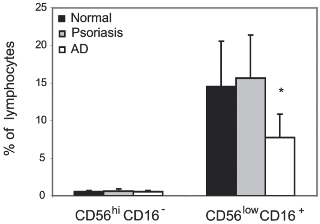
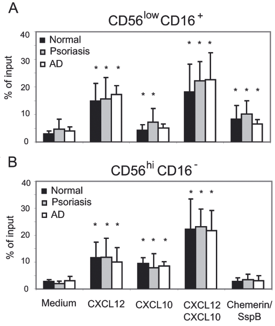
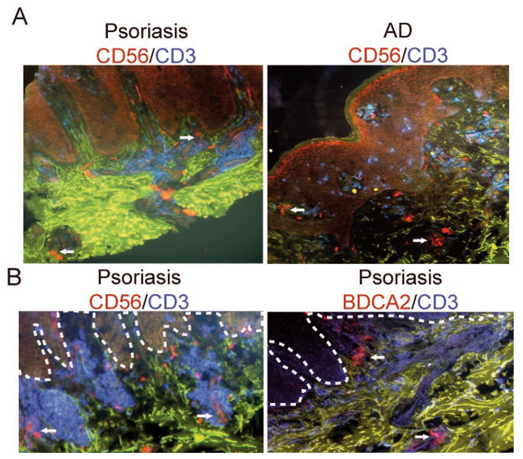
Natural killer (NK) cells play a major role in the initial control of many viral pathogens and in the rejection of tumors. Consistent with their roles as immune sentinels, NK cells are found in inflamed skin, including lichen planus, psoriasis and atopic dermatitis (AD) lesions. In oral lichen planus lesions, the recruitment as well as intradermal colocalization of NK cells and pDC (plasmacytoid dendritic cells) appear to be mediated by chemerin, a recently identified protein ligand for chemokine-like receptor 1 (CMKLR1), a chemoattractant receptor expressed by both cell types. Dendritic cells can regulate NK cell activity, and NK cells can regulate DC-mediated responses. Since chemerin was recently implicated in recruitment of pDC to psoriatic skin, in this work we determined whether chemerin facilitates interactions between NK and pDC in psoriatic plaques through controlling influx of NK cells to diseased skin. We demonstrate that circulating NK cells from normal donors as well as psoriasis and AD patients respond similarly in functional migration assays to chemerin. However, differences in the distribution of NK cells and pDC in skin lesions suggest that recruitment of both NK cells and pDC is unlikely to be controlled solely by chemerin.


Figures

Figure 1.
Figure 1.. Circulating NK cells are present at a lower frequency in AD patients compared with psoriasis patients and normal donors.
PBMC were isolated from the blood of indicated donors and stained for NK cells using anti- CD56 and CD16 mAbs. The cells were then analyzed by flow cytometry. Data are shown as percentage of NK cell subsets among the cells falling in a lymphocyte light scatter gate (mean ±S.D. for the indicated number of donors; normal n = 6; psoriasis n = 10; AD n = 8). Statistically significant difference between normal and AD NK cell levels is indicated by asterisk (*P<0.05; Student’s t-test).
Figure 2.
Figure 2.. CD56low CD16+ blood NK cells from normal donors as well as psoriasis and AD patients respond to active chemerin.
Total PBMC were tested in transwell chemotaxis, and the migrated cells were stained for CD56 and CD16 to define two circulating NK cell subsets, CD56low CD16+ (A) and CD56hi CD16 (B). The following chemoattractants were used as indicated: CXCL12 (10 nM), CXCL10 (115 nM) and chemerin/SspB (50 pM). Migration to chemotaxis medium served as a negative control (Medium). NK cell subset migration is displayed as the mean of six independent donors in each group ±S.D. Statistically significant differences in the migration to the negative control (chemotaxis medium) vs. various chemoattractants in pairwise comparisons within patient groups was determined by Student’s t-test (P<0.05) and indicated by *.
Figure 3.
Figure 3.. NK cell skin infiltration does not correlate with pDC influx.
Frozen sections of skin biopsies from indicated patients were stained to detect T cells (CD3+, blue), NK cells (CD56+, red; CD3) or pDC (BDCA2+, red). Autofluorescent components of extracellular matrix are shown in green. The slides were examined by fluorescence microscopy. Original magnification: ×10. Dotted lines in B indicate location of epidermis. Arrows indicate NK cells (upper panel and lower panel, left) and pDC (lower panel, right). Data shown in B represent serial sections. Data are representative of three different donors in each staining combination.

References

    1. Albanesi C, Scarponi C, Pallotta S, Daniele R, Bosisio D, Madonna S, Fortugno P, Gonzalvo-Feo S, Franssen JD, Parmentier M, De Pità O, Girolomoni G, Sozzani S (2009) Chemerin expression marks early psoriatic skin lesions and correlates with plasmacytoid dendritic cell recruitment. J Exp Med 206: 249–258. - PMC - PubMed
    1. Buentke E, Heffler LC, Wilson JL, Wallin RP, Löfman C, Chambers BJ, Ljunggren HG, Scheynius A (2002) Natural killer and dendritic cell contact in lesional atopic dermatitis skin — Malassezia-intluenced cell interaction. J Invest Dermatol 119: 850–857. - PubMed
    1. Cash JL, Hart R, Russ A, Dixon JP, Colledge WH, Doran J, Hendrick AG, Carlton MB, Greaves DR (2008) Synthetic chemerin-derived peptides suppress inflammation through ChemR23. J Exp Med 205: 767–775. - PMC - PubMed
    1. Colonna M, Trinchieri G, Liu YJ (2004) Plasmacytoid dendritic cells in immunity. Nat Immunol 5: 1219–1226. - PubMed
    1. Cooper MA, Elliott JM, Keyel PA, Yang L, Carrero JA, Yokoyama WM (2009) Cytokine-induced memory-like natural killer cells. Proc Natl Acad Sci USA 106: 1915–1919. - PMC - PubMed

Publication types

MeSH terms