. 2010 Jan 1;58(1):43-54.
doi: 10.1002/glia.20900.

Maintaining retinal astrocytes normalizes revascularization and prevents vascular pathology associated with oxygen-induced retinopathy

Affiliations

Maintaining retinal astrocytes normalizes revascularization and prevents vascular pathology associated with oxygen-induced retinopathy

Michael I Dorrell et al. Glia. .

Abstract

Astrocytes are well known modulators of normal developmental retinal vascularization. However, relatively little is known about the role of glial cells during pathological retinal neovascularization (NV), a leading contributor to vision loss in industrialized nations. We demonstrate that the loss of astrocytes and microglia directly correlates with the development of pathological NV in a mouse model of oxygen-induced retinopathy (OIR). These two distinct glial cell populations were found to have cooperative survival effects in vitro and in vivo. The intravitreal injection of myeloid progenitor cells, astrocytes, or astrocyte-conditioned media rescued endogenous astrocytes from degeneration that normally occurs within the hypoxic, vaso-obliterated retina following return to normoxia. Protection of the retinal astrocytes and microglia was directly correlated with accelerated revascularization of the normal retinal plexuses and reduction of pathological intravitreal NV normally associated with OIR. Using astrocyte-conditioned media, several factors were identified that may contribute to the observed astrocytic protection and subsequent normalization of the retinal vasculature, including vascular endothelial growth factor (VEGF) and basic fibroblast growth factor (bFGF). Injection of VEGF or bFGF at specific doses rescued the retinas from developing OIR-associated pathology, an effect that was also preceded by protection of endogenous glia from hypoxia-induced degeneration. Together, these data suggest that vascular-associated glia are also required for normalized revascularization of the hypoxic retina. Methods developed to target and protect glial cells may provide a novel strategy by which normalized revascularization can be promoted and the consequences of abnormal NV in retinal vascular diseases can be prevented.


Figures

Figure 1
Figure 1
BALB/cByJ and C57Bl/6J mouse strains react differently to OIR. A) Mouse model of OIR: 7 day old pups (post-natal day 7; P7) and their mothers are placed in 75% oxygen chambers for 5 days followed by return to ambient room air at P12. The retinas are analyzed at P17 when maximum pathology is observed in C57BL/6J mice. B) Normal P12 retinas have a characteristic retinal vasculature that extends from the central retinal artery to the periphery and includes both superficial and deep vascular plexuses. C) Exposure to hyperoxia from P7 to P12 results in attenuation of normal retinal vascular growth and obliteration of the central retinal vessels in mice. D) Upon return to normoxia, the under-vascularized central retina becomes hypoxic leading to abnormal, pathological NV in C57BL/6J mouse retinas by P17. E) The extent of normal revascularization can be quantified by measuring the remaining area of vascular obliteration at P17 (yellow) and the extent of pathological pre-laminar neovascularization (red). F) BALB/cByJ and C57BL/6J mouse strains have different responses to the OIR model. Following five days of hyperoxia (P12), the extent of vaso-obliteration in the central retina is similar (top). However, by P17, normalized revascularization is completed in BALB/cByJ mouse retinas, while large areas of vaso-obliteration remain and increased pathology is observed in retinas from C57BL/6J mice (bottom). (Size bars = 500 μm)
Figure 2
Figure 2
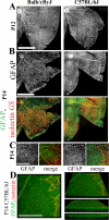
Astrocytes degenerate in the avascular, hypoxic areas of the central retina following return of the OIR retinas to normoxia. A) Staining with GFAP demonstrates the presence of astrocytes in the vaso-obliterated areas of retinas from both C57Bl/6J and BALB/cByJ strains of mice at P12, immediately after return from hyperoxia to normoxia. B) Astroctyes are maintained within the vaso-obliterated areas of BALB/cByJ mice at P14, but have degenerated by P14 in the vaso-obliterated zone of C57BL/6J mice. The punctate GFAP staining within the obliterated zone of C57BL/6J mice represents the inner endfeet of activated Müller glia. C) Higher magnification images of the interface between the vascularized and vaso-obliterated zones showing strain differences in astrocyte persistence and Müller glia activation at P14 of the OIR model. D) 3-dimentional renderings demonstrate GFAP staining of the characteristic trans-retinal processes of activated Müller glia and show association with the punctate staining observed within the superficial plexus of C57BL/6J retinae. All four images are of the same region, but rotated around the Y axis by 0° (left), 30° (top right), 60° (middle right), and 90° (bottom right). (Size bars = 500 μm (A, B), 100 μm (C, D))
Figure 3
Figure 3
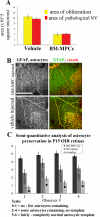
Injected bone marrow-derived myeloid progenitor cells (BM-MPCs) protect retinal astrocytes and rescue the retinal vasculature following OIR. A) Injected BM-MPCs rescue the OIR phenotype by reducing the area of obliteration in C57BL/6J retinas at P17 (demonstrating accelerated revascularization of the normal retinal plexuses), while reducing the formation of pathological neovascular tufts (Cummulative data; N's > 60; p values < 0.001). B) The vascular rescue is associated with protection of the endogenous astrocytes within the vascular obliterated zones at P14. C) Injection of BM-MPCs significantly increases astrocyte persistence compared with vehicle or non-injected retinas (N = 12 for each condition; p values comparing each treatment to vehicle injected or non-injected levels are as indicated (stars indicate p values < 0.0001)). (Size bars = 100 μm)
Figure 4
Figure 4
Injection of astrocytes or astrocyte-conditioned media results in normalized revascularization in the OIR model. A) Astrocytes facilitate growth and survival of BM-MPCs in culture. B) Injection of astrocyte-conditioned media results in persistence of isolectin GS-labeled activated macrophages (left; red arrows), GFAP positive astrocytes (middle) and CD11b positive microglia (right) within the hypoxic, vascular obliterated regions of C57BL/6J retinas compared with injection of control, non-conditioned media. C) Astrocyte-conditioned media significantly protects endogenous astrocytes in the OIR model (N = 16: astrocyte-conditioned media and no-injection controls; N = 12: vehicle injected controls; p values comparing each treatment to vehicle injected or non-injected levels are as indicated (stars indicate p values < 0.0001)). D-E) At P17, injection of astrocytes or astrocyte-conditioned media accelerates normalized revascularization and prevents pathological neovascular tuft formation at levels equivalent or better than the injected BM-MPCs (N = 12 for each condition; all p-values < 0.001 for each treatment compared to the appropriate vehicle control). (Size bars = 100 μm)
Figure 5
Figure 5
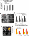
Injection of small quantities of bFGF or VEGF can protect retinal astrocytes and rescue the retinal vasculature in the OIR model. A) bFGF and VEGF is secreted by astrocytes and is present in the astrocyte-conditioned, serum-free media. B-C) Injection of bFGF (B) or VEGF (C) can rescue the retinal vasculature in the OIR model in a dose dependent manner (N ≥ 10 for each condition). D) The observed vascular rescue effects are preceded by protection of the endogenous astrocytes from hypoxia-induced degeneration within the vascular obliterated zones at P14 E) Quantification of the observed astrocytic rescue effects (N ≥ 10 for each condition; for all observers p values ≤ 0.001 for bFGF or VEGF compared to vehicle injection). F) Filopodia are extended from the endothelial cells planar to the protected astrocytes within the vaso-obliterated retina. These extensions can mediate potential guidance along the astrocytic template and suggest an explanation for the observed normalized revascularization associated with astrocyte protection in the vaso-obliterated zone. Each slightly opaque square indicates the area shown in the adjacent, higher magnification image. (Size bars = 100 μm)

References

    1. Adamis AP, Aiello LP, D'Amato RA. Angiogenesis and ophthalmic disease. Angiogenesis. 1999;3(1):9–14. - PubMed
    1. Aiello LP, Pierce EA, Foley ED, Takagi H, Chen H, Riddle L, Ferrara N, King GL, Smith LE. Suppression of retinal neovascularization in vivo by inhibition of vascular endothelial growth factor (VEGF) using soluble VEGF-receptor chimeric proteins. Proc Natl Acad Sci U S A. 1995;92(23):10457–61. - PMC - PubMed
    1. Araujo DM, Cotman CW. Basic FGF in astroglial, microglial, and neuronal cultures: characterization of binding sites and modulation of release by lymphokines and trophic factors. J Neurosci. 1992;12(5):1668–78. - PMC - PubMed
    1. Avery RL, Pieramici DJ, Rabena MD, Castellarin AA, Nasir MA, Giust MJ. Intravitreal bevacizumab (Avastin) for neovascular age-related macular degeneration. Ophthalmology. 2006;113(3):363–372. e5. - PubMed
    1. Banin E, Dorrell MI, Aguilar E, Ritter MR, Aderman CM, Smith AC, Friedlander J, Friedlander M. T2-TrpRS inhibits preretinal neovascularization and enhances physiological vascular regrowth in OIR as assessed by a new method of quantification. Invest Ophthalmol Vis Sci. 2006;47(5):2125–34. - PubMed

Publication types

MeSH terms