. 2009 Aug;27(8):1879-86.
doi: 10.1002/stem.96.

Low oxygen enhances primitive and definitive neural stem cell colony formation by inhibiting distinct cell death pathways

Affiliations
Free PMC article

Low oxygen enhances primitive and definitive neural stem cell colony formation by inhibiting distinct cell death pathways

Laura Clarke et al. Stem Cells. 2009 Aug.
Free PMC article

Abstract

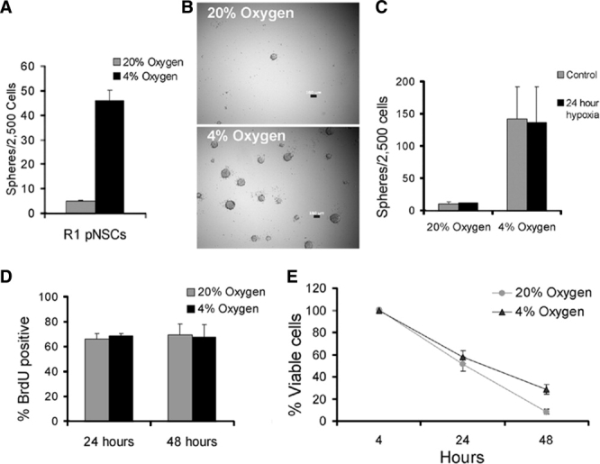
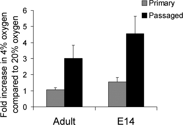
Neural stem cells (NSCs) can be derived from single mouse embryonic stem cells (ESCs) in the absence of instructive factors. Clonal primitive NSC (pNSC) colonies are formed first, and then give rise to clonal, fibroblast growth factor-dependent definitive neural stem cells (dNSCs). We tested low-oxygen culture as a potential method of alleviating the extensive cell death seen in pNSCs and dNSCs. Culture in low (4%) oxygen promoted survival of pNSCs by inhibiting apoptosis-inducing factor (AIF)-dependent cell death, although pNSCs undergo both AIF- and caspase-mediated cell death in 20% oxygen. In contrast, survival of dNSCs in low oxygen was increased by inhibition of caspase-dependent cell death. In normoxia, AIF is implicated in promoting dNSC survival. Neither survival effect was dependent on the main transcriptional effector of hypoxia, hypoxia-inducible factor 1. Low-oxygen concentrations may be involved in expansion of early NSC populations by inhibiting cell death through different pathways in these sequential pNSC and dNSC populations.


Figures

Figure 1
Figure 1
Low oxygen enhances pNSC colony formation by promoting pNSC survival. (A, B): The number of pNSC colonies that formed in 4% oxygen culture was approximately 10-fold greater than in control, 20% oxygen culture, t4 = 34.9, p < .05, n = 3. Bar in (B) = 100 μm. (C): Embryonic stem cells (ESCs) pre-exposed to 4% oxygen before being placed into the default assay did not show a difference in colony number, suggesting that oxygen does not act by driving ESCs toward a pNSC fate, F1,7 = 0.007, p > .05, n = 2. (D): Twenty-four hours (t4 = 0.63, p > .05, n = 3) or 48 hours (t4 = 0.179, p > .05, n = 3) after being placed into the neural default assay, ESCs that had transitioned into pNSCs proliferated at similar rates in high and low oxygen, assayed by 2-hour BrdU incorporation. (E): Increased pNSC colony formation in the low-oxygen condition is due to improved cell survival. The number of viable cells present in 4% oxygen was significantly higher than in 20% oxygen by 48 hours after being placed into the default assay as measured by trypan blue exclusion assay, F1,17 = 11.1, p < .05, n = 3. All data are mean ± SEM. Abbreviations: BrdU, bromodeoxyuridine; pNSC, primitive neural stem cell.
Figure 2
Figure 2
Increased pNSC survival in low-oxygen culture is independent of hypoxia-inducible factor 1 and dependent upon inhibition of apoptosis-inducing factor (AIF)-mediated cell death, whereas increased dNSC survival involves inhibition of caspase9. (A): hif1α−/− embryonic stem cells (ESCs) showed increased pNSC colony formation in 4% oxygen, similar to wild-type R1 ESCs, F1,15 = 303.8, p < .05, n = 4. (B): In the normoxic condition, caspase9−/− ESCs showed the most enhanced colony formation over wild-type E14K cells in normoxia, and a smaller increase was seen for aif-/Y ESCs, F2,12 = 31.9, p < .05 for each, n = 4. (C): E14K and caspase9−/− pNSC colony numbers were improved by low oxygen, however increased colony formation in low oxygen was blocked by the absence of AIF, F2,10 = 44.0, p < .05, n = 3. (D): There was a near 10-fold increase in dNSC colonies in low-oxygen culture, t4 = 8.25, p < .05, n = 3. (E): Caspase9−/− pNSC colonies gave rise to more dNSC colonies than the wild-type E14K line. However, very few dNSC colonies arose from aif-/Y pNSC colonies, F2,11 = 171.8, p < .05 versus E14K for each, n = 4. (F): Both E14K and aif-/Y dNSC colony numbers were increased by low-oxygen culture, however caspase9−/− dNSCs showed a significantly smaller enhancement in low oxygen, F2,11 = 6.26, p < .05 versus E14K for caspase9−/−, p > .05 versus E14K for aif-/Y, n = 4. Data in (A, B, D, E) are mean sphere number ± SEM; data in (C, F) are expressed as mean fold increases ± SEM in colony number in 4% oxygen over 20% oxygen. Abbreviations: dNSC, definitive neural stem cell; pNSC, primitive neural stem cell.
Figure 3
Figure 3
Derivation of pNSCs in 4% oxygen does not alter acquisition of a neural fate. pNSCs grown in 4% oxygen for 7 days upregulate the neural markers sox1, sox2, nestin, and betaIII tubulin and downregulate the mesoderm marker brachyury relative to starting ESC populations. Levels of oct4 and nanog were unchanged between pNSCs in 20% and 4% oxygen, n = 4 (A). (B): Differentiation of pNSCs grown in high and low oxygen showed that cells from both conditions produced beta-III tubulin-positive neurons at the same frequency, t4 = 0.34, p > .05, n = 3. Bar in (B) = 100 μm. Abbreviations: ESCs, embryonic stem cells; pNSC, primitive neural stem cell.
Figure 4
Figure 4
Brain-derived definitive neural stem cell (dNSC) colony numbers are increased by low-oxygen culture after exposure to normoxic conditions. Primary dNSCs from embryonic (E14) ganglionic eminence or adult lateral ventricle showed little response to low oxygen, adult t6 = 0.20, p > .05, n = 4, embryonic t4 = 5.24, p > .05. Passaged dNSC colonies, which had been exposed to 20% oxygen for at least 7 days, were positively influenced by low-oxygen culture. Colony number in 4% oxygen was significantly greater than in 20% oxygen: adult: t8 = 3.06, embryonic t6 = 3.68, p < .05 for each, n = 4,5. The average fold increases ± SEM in primary dNSC colony number in 4% oxygen over that in 20% oxygen are shown.
Figure 5
Figure 5
Double dissociation of cell death pathways in response to variable oxygen between pNSCs and dNSCs. Single ESCs acquire a neural identity when plated at low density in minimal media. ESCs first give rise to pNSCs, of which few survive as they undergo both caspase9-dependent and AIF-dependent cell death. pNSCs can be passaged to give rise to FGF-dependent dNSCs. dNSCs are also subject to cell death, but primarily through caspase-dependent processes. AIF has a prosurvival role in dNSCs, opposite to its actions in pNSCs. When pNSCs are derived in 4% oxygen, AIF-dependent but not caspase9-dependent cell death is attenuated and many more pNSCs survive to give rise to LIF-dependent colonies. Low-oxygen culture diminishes caspase-dependent apoptosis in dNSCs and AIF-dependent cell death is unaffected. Response to low oxygen double dissociates cell death pathways in these two early NSC populations. Abbreviations: AIF, apoptosis-inducing factor; dNSC, definitive neural stem cell; ESC, embryonic stem cell; FGF, fibroblast growth factor; LIF, leukemia inhibitory factor; pNSC, primitive neural stem cell.

References

    1. Hitoshi S, Seaberg R, Koscik C, et al. Primitive neural stem cells from the mammalian epiblast differentiate to definitive neural stem cells under the control of Notch signaling. Genes Dev. 2004;18:1806–1811. - PMC - PubMed
    1. Tropepe V, Hitoshi S, Sirard C, et al. Direct neural fate specification from embryonic stem cells: a primitive mammalian neural stem cell stage acquired through a default mechanism. Neuron. 2001;30:65–78. - PubMed
    1. Smukler S, Runciman S, Xu S, et al. Embryonic stem cells assume a primitive neural stem cell fate in the absence of extrinsic influences. J Cell Biol. 2006;172:79–90. - PMC - PubMed
    1. Csete M. Oxygen in the cultivation of stem cells. Ann Ny Acad Sci. 2005;1049:1–8. - PubMed
    1. Parmar K, Mauch P, Vergilio J, et al. Distribution of hematopoietic stem cells in the bone marrow according to regional hypoxia. Proc Natl Acad Sci U S A. 2007;104:5431–5436. - PMC - PubMed

Publication types