. 2009 Jul;27(7):1538-47.
doi: 10.1002/stem.86.

ES cell cycle progression and differentiation require the action of the histone methyltransferase Dot1L

Affiliations

ES cell cycle progression and differentiation require the action of the histone methyltransferase Dot1L

Evan R Barry et al. Stem Cells. 2009 Jul.

Abstract

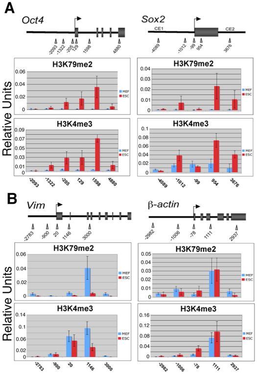
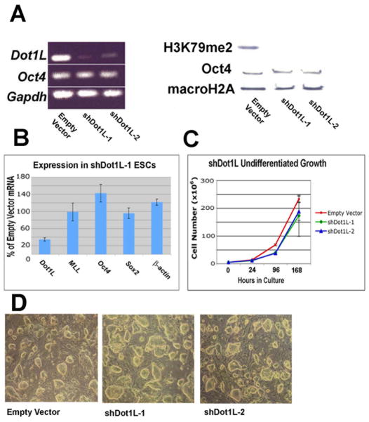
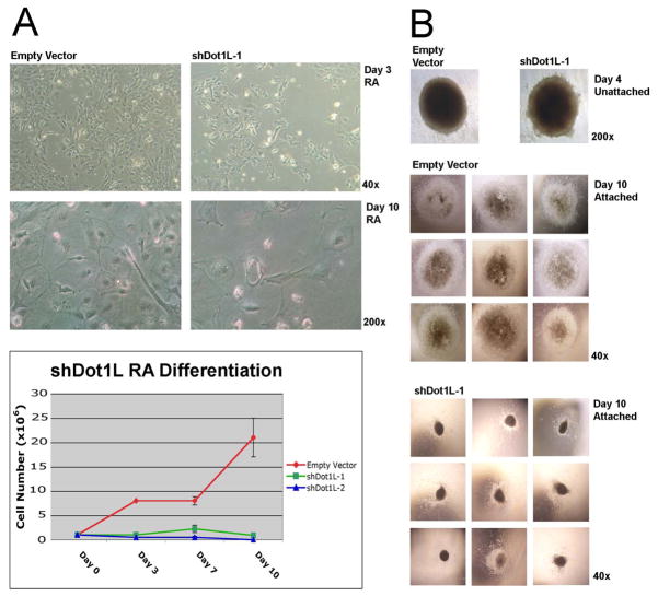
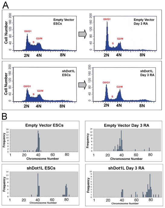
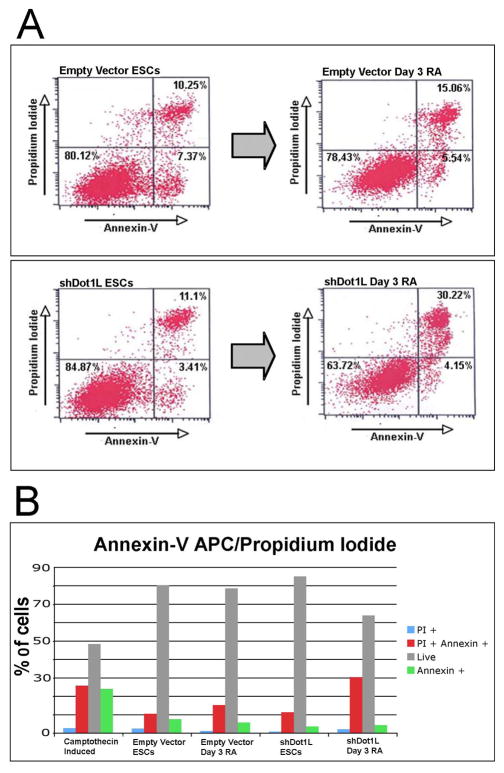
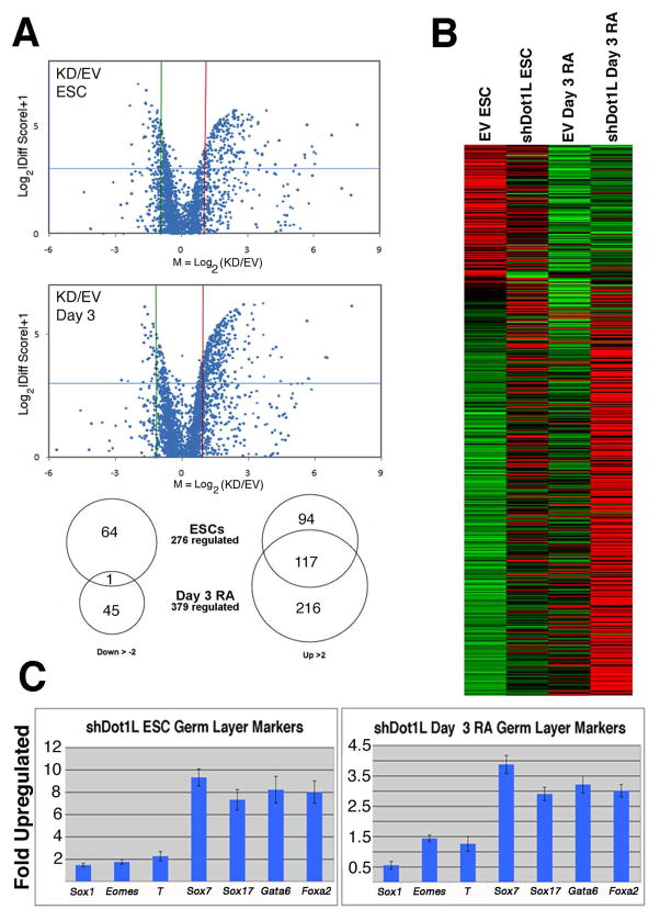
Mouse embryonic stem cells (ESCs) proliferate with rapid cell cycle kinetics but without loss of pluripotency. The histone methyltransferase Dot1L is responsible for methylation of histone H3 at lysine 79 (H3K79me). We investigated whether ESCs require Dot1L for proper stem cell behavior. ESCs deficient in Dot1L tolerate a nearly complete loss of H3K79 methylation without a substantial impact on proliferation or morphology. However, shortly after differentiation is induced, Dot1L-deficient cells cease proliferating and arrest in G2/M-phase of the cell cycle, with increased levels of aneuploidy. In addition, many aberrant mitotic spindles occur in Dot1L-deficient cells. Surprisingly, these mitotic and cell cycle defects fail to trigger apoptosis, indicating that mouse ESCs lack stringent cell cycle checkpoint control during initial stages of differentiation. Transcriptome analysis indicates that Dot1L deficiency causes the misregulation of a select set of genes, including many with known roles in cell cycle control and cellular proliferation as well as markers of endoderm differentiation. The data indicate a requirement for Dot1L function for early stages of ESC differentiation where Dot1L is necessary for faithful execution of mitosis and proper transcription of many genes throughout the genome.


Figures

Figure 1
Figure 1
Distribution of H3K79me2 and H3K4me3 across transcribed genes in ESCs and MEFs. ChIP samples were collected from ESCs and MEFs using antibodies specific for H3K79me2 and H3K4me3 then assayed by quantitative real-time PCR using primer pairs tiled across the Oct4 and Sox2 promoters and flanking regions. (A) Oct4 chromatin is enriched for H3K79me2 and H3K4me3 ~1.5kb downstream of the TSS in ESCs (red bars), with virtually no enrichment in MEFs (blue bars). Sox2 chromatin was assayed by ChIP-QPCR for the same histone modifications. (B) H3K79me2 and H3K4me3 distribution across the promoter and flanking regions of the Vimentin gene (which is highly expressed in MEFs as compared to ESCs) in MEFs and ESCs. H3K79me2 and H3K4me3 distribution across the promoter and flanking regions of the β-actin housekeeping gene, which is constitutively expressed in all cell-types. Data points represent the mean of 9 PCR reactions, +/− the standard deviation.
Figure 2
Figure 2
Analysis of Dot1L deficiency in undifferentiated ESCs harboring stably-integrated shRNA vectors. (A) RT-PCR of RNA from ESC lines shDot1L-1 and shDot1L-2 exhibited robust knockdown of Dot1L, but not Oct4 or Gapdh mRNAs. Also, Western blot analysis shows greatly diminished H3K79 methylation in ESC lines shDot1L-1 and shDot1L-2 without substantial alteration of Oct4 protein levels. The histone macroH2A was used as a loading control. (B) Quantitative real-time PCR was used to assay the expression of several genes in Dot1L-deficient ESCs. Dot1L mRNA is decreased in Dot1L knockdown ESCs compared to the empty vector control. MLL, a gene which encodes a histone methyltransferase specific for H3K4, is unaffected, as well as mRNAs expressed from Oct4, Sox2, or β-actin, which were not downregulated. Data is shown as the mean of 6 PCR reactions +/− the standard deviation. (C) Growth curve analysis of two Dot1L-deficient ESC lines as compared to a wild-type ESC control line harboring an integrated empty vector. The plotted data is the mean of 3 cell culture counts; error bars indicate standard deviation. (D) Cellular morphology of shDot1L ESCs compared to empty vector ESCs.
Figure 3
Figure 3
Differentiation of ESCs deficient for Dot1L. (A) Empty vector control and shDot1L (Dot1L-deficient) ESCs were subjected to RA-induced differentiation in the absence of LIF and feeder cells. Equal numbers of both cell lines were plated prior to RA-induced differentiation. Representative images after 3 and 10 days of RA-induced differentiation. RA differentiation experiments were performed in triplicate for two stable knockdown ESC lines (shDot1L-1 and shDot1L-2) and empty vector ESCs, and cells were counted over the course of 10 days to yield growth curves. (B) shDot1L-1 and empty vector ESCs subjected to a protocol designed to yield EBs of standardized size. Each EB was formed by aggregating 2×10⁴ ESCs and imaged after 4 days in suspension culture, and again after attachment and 10 additional days in culture.
Figure 4
Figure 4
Cell cycle and karyotype analysis of empty vector and shDot1L cell lines. (A) Undifferentiated empty vector and shDot1L ESCs had a similar proportion of cells in each stage of the cell cycle (left panels). shDot1L ESCs exhibited a relatively normal cell cycle, except for a small but detectable population of hyperploid (>4N) cells. After 3 days of RA differentiation, shDot1L cultures contained increased cells in G2/M-phase, and the number of hyperploid (>4N) cells also increased. (B) Karyotype Analysis. Undifferentiated empty vector ESCs had a modal number of 40 chromosomes, as did empty vector cells after 3 days of RA differentiation. Undifferentiated shDot1L ESCs yielded bimodally-distributed karyotypes (40, diploid; 80, tetraploid) when grown in standard ESC conditions. After 3 days of RA differentiation of shDot1L ESCs, the modal count changed to 80, with many spreads containing 60–90 chromosomes.
Figure 5
Figure 5
Annexin-V flow cytometry assays for early apoptotic cells. (A) Undifferentiated and day 3 RA differentiated empty vector and shDot1L cells were stained with Annexin-V (AV) and propidium iodide (PI) and analyzed by flow cytometry. Three classes of cells were observed in each sample: low AV, low PI (live non-apoptotic cells), high AV, low PI (early apoptotic cells), and high AV, high PI (disrupted, presumably dead cells). Undifferentiated empty vector and shDot1L ESCs had similar profiles. (B) Flow cytometry results in graph form for each cytometry quadrant shown.
Figure 6
Figure 6
Transcriptome analysis of normal and Dot1L-deficient ESCs and cells after 3 days of RA-induced differentiation. (A) Volcano plots of Illumina transcriptome array results. The upper plot shows genes whose expression is affected by disruption of Dot1L in undifferentiated ESCs as compared to wild-type empty vector ESCs. The lower plot shows genes de-regulated by disruption of Dot1L in cells subjected to 3 days of RA-induced differentiation. Each dot indicates the log of the knockdown/wild-type expression ratio. Genes greater than 2-fold up-regulated lie to the right of the vertical red line, and genes greater than 2-fold down-regulated lie to the left of the vertical green line. Genes with statistically-significant responses to Dot1L disruption lie above the horizontal blue line. Also, a Venn diagram of Dot1L-regulated genes was derived from the volcano plot analysis. (B) A heat map that shows the expression (indicated by graded color hues) of the 534 genes identified as regulated by Dot1L. Red is highly expressed and green is lowly expressed. (C) Taqman QPCR validation of upregulated endoderm markers in shDot1L ESCs and RA differentiated cells.

References

    1. Rasmussen T. Developmentally-poised chromatin of embryonic stem cells. Front Biosci. 2008;13:1568–1577. - PubMed
    1. Bernstein BE, Mikkelsen TS, Xie X, et al. A bivalent chromatin structure marks key developmental genes in embryonic stem cells. Cell. 2006;125(2):315–326. - PubMed
    1. Feldman N, Gerson A, Fang J, et al. G9a-mediated irreversible epigenetic inactivation of Oct-3/4 during early embryogenesis. Nat Cell Biol. 2006;8(2):188–194. - PubMed
    1. Feng Q, Wang H, Ng HH, et al. Methylation of H3-lysine 79 is mediated by a new family of HMTases without a SET domain. Curr Biol. 2002;12(12):1052–1058. - PubMed
    1. Krogan NJ, Dover J, Wood A, et al. The Paf1 complex is required for histone H3 methylation by COMPASS and Dot1p: linking transcriptional elongation to histone methylation. Mol Cell. 2003;11(3):721–729. - PubMed

Publication types

Substances