. 2009 Jul;27(7):1592-603.
doi: 10.1002/stem.77.

Regulation of boundary cap neural crest stem cell differentiation after transplantation

Affiliations
Free PMC article

Regulation of boundary cap neural crest stem cell differentiation after transplantation

Hakan Aldskogius et al. Stem Cells. 2009 Jul.
Free PMC article

Abstract

Success of cell replacement therapies for neurological disorders will depend largely on the optimization of strategies to enhance viability and control the developmental fate of stem cells after transplantation. Once transplanted, stem/progenitor cells display a tendency to maintain an undifferentiated phenotype or differentiate into inappropriate cell types. Gain and loss of function experiments have revealed key transcription factors which drive differentiation of immature stem/progenitor cells toward more mature stages and eventually to full differentiation. An attractive course of action to promote survival and direct the differentiation of transplanted stem cells to a specific cell type would therefore be to force expression of regulatory differentiation molecules in already transplanted stem cells, using inducible gene expression systems which can be controlled from the outside. Here, we explore this hypothesis by employing a tetracycline gene regulating system (Tet-On) to drive the differentiation of boundary cap neural crest stem cells (bNCSCs) toward a sensory neuron fate after transplantation. We induced the expression of the key transcription factor Runx1 in Sox10-expressing bNCSCs. Forced expression of Runx1 strongly increased transplant survival in the enriched neurotrophic environment of the dorsal root ganglion cavity, and was sufficient to guide differentiation of bNCSCs toward a nonpeptidergic nociceptive sensory neuron phenotype both in vitro and in vivo after transplantation. These findings suggest that exogenous activation of transcription factors expression after transplantation in stem/progenitor cell grafts can be a constructive approach to control their survival as well as their differentiation to the desired type of cell and that the Tet-system is a useful tool to achieve this.


Figures
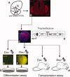
Figure 1
Figure 1
Overview of experiments. Sox10+/rtTA embryos at E11.5 (A): express Sox10 (red) in the dorsal root ganglia (B) as well as in newly formed boundary cap neural crest stem cell (bNCSC) neurospheres (C). The isolated bNCSCs are transfected with the TREbi-Runx1-eYFP vector (D) containing a bidirectional promoter, which on DOX treatment induces expression of both Runx1 and EYFP. DOX-activated expression of Runx1/EYFP in bNCSC neurospheres (E) two days after transfection and 1 day after DOX activation before differentiation assay (F) and transplantation (G). Nuclear labeling with Hoechst is shown in blue. Abbreviation: DOX, doxycycline.
Figure 2
Figure 2
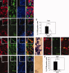
DOX-activated Runx1 expression induced differentiation of boundary cap neural crest stem cells to dorsal root ganglion neurons in vitro. DOX-induced expression of Runx1 leads to significantly increased neurite outgrowth. DOX-treated (A) and control (B) cultures, quantified in (C; p = 3.6 × 10⁻⁸). Panels (D), (F), and (H) show DOX-treated cultures to the left and DOX-untreated cultures to the right of the graphs. The quantitative analyses of DOX-treated and DOX-untreated cultures are shown in graphs (E), (G), and (I). RET expression is induced in βIII-tubulin (bTUB)+ cells in DOX-treated, but not in DOX-untreated cultures (panel D; quantified in E) (p = 1.8 × 10⁻⁴). Also isolectin B4 binding by bTUB+ cells was induced in DOX-treated, but not in DOX-untreated cultures (panel F; quantified in G) (p = 3.6 × 10⁻⁶). Conversely, there was no significant difference in calcitonin gene-related peptide-labeled cells in DOX-treated compared with DOX-untreated cultures (panel H; quantified in I) (p = .21). Scale bar = 100 μm. Abbreviation: DOX, doxycycline.
Figure 3
Figure 3
bNCSC grafts one month after transplantation to the L4 dorsal root ganglion cavity. (A–F): Immunostainings for βIII-tubulin (bTUB; red) and GFAP (green) together with nuclear marker Hoechst (blue). (A, B): bTUB staining shows slightly more positive cells in the DOX-treated animals. (C, D): Increased GFAP staining in the DOX-treated animals. Note the difference in morphology of positive cells and the overall structure between the two treatments. (G–J): Growth of bTUB+ fibers from EGFP expressing bNCSCs is extensive within the graft (G) and in the associated dorsal root (I) of DOX-treated (G, I) compared with DOX-untreated (H, J) transplants. bTUB+ neurites from DOX-treated bNCSC transplants intermingle with bTUB+ host fibers in the dorsal root (I). (K, L): Immunostaining for CGRP (blue), RT97 (red), and isolectin B4 (IB4; green). A small number of CGRP+ cells occur in both conditions. There was a decreased number of RT97+ cells while IB4 binding was increased by DOX-activated Runx1 expression. (M): Quantification of bTUB (p = .085) and GFAP stainings (p = 4.7 × 10⁻⁴). (N): Quantification of CGRP (p = .32), IB4 (p = .0048), and RT97 (p = .0022) stainings. Scale bar = 50 μm. (A–H; K, L); 25 μm (I, J). Abbreviations: bTUB, βIII-tubulin; CGRP, calcitonin gene-related peptide; DOX, doxycycline; GFAP, glial fibrillary acidic protein.
Figure 4
Figure 4
Immunostainings and quantification of TrkA and RET in transplants. Staining for bTUB (red) and TrkA (green) show an increased number of TrkA+ cells after DOX treatment (panel A) when compared with the control (panel B). Some TrkA is expressed in bTUB+ cells but most TrkA staining is associated with bTUB negative cells. (C): Quantification of the increase of TrkA positive cells among the bTUB+ population (p = .0034). Staining of RET (red) in DOX-treated (panel D) and DOX-untreated (panel E) transplants. In DOX-treated transplants, the majority of RET expression (panel D; red) is associated with bTUB+ (green) cells. In situ hybridization for RET mRNA shows extensive labeling in DOX-treated (panel D), and absence of labeling in DOX-untreated (panel E) transplants. BrdU-labeled cells, some of which express RET are shown in panel D (right). (F): Quantification of immunolabeled cells (D, E) showing the almost complete absence of RET positive cells in the control (p = 3.4 × 10⁻⁷). Nuclear labeling with Hoechst is shown in blue. Scale bar (E) = 50 μm. Abbreviations: bTUB, βIII-tubulin; BrdU, bromodeoxyuridine; DOX, doxycycline.
Figure 5
Figure 5
Immunostainings and quantification of the ATP receptor P2X3 in transplants. Staining for bTUB (red) and P2X3 (green) shows an increased number of P2X3+ cells after DOX treatment (A) when compared with the control (B). (C): Quantification of the increase of positive cells among the bTUB+ population (p = .0034; n = 6). Scale bar (B) = 50 μm. Abbreviations: bTUB, βIII-tubulin; DOX, doxycycline.
Figure 6
Figure 6
Increased graft size and cell survival after DOX-induced Runx1 expression. (A): Example of three-dimensional reconstructions of grafts of C57BL/6-β-actin (CAG)-eGFP:Sox10+/rrTA boundary cap neural crest stem cells. (B): Quantification of graft size showing the relative increase in size after Runx1 overexpression (p = 5.5 × 10⁻⁴; n = 4). (C): Quantification of eGFP+ graft cell numbers showing an increased survival after 1 month in treated versus control (p = 1.4 × 10⁻⁴, n = 4). Abbreviations: DOX, doxycycline; eGFP, enhanced green fluorescent protein.

References

    1. Bithell A, Williams BP. Neural stem cells and cell replacement therapy: Making the right cells. Clin Sci (Lond) 2005;108:13–22. - PubMed
    1. Brederlau A, Correia AS, Anisimov SV, et al. Transplantation of human embryonic stem cell-derived cells to a rat model of Parkinson's disease: Effect of in vitro differentiation on graft survival and teratoma formation. Stem Cells. 2006;24:1433–1440. - PubMed
    1. Martino G, Pluchino S. The therapeutic potential of neural stem cells. Nat Rev Neurosci. 2006;7:395–406. - PubMed
    1. Kozlova EN, Rosario CM, Stromberg I, et al. Peripherally grafted human foetal dorsal root ganglion cells extend axons into the spinal cord of adult host rats by circumventing dorsal root entry zone astrocytes. Neuroreport. 1995;6:269–272. - PubMed
    1. Kozlova EN, Seiger A, Aldskogius H. Human dorsal root ganglion neurons from embryonic donors extend axons into the host rat spinal cord along laminin-rich peripheral surroundings of the dorsal root transitional zone. J Neurocytol. 1997;26:811–822. - PubMed

Publication types

MeSH terms

Substances