. 2009 Jul;39(7):1870-8.
doi: 10.1002/eji.200838956.

Role of T cell TGF-beta signaling in intestinal cytokine responses and helminthic immune modulation

Affiliations

Role of T cell TGF-beta signaling in intestinal cytokine responses and helminthic immune modulation

M Nedim Ince et al. Eur J Immunol. 2009 Jul.

Abstract

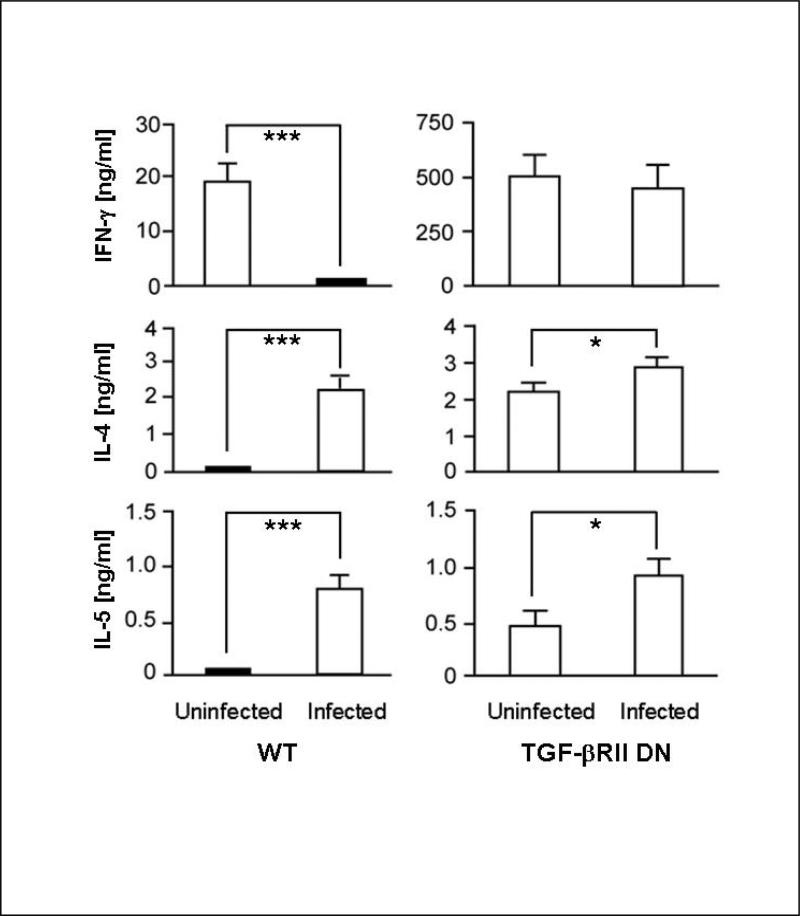
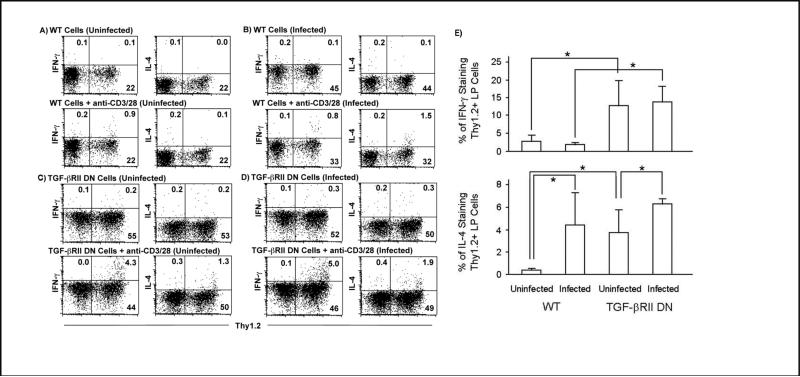
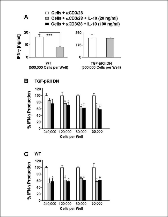
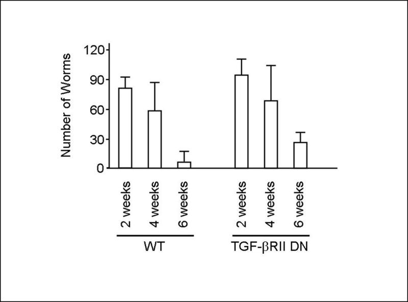
Colonization with helminthic parasites induces mucosal regulatory cytokines, like IL-10 or TGF-beta, that are important in suppressing colitis. Helminths induce mucosal T cell IL-10 secretion and regulate lamina propria mononuclear cell (LPMC) Th1 cytokine generation in an IL-10-dependent manner in WT mice. Helminths also stimulate mucosal TGF-beta release. As TGF-beta exerts major regulatory effects on T lymphocytes, we investigated the role of T lymphocyte TGF-beta signaling in helminthic modulation of intestinal immunity. T cell TGF-beta signaling is interrupted in TGF-beta receptor II dominant negative (TGF-betaRII DN) mice by T-cell-specific over-expression of a TGF-betaRII DN. We studied LPMC responses in WT and TGF-betaRII DN mice that were uninfected or colonized with the nematode, Heligmosomoides polygyrus. Our results indicate an essential role of T cell TGF-beta signaling in limiting mucosal Th1 and Th2 responses. Furthermore, we demonstrate that helminthic induction of intestinal T cell IL-10 secretion requires intact T cell TGF-beta-signaling pathway. Helminths fail to curtail robust, dysregulated intestinal Th1 cytokine production and chronic colitis in TGF-betaRII DN mice. Thus, T cell TGF-beta signaling is essential for helminthic stimulation of mucosal IL-10 production, helminthic modulation of intestinal IFN-gamma generation and H. polygyrus-mediated suppression of chronic colitis.


Figures

Figure 1
Figure 1
TCR-triggered TGF-β production in LPMC from uninfected control or H. polygyrus-infected WT and TGF-βRII DN mice. LPMC from uninfected or helminth-infected WT or TGF-βRII DN mice were cultured in vitro alone (Cells) or with anti-CD3/CD28 mAb (Cells + αCD3/28) for 48 h in TGF-β culture medium. Supernatants then were analyzed for TGF-β by ELISA. Data show mean ± SD from three independent experiments with each experiment containing multiple determinations. (WT: unstimulated LPMC Uninfected vs. Infected, p>0.05, NS; anti-CD3/CD28 stimulated Uninfected vs. Infected, **p<0.01).
Figure 2
Figure 2
Helminths cannot modulate the Th1 response and only weakly promote the Th2 response in LPMC from TGF-βRII DN mice. LPMC from uninfected or helminth-infected WT or TGF-βRII DN mice were stimulated in vitro with anti-CD3/CD28 mAb for 48 mAb. Supernatants were harvested and analyzed for IFN-γ, IL-4 and IL-5 by ELISA. Data show mean ± SD from at least three independent experiments with each experiment containing multiple determinations. (IFN-γ: WT Uninfected vs. Infected, ***p<0.001; TGF-βRII DN Uninfected vs. Infected, p>0.05, NS. IL-4: WT Uninfected vs. Infected, ***p<0.001; TGF-βRII DN Uninfected vs. Infected, *p<0.05. IL-5: WT Uninfected vs. Infected, ***p<0.001; TGF-βRII DN Uninfected vs. Infected, *p<0.05).
Figure 3
Figure 3
Helminth-associated changes in cytokine producing LP T cells from WT and TGF-βRII DN mice. Representative dot plots of LPMC from (A) uninfected WT, (B) helminth-infected WT, (C) uninfected TGF-βRII DN or (D) helminth-infected TGF-βRII DN mice that were stimulated overnight in vitro with anti-CD3/28. Brefeldin A was added to culture supernatants for the last 12 hours. Subsequently, cells were harvested and stained for Thy1.2, IFN-γ and IL-4 for FACS analysis. (E) Data show mean ± SD of the percentage IFNγ (upper panel) or IL-4 (lower panel) producing Thy1.2 positive LP cells from four (WT) and three (TGFβ RII DN) independent experiments. (IFN-γ: WT Uninfected vs. Infected, p>0.05, NS; TGF-βRII DN Uninfected vs. Infected, p>0.05, NS. IFN-γ: WT Uninfected vs. TGFβ RII DN Uninfected, *p<0.05; WT Infected vs. TGFβ RII DN Infected, *p<0.05. IL-4: WT Uninfected vs. Infected, *p<0.05; TGF-βRII DN Uninfected vs. Infected, p<0.05; WT Uninfected vs. TGFβ RII DN Uninfected *p<0.05; WT Infected vs. TGFβ RII DN Infected, p>0.05, NS).
Figure 4
Figure 4
Helminthic induction of IL-10 in LPMC is dependent on T cell TGF-β signaling. LPMC from uninfected or helminth-infected WT or TGF-βR DN mice were stimulated in vitro with anti-CD3/CD28 mAb for 48 hr. Supernatants were harvested and analyzed for IL-10 by ELISA. Data show mean ± SD from three independent experiments with each experiment containing multiple determinations. (WT Uninfected vs. Infected, ***p<0.001; TGF-βRII DN Uninfected vs. Infected, p>0.05, NS).
Figure 5
Figure 5
Effect of exogenous IL-10 on LPMC IFN-γ production from WT and TGF-βRII DN mice. LPMC from uninfected WT (A and C) or TGF-βRII DN (A and B) mice were stimulated in vitro with anti-CD3/CD28 (white bars) for 48 hr, in indicated cell numbers per well, with parallel cultures also containing 20 ng/ml (gray bars) or 100 ng/ml recombinant IL-10 (black bars). Supernatants were harvested and analyzed for IFN-γ by ELISA. Data show mean ± SD of (A) IFNγ secretion (two independent experiments with each experiment containing multiple determinations) or (B-C) the percentile ratio of LPMC IFNγ secretion in each condition to IFN-γ output of anti-CD3/28-stimulated culture (three independent experiments with each experiment containing multiple determinations). ((A) WT Uninfected vs. Infected, ***p<0.001; TGF-βRII DN Uninfected vs. Infected, p>0.05, NS). (B-C) (*p<0.05 for IFN-γ secretion between IL-10 containing and IL-10 naïve cultures in incubations with the corresponding cell numbers).
Figure 6
Figure 6
The reduction in worm population in WT and TGF-βRII DN mice is similar. The number of duodenal helminths was counted in 6 WT and 6 TGF-βRII DN mice at each time point, 2, 4 and 6 weeks after worm colonization. Data from 18 WT and 18 TGF-βRII DN mice in 3 different time points were accumulated in 3 independent experiments. Data show mean ± SD at each time point. (2 wk: WT vs. TGF-βRII DN, p>0.05; 4 wk: WT vs. TGF-βRII DN, p>0.05; 6 wk: WT vs. TGF-βRII DN, p>0.05, NS).
Figure 7
Figure 7
H. polygyrus colonization does not reverse spontaneous colitis in TGF-βRII DN mice. (A) Representative example of dissected colons from 12-14 wk old WT, uninfected control TGF-βRII DN and H. polygyrus-infected TGF-βRII DN mouse. (B-D) Representative example of colonic histology of 12-14 wk old WT (B), uninfected control TGF-βRII DN (C) and H. polygyrus-infected TGF-βRII DN (D) mouse (4x magnified). (E) Colonic tissues of uninfected or helminth-infected TGF-βRII DN mice were analyzed by histopathology. Data show mean ± SD of histologic colitis scores from 10 uninfected and 13 H. polygyrus-infected mice in 3 independent experiments (TGF-βRII DN Uninfected vs. Infected, p>0.05, NS).

Similar articles

Cited by

References

    1. Elliott DE, Summers RW, Weinstock JV. Helminths as governors of immune-mediated inflammation. Int.J.Parasitol. 2007;37:457–464. - PubMed
    1. Summers RW, Elliott DE, Urban JF, Jr., Thompson R, Weinstock JV. Trichuris suis therapy in Crohn's disease. Gut. 2005;54:87–90. - PMC - PubMed
    1. Summers RW, Elliott DE, Urban JF, Jr., Thompson RA, Weinstock JV. Trichuris suis therapy for active ulcerative colitis: a randomized controlled trial. Gastroenterology. 2005;128:825–832. - PubMed
    1. Finkelman FD, Urban JF., Jr. The other side of the coin: the protective role of the TH2 cytokines. J.Allergy Clin.Immunol. 2001;107:772–780. - PubMed
    1. Sacco R, Hagen M, Sandor M, Weinstock JV, Lynch RG. Established T(H1) granulomatous responses induced by active Mycobacterium avium infection switch to T(H2) following challenge with Schistosoma mansoni. Clin.Immunol. 2002;104:274–281. - PubMed

Publication types

MeSH terms

Substances