. 2009 May;36(5):1700-10.
doi: 10.1118/1.3112363.

Investigation of lung nodule detectability in low-dose 320-slice computed tomography

Affiliations

Investigation of lung nodule detectability in low-dose 320-slice computed tomography

J D Silverman et al. Med Phys. 2009 May.

Abstract

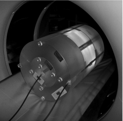
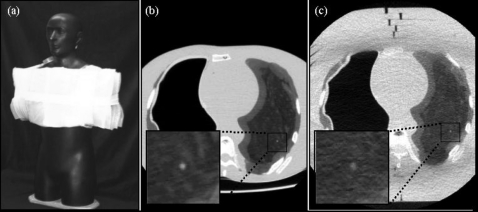
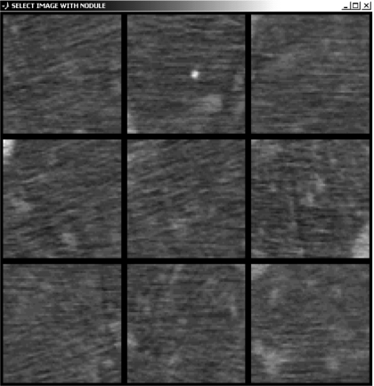
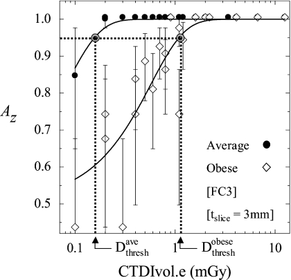
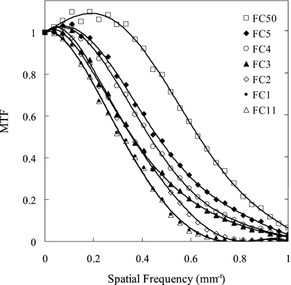
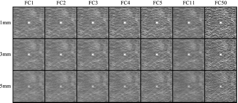
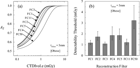
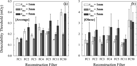
Low-dose imaging protocols in chest CT are important in the screening and surveillance of suspicious and indeterminate lung nodules. Techniques that maintain nodule detectability yet permit dose reduction, particularly for large body habitus, were investigated. The objective of this study was to determine the extent to which radiation dose can be minimized while maintaining diagnostic performance through knowledgeable selection of reconstruction techniques. A 320-slice volumetric CT scanner (Aquilion ONE, Toshiba Medical Systems) was used to scan an anthropomorphic phantom at doses ranging from approximately 0.1 mGy up to that typical of low-dose CT (LDCT, approximately 5 mGy) and diagnostic CT (approximately 10 mGy). Radiation dose was measured via Farmer chamber and MOSFET dosimetry. The phantom presented simulated nodules of varying size and contrast within a heterogeneous background, and chest thickness was varied through addition of tissue-equivalent bolus about the chest. Detectability of a small solid lung nodule (3.2 mm diameter, -37 HU, typically the smallest nodule of clinical significance in screening and surveillance) was evaluated as a function of dose, patient size, reconstruction filter, and slice thickness by means of nine-alternative forced-choice (9AFC) observer tests to quantify nodule detectability. For a given reconstruction filter, nodule detectability decreased sharply below a threshold dose level due to increased image noise, especially for large body size. However, nodule detectability could be maintained at lower doses through knowledgeable selection of (smoother) reconstruction filters. For large body habitus, optimal filter selection reduced the dose required for nodule detection by up to a factor of approximately 3 (from approximately 3.3 mGy for sharp filters to approximately 1.0 mGy for the optimal filter). The results indicate that radiation dose can be reduced below the current low-dose (5 mGy) and ultralow-dose (1 mGy) levels with knowledgeable selection of reconstruction parameters. Image noise, not spatial resolution, was found to be the limiting factor in detection of small lung nodules. Therefore, the use of smoother reconstruction filters may permit lower-dose protocols without trade-off in diagnostic performance.


Figures

Figure 1
Figure 1
Photograph of setup for dose measurements. Two Farmer chambers were inserted in the center and periphery of 32 cm diameter acrylic cylinders, stacked to 48 cm length.
Figure 2
Figure 2
Anthropomorphic phantom. (a) Photograph of the phantom with 10 cm SuperFlab™ secured to the torso to simulate an obese habitus. Axial images of the phantom in (b) the average habitus configuration (without SuperFlab™) and (c) the obese habitus configuration. Magnified views of a simulated 3.2 mm nodule are shown in each case. Imaging techniques for example images (b) and (c) were 100 kVp, 105 mAs (CTDIvol.e=6.7 mGy), FC3, and tslice=3 mm.
Figure 3
Figure 3
Illustration of 9AFC test for nodule detection. In this example [120 kVp, 14 mAs (CTDIvol.e=1.6 mGy), FC2 filter, tslice=3 mm, average body habitus] the stimulus is in the top-center subimage. The streaks in several ROIs are believed to be due to photon starvation and beam-hardening artifacts associated with the spine, ribs, sternum, and mediastinum (shown in Fig. 2).
Figure 4
Figure 4
Comparison of CTDIvol.e (as reported by the scanner) and Dcenter, or CTDIw (as measured by Farmer chambers and MOSFETs in a long 32 cm diameter CTDI phantom for one x-ray tube revolution). For Dcenter and CTDIw, the mean and standard deviation over five trials are shown.
Figure 5
Figure 5
Observer performance plotted as a function of dose for average and obese body habitus. Logistic functions were used to fit the measurement to a sigmoid (solid curves). Calculation of Dthresh is illustrated graphically as the dose at which observer performance falls to 0.95. Examples shown are for fixed reconstruction filter (FC3) and slice thickness (tslice=3 mm).
Figure 6
Figure 6
MTF corresponding to the seven reconstruction filters investigated in this work. The response is plotted up to the Nyquist frequency.
Figure 7
Figure 7
Example images in a region about a 3.2 mm simulated lung nodule for all combinations of reconstruction filter and slice thickness investigated. Examples were acquired at 100 kVp, 105 mAs (CTDIvol.e=6.7 mGy) in the obese phantom configuration. For purposes of illustration, the nodule is shown at the center of each image.
Figure 8
Figure 8
Effect of reconstruction filter on Dthresh (tslice=3 mm; obese configuration). (a) Observer performance plotted as a function of dose for seven reconstruction filters. (b) Comparison of Dthresh across seven reconstruction filters.
Figure 9
Figure 9
Effect of slice thickness on Dthresh for average and obese habitus. The cases shown correspond to the FC11 reconstruction filter.
Figure 10
Figure 10
Effect of reconstruction techniques on Dthresh for (a) average and (b) obese body habitus. Note the order-of-magnitude scale factor between y axes of (a) and (b), discussed below.
Figure 11
Figure 11
Example images at similar CTDIvol.e [(a) and (b)] and measured dose Dlung [(c) and (d)] for average [(a) and (c)] and obese [(b) and (d)] configurations (reconstructed with the FC3 filter at tslice=3 mm). Images in (a) and (b) were acquired at (100 kVp, 35 mAs), giving CTDIvol.e=2.2 mGy and measured doses of (a) Dlung=3.6 mGy and (b) Dlung=1.8 mGy. By comparison, images (c) and (d) were acquired at (c) Dlung=1.4 mGy, CTDIvol.e=0.8 mGy (120 kVp, 7 mAs) and (d) Dlung=1.5 mGy, CTDIvol.e=2.0 mGy (120 kVp, 17.5 mAs).

References

    1. Remy-Jardin M., Remy J., Giraud F., and Marquette C. H., “Pulmonary nodules: Detection with thick-section spiral CT versus conventional CT,” Radiology 187, 513–520 (1993). - PubMed
    1. Piyaviset N. et al., “Small incidental pulmonary nodules: How useful is short term interval CT?,” J. Thorac. Imaging 20, 5–9 (2005). - PubMed
    1. Paul N. S., Siewerdsen J. H., Patsios D., and Chung T. B., “Investigating the low-dose limits of multidetector CT in lung nodule surveillance,” Med. Phys. MPHYA610.1118/1.2768866 34, 3587–3595 (2007). - DOI - PubMed
    1. Yankelevitz D. F. et al., “Small pulmonary nodules: Evaluation with repeat CT—Preliminary experience,” Radiology 212, 561–566 (1999). - PubMed
    1. Revel M. P. et al., “Pulmonary nodules: Preliminary experience with three-dimensional evaluation,” Radiology RADLAX10.1148/radiol.2312030241 231, 459–466 (2004). - DOI - PubMed

Publication types

MeSH terms