

Exploration of the potential of liquid scintillators for real-time 3D dosimetry of intensity modulated proton beams
- PMID: 19544791
- PMCID: PMC2832031
- DOI: 10.1118/1.3117583
Exploration of the potential of liquid scintillators for real-time 3D dosimetry of intensity modulated proton beams
Abstract
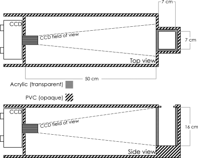
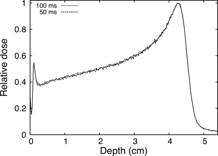
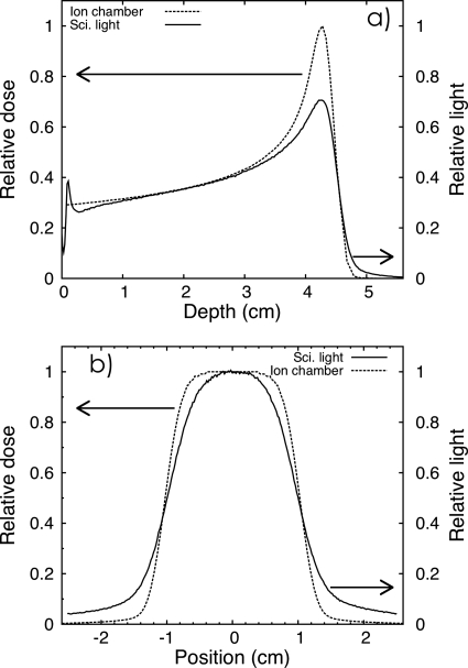
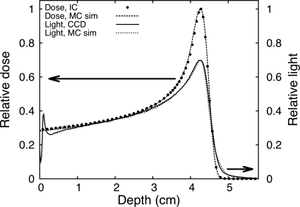
In this study, the authors investigated the feasibility of using a 3D liquid scintillator (LS) detector system for the verification and characterization of proton beams in real time for intensity and energy-modulated proton therapy. A plastic tank filled with liquid scintillator was irradiated with pristine proton Bragg peaks. Scintillation light produced during the irradiation was measured with a CCD camera. Acquisition rates of 20 and 10 frames per second (fps) were used to image consecutive frame sequences. These measurements were then compared to ion chamber measurements and Monte Carlo simulations. The light distribution measured from the images acquired at rates of 20 and 10 fps have standard deviations of 1.1% and 0.7%, respectively, in the plateau region of the Bragg curve. Differences were seen between the raw LS signal and the ion chamber due to the quenching effects of the LS and due to the optical properties of the imaging system. The authors showed that this effect can be accounted for and corrected by Monte Carlo simulations. The liquid scintillator detector system has a good potential for performing fast proton beam verification and characterization.
Figures
References
-
- Lomax A. J., Böhringer T., Bolsi A., Coray D., Emert F., Goitein G., Jermann M., Lin S., Pedroni E., Rutz H., Stadelmann O., Timmermann B., Verwey J., and Weber D. C., “Treatment planning and verification of proton therapy using spot scanning: initial experiences,” Med. Phys. MPHYA6 31, 3150–3157 (2004).10.1118/1.1779371 - DOI - PubMed
-
- Nohtomi A., Sakae T., Terunuma T., Tsunashima Y., Hosonoa K., and Hayakawa Y., “Measurement of depth-dose distribution of protons by an imaging plate,” Nucl. Instrum. Methods Phys. Res. A NIMAER 511, 382–387 (2003).
-
- Cirio R., Garelli E., Schulte R., Amerio S., Boriano A., Bourhaleb F., Coutrakon G., Donetti M., Giordanengo S., Koss P., Madon E., Marchetto F., Nastasi U., Peroni C., Santuari D., Sardo A., Scielzo G., Stasi M., and Trevisiol E., “Two-dimensional and quasi-three-dimensional dosimetry of hadron and photon beams with the magic cube and the pixel ionization chamber,” Phys. Med. Biol. PHMBA7 49, 3713–3724 (2004).10.1088/0031-9155/49/16/017 - DOI - PubMed
Publication types
MeSH terms
Substances
Grants and funding
LinkOut - more resources
Full Text Sources
Other Literature Sources
Miscellaneous
